. 2021 Oct 6;12(1):5862.
doi: 10.1038/s41467-021-26142-w.

NLRP3 phosphorylation in its LRR domain critically regulates inflammasome assembly

Affiliations

NLRP3 phosphorylation in its LRR domain critically regulates inflammasome assembly

Tingting Niu et al. Nat Commun. .

Abstract

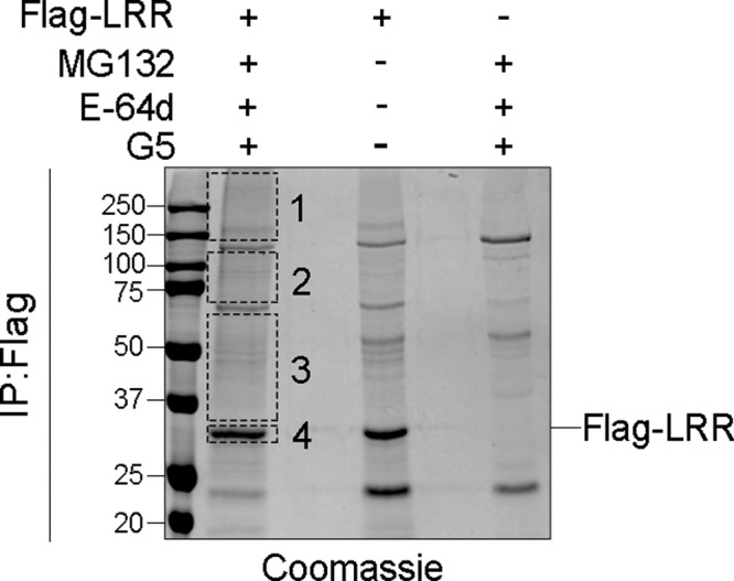
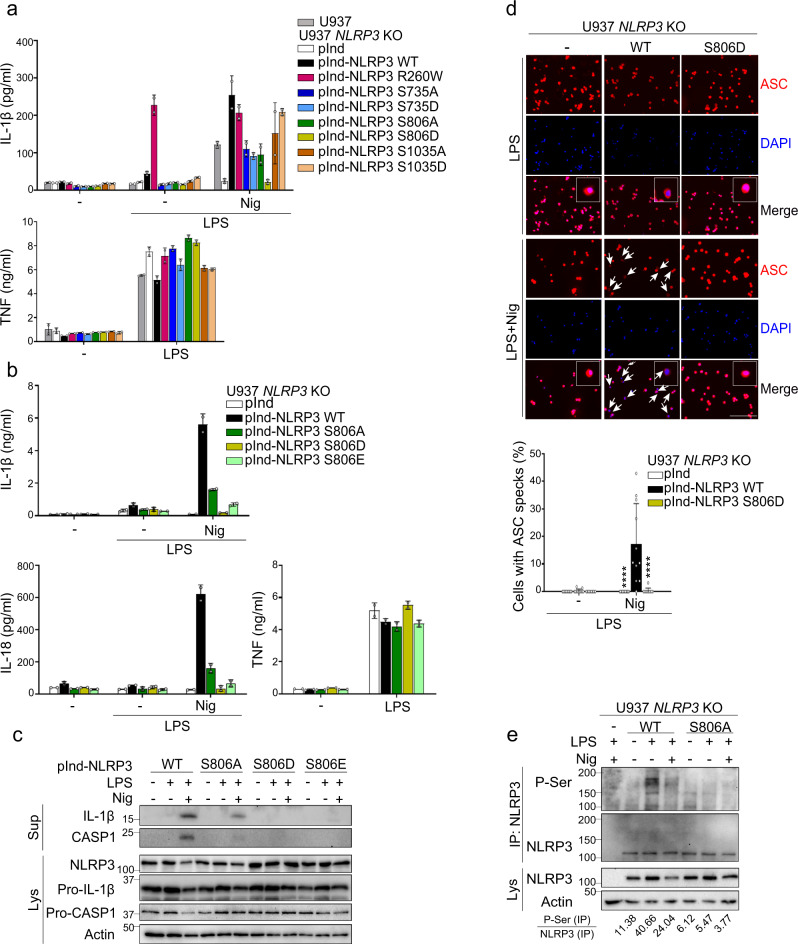
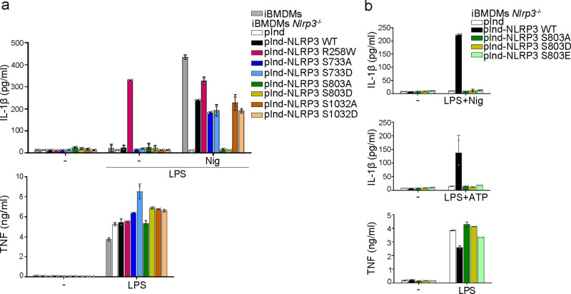
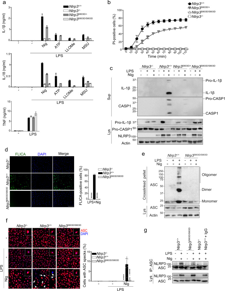
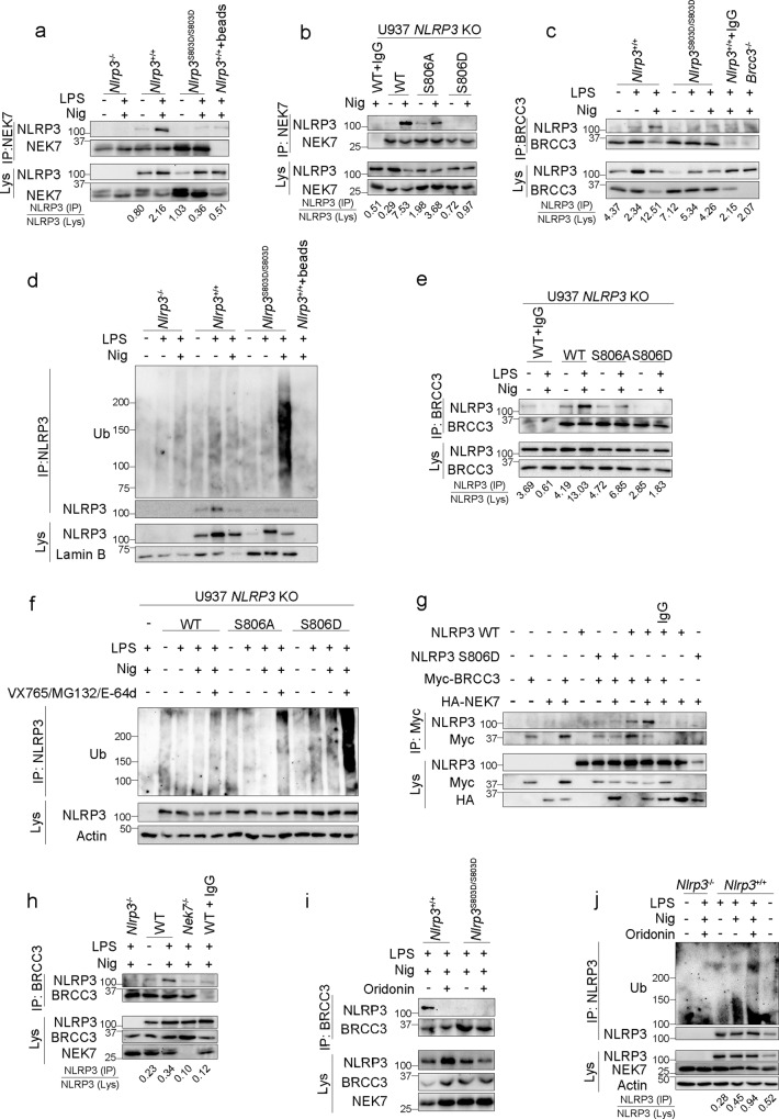
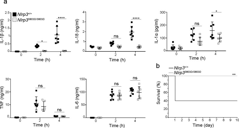
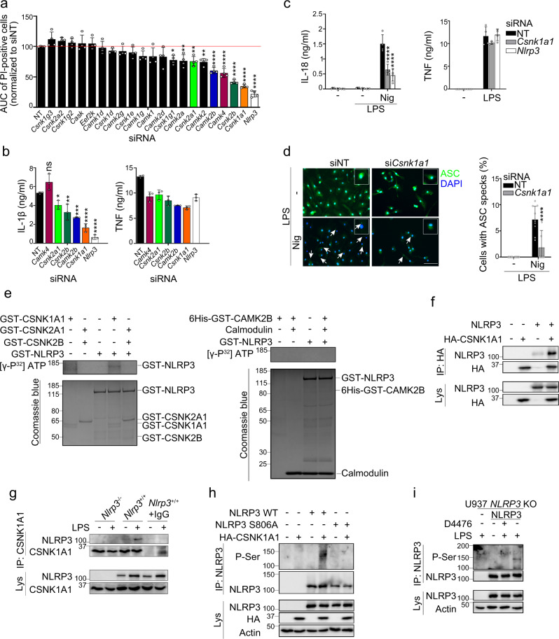
NLRP3 controls the secretion of inflammatory cytokines IL-1β/18 and pyroptosis by assembling the inflammasome. Upon coordinated priming and activation stimuli, NLRP3 recruits NEK7 within hetero-oligomers that nucleate ASC and caspase-1 filaments, but the apical molecular mechanisms underlying inflammasome assembly remain elusive. Here we show that NEK7 recruitment to NLRP3 is controlled by the phosphorylation status of NLRP3 S803 located within the interaction surface, in which NLRP3 S803 is phosphorylated upon priming and later dephosphorylated upon activation. Phosphomimetic substitutions of S803 abolish NEK7 recruitment and inflammasome activity in macrophages in vitro and in vivo. In addition, NLRP3-NEK7 binding is also essential for NLRP3 deubiquitination by BRCC3 and subsequently inflammasome assembly, with NLRP3 phosphomimetic mutants showing enhanced ubiquitination and degradation than wildtype NLRP3. Finally, we identify CSNK1A1 as the kinase targeting NLRP3 S803. Our findings thus reveal NLRP3 S803 phosphorylation status as a druggable apical molecular mechanism controlling inflammasome assembly.


Conflict of interest statement

The authors declare no competing interests.

Figures

Fig. 1
Fig. 1. NLRP3 is ubiquitinated at K878, K927, K973, and phosphorylated at S735, S806, S1035.
293T cells were transfected with plasmids coding for Flag-LRR. One day later, cells were treated with MG132 (10 μM), E-64d (20 μg/ml), and G5 (1 μM) for 30 min. Flag-LRR was purified by anti-Flag immunoprecipitation and analyzed by SDS-PAGE stained with Coomassie solution. Smear corresponding to ubiquitinated Flag-LRR (sections 1–4, excluding major unspecific bands) was analyzed by mass spectrometry. Data correspond to the one selected experiment used to perform the mass spectrometry analysis, out of 8 independent repeats. Molecular weights are indicated in kDa. IP:Flag anti-Flag immunoprecipitates.
Fig. 2
Fig. 2. S806 is critical for NLRP3 inflammasome activity in human U937 monocytes.
U937 and NLRP3-deficient U937 cells reconstituted with doxycycline-inducible NLRP3 mutants were sequentially treated with PMA, doxycycline, LPS, and nigericin as indicated. a IL-1β and TNF secretions were measured by ELISA. NLRP3 R260W was used as a control of constitutively active mutant. b IL-1β, IL-18, and TNF secretions were measured by ELISA. c Expression, cleavage, and secretion of IL-1β and caspase-1 were assessed in cell supernatants and lysates by WB. d ASC specks were visualized and counted by immunofluorescence (arrows, scale bar 50 μm). Cells transduced with empty lentivector were used as control (−). Quantification of >20 cells per replicates is shown (total cells n = 408 pInd, 292 pInd-NLRP3 WT, 377 pInd-NLRP3 S806D). e NLRP3-deficient U937 cells reconstituted with doxycycline-inducible NLRP3 WT or S806A mutant were treated with PMA and doxycycline for 16 h, and then with LPS (5 h) and nigericin (20 min). Anti-NLRP3 immunoprecipitates were analyzed for Serine phosphorylation by WB. Means and 1 SD of biological duplicates (a, b) and 10–11 technical replicates (d) are represented. Data are representative of 2 (c), 3 (a, e), and 4 (b, d) independent experiments. Ordinary two-way ANOVA with Tukey’s multiple comparisons of each condition with corresponding WT control, ****p < 0.0001. Molecular weights are indicated in kDa (c, d). pInd pInducer21, Sup supernatants, Lys lysates, IP:NLRP3 anti-NLRP3 immunoprecipitates.
Fig. 3
Fig. 3. S803 is critical for NLRP3 inflammasome activity in immortalized BMDMs.
a, b Immortalized WT and Nlrp3−/− BMDMs reconstituted with doxycycline-inducible NLRP3 mutants were sequentially treated with doxycycline, LPS, and nigericin or ATP as indicated. IL-1β and TNF secretions were measured by ELISA. NLRP3 R258W was used as a constitutively active control. Means and 1 SD are represented. Data are biological duplicates representative of 4 (a) and 3 (b) independent experiment. pInd pInducer21, iBMDMs immortalized BMDMs.
Fig. 4
Fig. 4. S803 is critical for NLRP3 inflammasome activity in primary BMDMs.
a BMDMs from Nlrp3S803D/S803D, Nlrp3S803D/+ and Nlrp3+/+ littermate mice were primed with LPS followed by nigericin, ATP, LLOMe, or MSU treatments as indicated. IL-1β, IL-18, and TNF secretions in the supernatant were measured by ELISA. BMDMs from Nlrp3−/− mice were used as controls. b Cell death of LPS-primed BMDMs was monitored by PI incorporation over time following nigericin treatment and quantified by high content microscopy. c BMDMs were primed with LPS (6 h) followed by nigericin (30 min) treatment as indicated. Secretion of mature IL-1β and cleaved caspase-1 in the supernatants, and expression of IL-1β, caspase-1, and NLRP3 were assessed by WB. d BMDMs were primed with LPS followed by nigericin treatment. Caspase-1 activity was visualized by FLICA staining and confocal microscopy. Scale bar, 50 μm. Quantification of >600 cells per biological triplicates is shown (total cells n = 1966 Nlrp3−/−, 1833 Nlrp3+/+, 2303 Nlrp3S803D/S803D). e BMDMs were treated with LPS and nigericin (30 min). Crosslinked ASC oligomers in the insoluble fraction of cell lysates were visualized by anti-ASC WB. f ASC subcellular localization was visualized by fluorescent microscopy (ASC, red; DAPI, blue). Arrow, ASC specks. Scale bar, 20 μm. Quantification of >60 cells per replicates is shown (total cells n = 1005 Nlrp3−/−, 656 Nlrp3+/+, 960 Nlrp3S803D/S803D). g BMDMs were treated with LPS (6 h) and nigericin (30 min). Anti-ASC immunoprecipitates from cell lysates were analyzed for NLRP3 by WB. Lysate of Nlrp3+/+ BMDMs incubated with A/G-beads and isotype control was used as a negative control (Nlrp3+/++IgG). Means and 1 SD are represented. Data are biological duplicates representative of three independent experiments (a), biological quadruplicates representative of two independent experiment (b), biological triplicates representative of two independent experiment (d), 10 technical replicates representative of two independent experiments (f), one representative of three (c, g) and four (e) independent experiments. Ordinary two-way ANOVA with Tukey’s multiple comparisons tests of each condition with corresponding WT control; ****p < 0.0001 (d, f). Molecular weights are indicated in kDa (c, e, g). Sup supernatants, Lys lysates, IP:ASC anti-ASC immunoprecipitates.
Fig. 5
Fig. 5. S803D phospho-mimetic mutation blocks NEK7 recruitment by NLRP3.
a Endogenous NEK7 immunoprecipitates from LPS-primed BMDMs treated with nigericin (30 min) were analyzed for NLRP3 (Nlrp3+/++beads, Nlrp3+/+ BMDM lysate incubated with A/G-beads without anti-NEK7). b NLRP3-deficient U937 cells reconstituted with NLRP3 mutants were treated with PMA, doxycycline, LPS, and nigericin. Endogenous NEK7 immunoprecipitates were analyzed for NLRP3 (WT + IgG, lysate of U937 expressing WT NLRP3 incubated with isotype control and A/G-beads). c, d BMDMs were treated with LPS (6 h) and nigericin (30 min). Endogenous BRCC3 immunoprecipitates were analyzed for NLRP3 (Nlrp3+/++IgG, BMDM lysate incubated with IgG isotype control and A-beads; Brcc3−/−, BRCC3 immunoprecipitates from Brcc3−/− BMDM lysate) (c). NLRP3 ubiquitination was assessed by NLRP3 immunoprecipitation followed by anti-Ub WB (Nlrp3+/++beads, Nlrp3+/+ BMDM lysate incubated with A/G-beads without anti-NLRP3) (d). e NLRP3-deficient U937 cells reconstituted with NLRP3 mutants were treated with PMA and doxycycline (2 μg/ml, 16 h) followed by LPS (5 h) and nigericin (20 min). Endogenous BRCC3 immunoprecipitates were analyzed for NLRP3 (WT + IgG, lysate of U937 cells reconstituted with NLRP3 WT incubated with isotype control and A-beads). f NLRP3-deficient U937 cells reconstituted with NLRP3 mutants were treated with PMA and doxycycline (2 μg/ml, 16 h) followed by LPS (50 ng/ml, 4 h) and MG132 (10 μM, 40 min), E-64d (20 μg/ml, 40 min) and VX765 (2.5 μM, 40 min) 10 min before nigericin (30 min). NLRP3 ubiquitination was assessed by NLRP3 immunoprecipitation followed by anti-Ub WB. Caspase-1 inhibitor VX765 was added to prevent pyroptosis. g NLRP3 WT and S806D mutant were expressed with Myc-BRCC3 in 293T cells in the presence or not of HA-NEK7. Myc-BRCC3 immunoprecipitates were analyzed for NLRP3 (IgG, lysate of 293T cells expressing NLRP3 WT, Myc-BRCC3 and HA-NEK7 incubated with isotype control and A/G-beads). h Immortalized WT and Nek7−/− BMDMs were treated with LPS (6 h) and nigericin (30 min) in the presence of VX765 (2.5 μM, 15 min before nigericin). BRCC3 immunoprecipitates were analyzed for NLRP3 by WB (WT + IgG, lysates of immortalized WT BMDMs incubated with isotype control and A-beads). i, j BMDMs were treated with LPS (6 h) and nigericin (30 min) with oridonin (2 μM, 30 min before nigericin). Endogenous BRCC3 immunoprecipitates were analyzed for NLRP3 (i). NLRP3 ubiquitination was assessed by NLRP3 immunoprecipitation followed by anti-Ub WB (j). Data are one representative of three independent experiments. Molecular weights are indicated in kDa. Lys lysates, IP immunoprecipitates, Ub ubiquitin.
Fig. 6
Fig. 6. Nlrp3S803D/S803D KI mice show an impaired response to endotoxic shock in vivo.
Nlrp3S803D/S803D and Nlrp3+/+ littermates were injected intraperitoneally with LPS. a Indicated cytokines were measured in the sera collected before as well as 2 and 4 h post-injection. Nlrp3S803D/S803D and Nlrp3+/+ (n = 7). Individual values, means and 1 SD are represented, repeated measure two-way ANOVA with Sidak’s multiple comparisons of each condition with corresponding WT control; ns, non significant; *p-value < 0.05; ****p-value < 0.0001. b Mice survival to endotoxic shock. Nlrp3S803D/S803D (n = 11), Nlrp3+/+ (n = 10), two-sided Mantel-Cox test, **p-value < 0.01. Data are biological replicates of one experiment.
Fig. 7
Fig. 7. CSNK1A1 phosphorylates NLRP3 on S806.
a BMDMs transfected with the indicated siRNAs were treated with LPS followed by nigericin. Cell death was monitored by PI incorporation over time (for 140 min following nigericin treatment) and quantified by high content microscopy. Means of area under the curve normalized to non-targeting siRNA control and 1 SD are represented. b IL-1β and TNF secretions were measured by ELISA. c, d BMDMs transfected with Csnk1a1 siRNA were treated with LPS (6 h) followed by nigericin. IL-18 and TNF secretions were measured by ELISA (c). ASC specks were visualized and counted by immunofluorescence (d). Arrow, ASC specks. Scale bar, 20 μm. Quantification of >10 cells per replicates is shown (total cells n = 571 NT, 158 Csnk1a1). Means and 1 SD are represented. e GST-NLRP3 was incubated with GST-CSNK1A1, GST-CSNK2A1/GST-CSNK2B, or 6-His-GST-CAMK2B. In vitro phosphorylation was revealed by SDS-PAGE and autoradiography. Coomassie stainings serve as controls. f NLRP3 was ectopically expressed with HA-CSNK1A1 in 293T. HA-CSNK1A1 immunoprecipitates were analyzed for NLRP3. g BMDMs were treated with LPS (6 h). CSNK1A1 immunoprecipitates were analyzed for NLRP3. Lysate of Nlrp3+/+ BMDMs incubated with isotype control and A/G-beads (Nlrp3+/++IgG) and lysates of Nlrp3−/− BMDMs were used as negative controls. h NLRP3 WT or S806A mutant were ectopically expressed with HA-CSNK1A1 in 293 T. NLRP3 immunoprecipitates were analyzed for phospho-Ser by WB. i NLRP3-deficient U937 cells reconstituted with NLRP3 were treated with PMA and doxycycline for 16 h followed by D4476 and LPS (4 h). NLRP3 immunoprecipitates were analyzed for phospho-Ser by WB. Data are biological quadruplicates representative of two independent experiment (a, c), biological triplicates representative of four independent experiment (b), 11 technical replicates representative of two independent experiments (d), one representative of two (e, h, i) and three (f, g) independent experiments. Ordinary one-way ANOVA with Dunnett’s multiple comparisons (a), ordinary two-way ANOVA with Tukey’s multiple comparisons (c, d) to corresponding non-targeting siRNA control; *p-value < 0.05; **p-value < 0.01; ***p-value <0.001; ****p-value < 0.0001. Molecular weights are indicated in kDa (ei). AUC area under the curve, NT non-targeting siRNA, Lys lysates, IP immunoprecipitates.

References

    1. Martinon F, Mayor A, Tschopp J. The inflammasomes: guardians of the body. Annu. Rev. Immunol. 2009;27:229–265. doi: 10.1146/annurev.immunol.021908.132715. - DOI - PubMed
    1. Bauernfeind FG, et al. Cutting edge: NF-kappaB activating pattern recognition and cytokine receptors license NLRP3 inflammasome activation by regulating NLRP3 expression. J. Immunol. 2009;183:787–791. doi: 10.4049/jimmunol.0901363. - DOI - PMC - PubMed
    1. Juliana C, et al. Non-transcriptional priming and deubiquitination regulate NLRP3 inflammasome activation. J. Biol. Chem. 2012;287:36617–36622. doi: 10.1074/jbc.M112.407130. - DOI - PMC - PubMed
    1. Py BF, Kim MS, Vakifahmetoglu-Norberg H, Yuan J. Deubiquitination of NLRP3 by BRCC3 critically regulates inflammasome activity. Mol. Cell. 2013;49:331–338. doi: 10.1016/j.molcel.2012.11.009. - DOI - PubMed
    1. Tang, J. et al. Sequential ubiquitination of NLRP3 by RNF125 and Cbl-b limits inflammasome activation and endotoxemia. J. Exp. Med. 217, e20182091 (2020). - PMC - PubMed

Publication types

MeSH terms