Review
. 2021 Sep 20:12:675528.
doi: 10.3389/fmicb.2021.675528. eCollection 2021.

Spread of Mink SARS-CoV-2 Variants in Humans: A Model of Sarbecovirus Interspecies Evolution

Affiliations
Review

Spread of Mink SARS-CoV-2 Variants in Humans: A Model of Sarbecovirus Interspecies Evolution

Christian A Devaux et al. Front Microbiol. .

Abstract

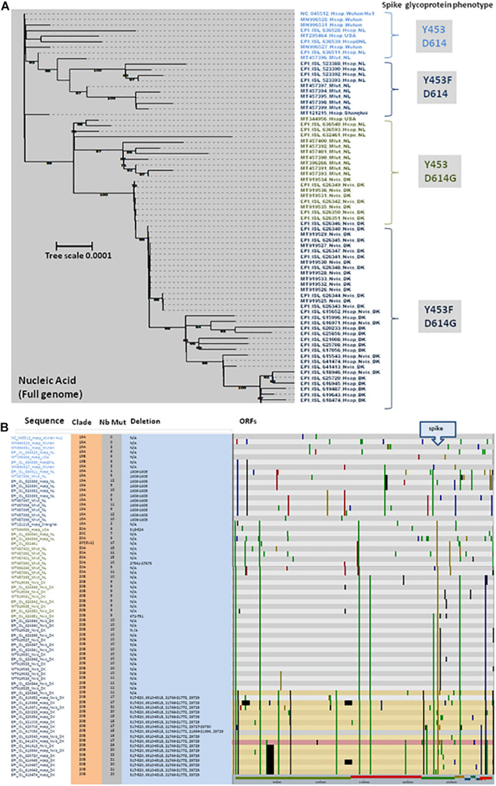
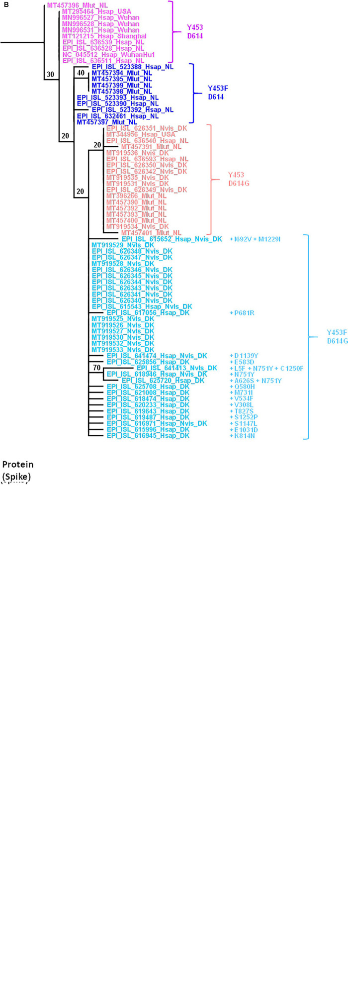
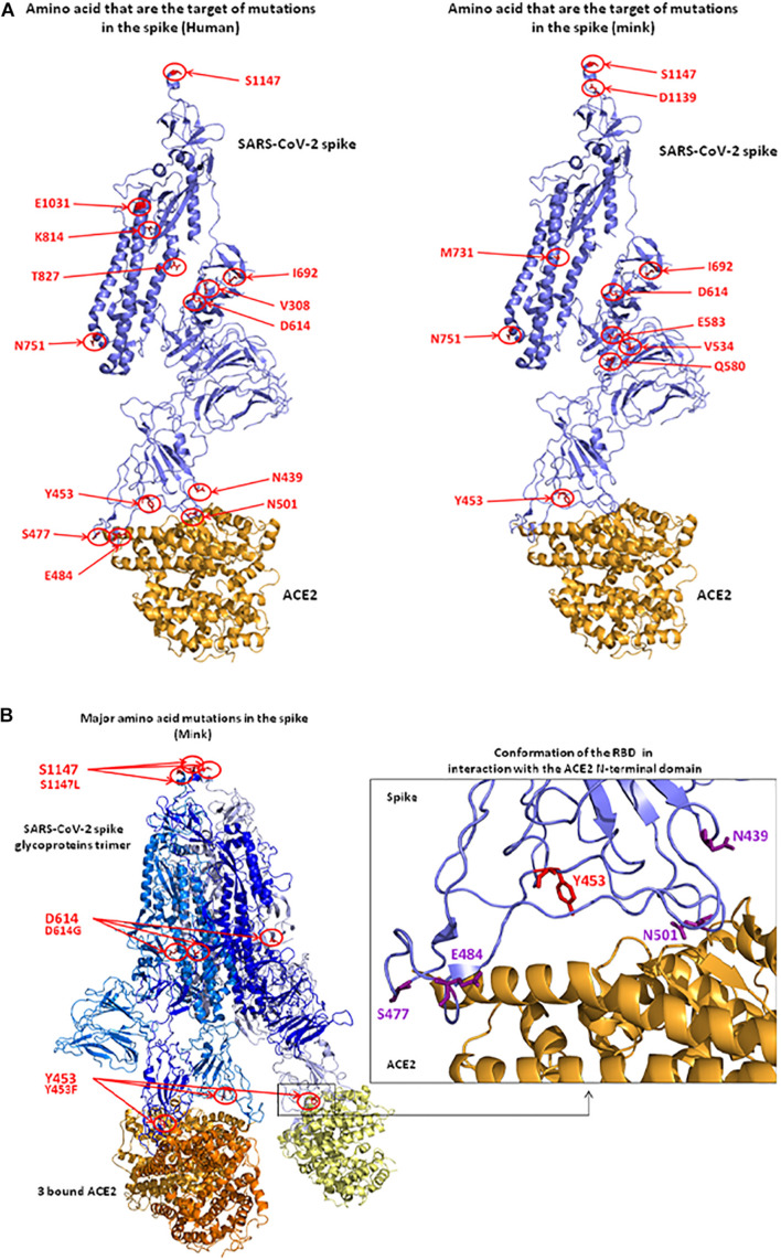
The rapid spread of SARS-CoV-2 variants has quickly spanned doubts and the fear about their ability escape vaccine protection. Some of these variants initially identified in caged were also found in humans. The claim that these variants exhibited lower susceptibility to antibody neutralization led to the slaughter of 17 million minks in Denmark. SARS-CoV-2 prevalence tests led to the discovery of infected farmed minks worldwide. In this study, we revisit the issue of the circulation of SARS-CoV-2 variants in minks as a model of sarbecovirus interspecies evolution by: (1) comparing human and mink angiotensin I converting enzyme 2 (ACE2) and neuropilin 1 (NRP-1) receptors; (2) comparing SARS-CoV-2 sequences from humans and minks; (3) analyzing the impact of mutations on the 3D structure of the spike protein; and (4) predicting linear epitope targets for immune response. Mink-selected SARS-CoV-2 variants carrying the Y453F/D614G mutations display an increased affinity for human ACE2 and can escape neutralization by one monoclonal antibody. However, they are unlikely to lose most of the major epitopes predicted to be targets for neutralizing antibodies. We discuss the consequences of these results for the rational use of SARS-CoV-2 vaccines.

Keywords: ACE2; COVID-19; COVID-19 vaccines; NRP-1; SARS-CoV-2; mink coronavirus; variant viruses.


Conflict of interest statement

The authors declare that the research was conducted in the absence of any commercial or financial relationships that could be construed as a potential conflict of interest.

Figures

FIGURE 1
FIGURE 1
Schematic representation of the experimental infection of ferrets and minks by SARS-CoV-2. All naïve animals became febrile and most collected specimens were positive for viral RNA. The isolation of infectious viral particles was sometimes possible from nasal washes. Transmission to naïve animals was observed following direct contact with infected animals (close but in different cages). Clinical sign and histological abnormalities are summarized.
FIGURE 2
FIGURE 2
The mink fur industry. Mink stocks according to reports from the European Commission (ECDC, 2020), the fur industry (Fur Europe, 2015) and associations. China, Denmark, Poland, and Netherlands were the main producers. Red box: 10–21 million minks/year; orange: 2–10 million minks/year; green: 1–2 million minks/year; and white: below 1 million. Mass culling of minks ordered by governments after SARS-CoV-2 outbreaks in mink farms in Europe: Denmark: 17,000,000; Netherlands: 900,000; Ireland: 100,000; Spain: 92,700; Italy: 28,000; Greece: 2,500; and France: 1,000. The number of mink farms in Europe [adapted from a report from the ECDC (2020)] is indicated by blue circles (it is possible to estimate the risk of cross contamination in mink farms as a function of the density of animal populations). Fur-farming ban (year): United Kingdom 2000; Austria 2004; Croatia 2006; Serbia 2009; Slovenia and New Zealand 2013; Japan 2016; Macedonia 2017; Luxembourg, the Czech Republic and Norway 2018; and Germany and Slovakia 2019. Scheduled shutdown of mink farms: Belgium and Denmark 2023; Netherlands: 2024 (early closure 2021); France: 2025; and Bosnia and Herzegovina 2027. Outside Europe, in China in 2018 farms were mainly concentrated in the northern provinces (Shandong, Liaoning, Heilongjiang, Jilin, and Henan), and produced 20.7 million minks. In North American, 245 farms produced 3.1 million pelts in the United States and 60 farms in Canada produced 1.7 million pelt. In Russia, 22 farms produced 1.2 million minks.
FIGURE 3
FIGURE 3
Schematic representation of the SARS-CoV-2 spread in mink farms. Infected animals reduced feed intake and lost body weight. The virus expansion in mink farms was favored by intensive breeding conditions with significant overcrowding in poorly ventilated rooms. Minks (M. lutreola in Netherlands and N. vison in Denmark) infected with SARS-CoV-2 were found to be either asymptomatic or to display signs of respiratory diseases (several animals died from interstitial pneumonia or sepsis). For fear of seeing minks-selected SARS-CoV-2 variants spreading more easily among people, of being more deadly, or having a negative impact on the deployment of anti-COVID-19 vaccines (interfering with vaccine effectiveness in humans), the Danish government decided to cull 17 million minks in more than 1,000 farms (Koopman, 2020; Larsen and Paludan, 2020; Oude Munnink et al., 2021). Countries who reported SARS-CoV-2 infected minks in farms, i.e., Netherlands, Ireland, Greece, Spain, Italy, France, United States, also started a mass slaughter.
FIGURE 4
FIGURE 4
Mink ACE2 sequences alignment. (A) Clustal Omega multiple sequence alignment (EMBL-EBI bioinformatic tool; Copyright EMBL 2020) was used to compare the ACE2 protein sequences of H. sapiens (Hsap) and minks N. vison (Nvis) and M. lutreola (Mlut). The amino acids that differ between human and minks are shown in red and those which differ between minks are shown in yellow. Some of the amino acids important for viral tropism are indicated in blue (previous studies showed that amino acid residues 31, 41, 90, and 353 are important for viral spike binding). (B) 3D model of ACE2 designed using the Phyre2 server (this model lacks the cytoplasmic tail of ACE2). An electrostatic potential surface (red: negative charge; blue: positive charge) was generated using PyMOL 1.8.0 and APBS tool plugin. The location of amino acids 31, 41, 90, and 353 is indicated by arrows.
FIGURE 5
FIGURE 5
Comparison between H. sapiens and different Mustelidae ACE2 proteins. (A) A schematic representation of the cell surface of the Hsap ACE2 molecule and its major domains is shown on the left side of the figure while the representation of the Mustelidae ACE2 is displayed on the right side of the figure. Amino acid changes compared to the human sequence are listed. The amino acid position is shown in black. The most important amino acids for SARS-CoV-2 interaction are in red. S, sugar; P, phosphorylation. The comparison of the H. sapiens (Hsap) ACE2 with five different ACE2 sequences from Mustelidae, i.e., N. vison (Nvis), M. nigripes (Mnig), M. putorius furo (Mput), M. lutreola (Mlut), M. erminea (Merm) was performed using Clustal Omega. The figure illustrates the homology (white or red) and difference (light blue or deep blue when there is a difference in amino acid usage between Mustelidae) in the three regions (30–42, 82–94, and 353–358) involved in SARS-CoV-2 binding (middle panel). (B) Local prediction of the 82–94 amino acids peptide secondary structure using the Phyre2 server and three-dimensional folding using PeP-FOLD algorithm.
FIGURE 6
FIGURE 6
(A) Maximum likelihood phylogenetic tree of different genomic SARS-CoV-2 sequences. Sample names are built with the GISAID or GenBank accession number followed by a four-letter code (Mlut, Nvis, and Hsap) identifying the species followed by a code indicating the geographical origin of the sample (Wuhan: Wuhan, China; NL: Netherlands; DK: Denmark; and USA: United States). This analysis compares the full genome sequence from minks (Mlut: M. lutreola or Nvis: N. vison) and human (Hsap). The main spike phenotype (amino acid substitution) of each group of SARS-CoV-2 is indicated on right side of the figure. Four different groups where identified: Y453/D614, Y453F/D614, Y453/D614G, and Y453F/D614G. (B) Location of genomic mutations and deletions on the complete nucleotide sequence from different isolates of SARS-CoV-2. Color code: red, mutation to A; blue, mutation to C; green, mutation to T; yellow, mutation to G; light gray, deletion; and dark gray, not covered. Nb Mut, number of mutations; ORFs, open reading frames.
FIGURE 7
FIGURE 7
(A) Maximum likelihood (GTR+R) phylogenetic tree of the SARS-CoV-2 spike gene from different isolates. Sample names are built with the GISAID or GenBank accession number followed by a four-letter code (Mlut, Nvis, and Hsap) identifying the species followed by a code indicating the geographical origin of the sample. The tree was rooted using the spike gene and protein sequence of the Wuhan Hu1 SARS-CoV-2 strain. (B) Maximum likelihood (LG) phylogenetic tree of SARS-CoV-2 spike glycoprotein from the different isolates.
FIGURE 8
FIGURE 8
3D structure of the SARS-CoV-2 spike protein complexed with ACE2. (A) Positions of observed mutant residues in human (left) and mink (right) SARS-CoV-2 spike proteins. In the published 3D structure (PDB: 7A98) (Benton et al., 2020), the spike glycoprotein is colored in blue and the bound ACE2 receptor is colored in orange. The main mutant amino acids are colored in red and their positions are indicated with red circles and arrows. (B) Representation of the SARS-CoV-2 spike trimer (SARS-CoV-2 sequence from mink) in interaction with ACE2 molecules (left). Representation of the conformation of RBD in interaction with the N-terminal domain of ACE2. The Y453 amino acid that was changed to F453 after the infection of minks (right).
FIGURE 9
FIGURE 9
Comparative antigenicity of human and mink SARS-CoV-2. (A) Schematic representation of the SARS-CoV-2 spike protein domains. SP, signal peptide; NTD, N-terminal domain; RBD, receptor (ACE2) binding domain; TM, transmembrane domain. (B) Prediction of protein hydrophobicity according to Kite and Doolittle using the ProtScale program. The score of hydrophobicity is indicated. (C) SVMTrip algorithm prediction of linear antigenic epitopes (12 amino acids scan) in the spike protein of SARS-CoV-2 (NC_045512_Hsap_WuhanHu1 isolate versus EPI_ISL_616971_Hsap_Nvis_DK). The location of the predicted linear epitopes is indicated as well as the score of antigenicity (1.000 being the highest score). The stars indicate the mutations found in the mink SARS-CoV-2 variant EPI_ISL_616971_Hsap_Nvis_DK. (D) Multiple sequence alignment of the first 960 amino acids of the spike protein sequence (S1 and the proximal S2) from the NC_045512_Hsap_WuhanHu1 isolate versus EPI_ISL_616971_Hsap_Nvis_DK and EPI_ISL_641413_Nvis_DK samples and location of the predicted linear B-cell epitopes. The amino acids differing between the three sequences are highlighted in yellow. The location of the variants Y453F and D164G is shown in red. NTD, N terminal domain; RBD, receptor binding domain; FP, fusion peptide.

Similar articles

Cited by

References

    1. Afelt A., Devaux C. A., Serra-Cobo J., Frutos R. (2018). Bats, bat-borne viruses, and environmental changes. IntechOpen chapter 8 113–131. 10.5772/intechopen.74377 - DOI
    1. Ahmed S. F., Quadeer A. A., McKay M. R. (2020). Preliminary identification of potential vaccine targets for the COVID-19 coronavirus (SARS-CoV-2) based on SARS-CoV immunological studies. Viruses 12:254. 10.3390/v12030254 - DOI - PMC - PubMed
    1. Andersen K. G., Rambaut A., Lipkin W. I., Holmes E. C., Garry R. F. (2020). The proximal origin of SARS-CoV-2. Nat. Med. 26 450–452. 10.1038/s41591-020-0820-9 - DOI - PMC - PubMed
    1. Baum A., Fulton B. O., Wloga E., Copin R., Pascal K. E., Russo V., et al. (2020). Antibody cocktail to SARS-CoV-2 spike protein prevents rapid mutational escape seen with individual antibodies. Science 369 1014–1018. 10.1126/science.abd0831 - DOI - PMC - PubMed
    1. Bazykin G. A. S., Danilenko D., Fadeev A., Komissarova K., Ivanova A., Sergeeva M., et al. (2021). Emergence of Y453F and D69-70HV Mutations In A Lymphoma Patient With Long-Term COVID-19.(2021). Available online at: https://virological.org/t/emergence-of-y453f-and-69-70hv-mutations-in-a-.... (accessed June 2021).