. 2021 Sep 20:12:719562.
doi: 10.3389/fimmu.2021.719562. eCollection 2021.

Interleukin-17A Drives IL-19 and IL-24 Expression in Skin Stromal Cells Regulating Keratinocyte Proliferation

Affiliations

Interleukin-17A Drives IL-19 and IL-24 Expression in Skin Stromal Cells Regulating Keratinocyte Proliferation

Xiaofei Xu et al. Front Immunol. .

Abstract

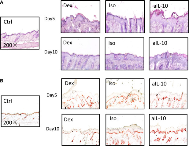
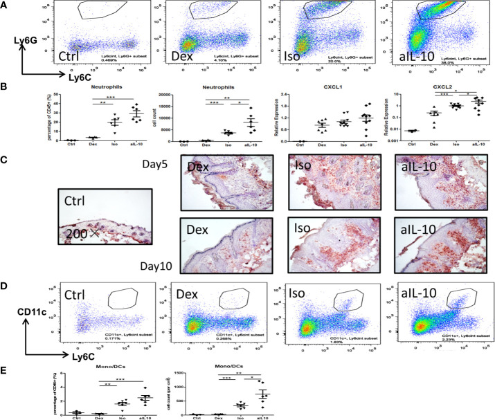
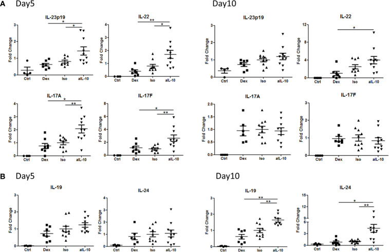
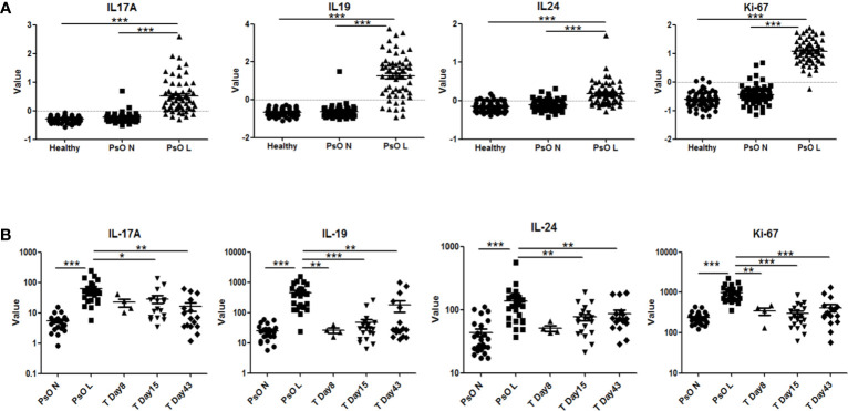
IL-17A has been shown to be up-regulated in psoriasis lesions and is central to psoriasis pathogenesis. IL-19, along with other IL-20 subfamily cytokines such as IL-20 and IL-24, is induced by IL-17A and contributes especially to epidermal hyperplasia in psoriasis. However, the regulation, cellular sources of IL-19 and whether targeting of IL-17A by biologics influence IL-19 expression is not completely understood. To investigate the regulation of IL-19 by IL-17A in psoriasis, the imiquimod-induced psoriasis mouse (IMQ) model was used. Enhanced expression of IL-17A in the IMQ model was achieved by anti-IL-10 antibody treatment. Assessments of skin inflammation macroscopically, by histology and flow cytometry, all confirmed increased psoriatic symptoms. Interestingly, depletion of IL-10 markedly upregulated IL-23/IL-17 pathway related cytokines followed by a significant increase in IL-19 and IL-24. The up-regulation of IL-19 and IL-24, but not IL-17A, coincided with increased keratinocyte proliferation. To investigate the cellular source and effects of biologics on IL-19, human skin fibroblasts from healthy controls and psoriasis patients were cultured alone or co-cultured with activated memory CD4+ T cells. Besides IL-1β, IL-17A induced direct expression of IL-19 and IL-24 in skin fibroblasts and keratinocytes. Importantly, intrinsic higher expression of IL-19 in psoriatic skin fibroblasts was observed in comparison to healthy skin fibroblasts. Neutralization of IL-17A in the human skin fibroblast-T cell co-culture system significantly suppressed IL-19 and IL-24 expression. Together, our data show that IL-17A-induced IL-19 and IL-24 expression in skin stromal cells contribute to keratinocyte proliferation.

Keywords: IL-17A; Th17; cytokines; inflammation; psoriasis; skin.


Conflict of interest statement

The authors declare that the research was conducted in the absence of any commercial or financial relationships that could be construed as a potential conflict of interest.

Figures

Figure 1
Figure 1
IL-10 neutralization worsens psoriatic symptoms and epidermal thickness in the IMQ-induced psoriasis mouse model. (A) At day 10, scores for skin scaling, thickness, redness and PASI in various groups following IMQ treatment. (B) Measured average epidermal thickness in psoriasis-like skin at days 5 and 10, and thickness ratios compared to isotype antibody controls at days 5 and 10. Data are shown as means ± SEMs. *P < 0.05, **P < 0.01, and ***P < 0.001.
Figure 2
Figure 2
IL-10 neutralization increases keratinocyte proliferation and neutrophil accumulation in the IMQ-induced psoriasis mouse model. (A) Representative H&E staining results of skin sections in IMQ-induced psoriasis mouse model at days 5 and 10. (B) Representative IHC staining of Ki-67+ proliferating keratinocytes in skin sections of IMQ-induced psoriasis mouse model at days 5 and 10. All images were taken with 200× magnification.
Figure 3
Figure 3
IL-10 neutralization increases neutrophil and monocyte/DC infiltration in the IMQ-induced psoriasis mouse model. (A) Flow cytometry staining of Ly6G+Ly6Cint neutrophils among pregated CD45+CD11b+ cells in lesional psoriasis-like skin at day10. (B) Percentages and cell numbers of neutrophils among total CD45+ immune cells at day 10 in flow cytometry analysis and levels of the neutrophil chemokines, CXCL1 and CXCL2, in lesional psoriasis-like skin. (C) Representative IHC staining of Gr-1+ neutrophils in skin sections of the IMQ-induced psoriasis mouse model at days 5 and 10. (D) Flow cytometry staining of CD11c+Ly6Cint monocyte-derived dendritic cells in lesional psoriasis-like skin at day10. (E) Percentages and cell numbers of monocyte-derived dendritic cells (mono/DCs) among total CD45+ immune cells at day 10 using flow cytometry. Data are shown as means ± SEMs. *P < 0.05, **P < 0.01, and ***P < 0.001.
Figure 4
Figure 4
Early and late up-regulation of IL-23/IL-17 cytokines and IL-20 subfamily cytokines in the skin during anti-IL-10 treatment in IMQ-induced psoriasis mouse model. (A) At days 5 and 10, mRNA expression of IL-23, IL-22, IL-17A and IL-17F in various groups following IMQ treatment. (B) At days 5 and 10, mRNA expression of IL-19 and IL-24 in various groups following IMQ treatment. Data are shown as means ± SEMs. *P < 0.05, **P < 0.01.
Figure 5
Figure 5
IL-17A induces IL-19 and IL-24 expression in human skin resident cells such as fibroblasts and keratinocytes, and IL-17A blockade reduces their expression. (A) IL-19 and IL-24 mRNA expression in fibroblasts of healthy donors (n=6) and psoriasis patients (n=4) without stimulation or stimulated with IL-1β, TNFα, IL-17A and IL-17F for 24 hours. (B) Healthy control skin fibroblasts and psoriatic fibroblasts (n=6 each) were co-cultured with unstimulated or anti-CD3/anti-CD28 stimulated memory CD4+ T cells from healthy controls for 72 hours. IL-19 and IL-24 mRNA expression in these co-cultures without treatment or treated with an anti-IL-17A antibody, anti-TNF antibody, their combination or an isotype control antibody. (C) Levels of human IL-19 and IL-8 protein in the supernatant of the above mentioned co-cultures without treatment or treated with an anti-IL-17A antibody, anti-TNF antibody, their combination or an isotype control antibody. Data are shown as means ± SEMs. *P < 0.05, **P < 0.01, and ***P < 0.001.
Figure 6
Figure 6
Expression of human IL-17A, IL-19, IL-24 and Ki-67 in psoriatic lesions and the effects of anti-IL-17RA therapy. (A) Expression of IL-17A, IL-19, IL-24 and Ki-67 in lesional and non-lesional skin biopsies from 58 psoriasis patients and in healthy skin biopsies from 64 normal controls in data set GSE13355. (B) Expression of IL-17A, IL-19, IL-24 and Ki-67 in lesional and non-lesional skin biopsies from 25 psoriasis patients before anti-IL-17RA treatment, 8, 15 and 43 days after treatments in data set GSE53552. Data are shown as means ± SEMs. *P < 0.05, **P < 0.01, and ***P < 0.001.

References

    1. Nestle FO, Kaplan DH, Barker J. Psoriasis. N Engl J Med (2009) 361:496–509. doi: 10.1056/NEJMra0804595 - DOI - PubMed
    1. Greb JE, Goldminz AM, Elder JT, Lebwohl MG, Gladman DD, Wu JJ, et al. . Psoriasis. Nat Rev Dis Primers (2016) 2:16082. doi: 10.1038/nrdp.2016.82 - DOI - PubMed
    1. Griffiths CE, Barker JN. Pathogenesis and Clinical Features of Psoriasis. Lancet (2007) 370:263–71. doi: 10.1016/S0140-6736(07)61128-3 - DOI - PubMed
    1. Nickoloff BJ. The Cytokine Network in Psoriasis. Arch Dermatol (1991) 127:871–84. doi: 10.1001/archderm.1991.01680050115015 - DOI - PubMed
    1. Johansen C, Usher PA, Kjellerup RB, Lundsgaard D, Iversen L, Kragballe K. Characterization of the Interleukin-17 Isoforms and Receptors in Lesional Psoriatic Skin. Br J Dermatol (2009) 160:319–24. doi: 10.1111/j.1365-2133.2008.08902.x - DOI - PubMed

Publication types

MeSH terms