

Neonatal biliary atresia combined with preduodenal portal vein: A case report
- PMID: 34616824
- PMCID: PMC8464463
- DOI: 10.12998/wjcc.v9.i25.7542
Neonatal biliary atresia combined with preduodenal portal vein: A case report
Abstract
Background: Congenital biliary atresia is a type of obstruction of the bile ducts inside and outside the liver, which can lead to cholestatic liver cirrhosis and eventually liver failure. The preduodenal portal vein (PD-PV) is a rare developmental malformation of the PV. The PV courses in front of the duodenum. However, very few cases of neonatal biliary atresia combined with PD-PV have been reported in the scientific literature.
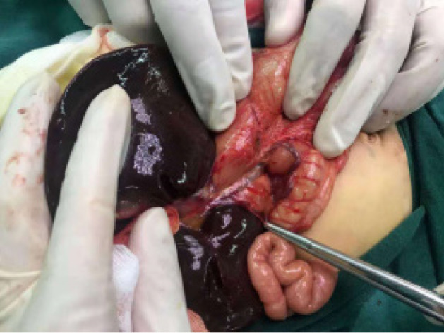
Case summary: A 1-mo-and-4-d-old child was admitted to the hospital in January because of yellowish skin. After surgical consultation, surgical intervention was recommended. The child underwent Hilar-jejunal anastomosis, duodenal rhomboid anastomosis, and abdominal drainage under general anesthesia. During the operation, the PV was located at the anterior edge of the duodenum.
Conclusion: Diagnoses: (1) Congenital biliary atresia; (2) PD-PV; and (3) Congenital cardiovascular malformations. Outcomes: Recommendation for liver transplantation. Lessons: The choice of treatment options for neonatal biliary atresia combined with PD-PV.
Keywords: Biliary atresia; Case report; Neonatal; Preduodenal portal vein; Treatment.
©The Author(s) 2021. Published by Baishideng Publishing Group Inc. All rights reserved.
Conflict of interest statement
Conflict-of-interest statement: The authors have no conflicts of interest to disclose.
Figures

References
-
- Petersen C, Ure BM. What's new in biliary atresia? Eur J Pediatr Surg. 2003;13:1–6. - PubMed
-
- Nio M, Ohi R, Miyano T, Saeki M, Shiraki K, Tanaka K Japanese Biliary Atresia Registry. Five- and 10-year survival rates after surgery for biliary atresia: a report from the Japanese Biliary Atresia Registry. J Pediatr Surg. 2003;38:997–1000. - PubMed
-
- Kouwenberg M, Kapusta L, van der Staak FH, Severijnen RS. Preduodenal portal vein and malrotation: what causes the obstruction? Eur J Pediatr Surg. 2008;18:153–155. - PubMed
-
- Singal AK, Ramu C, Paul S, Matthai J. Preduodenal portal vein in association with midgut malrotation and duodenal web-triple anomaly? J Pediatr Surg. 2009;44:e5–e7. - PubMed
Publication types
LinkOut - more resources
Full Text Sources
