

Review of imaging biomarkers for the vulnerable carotid plaque
- PMID: 34617065
- PMCID: PMC8489200
- DOI: 10.1016/j.jvssci.2021.03.001
Review of imaging biomarkers for the vulnerable carotid plaque
Abstract
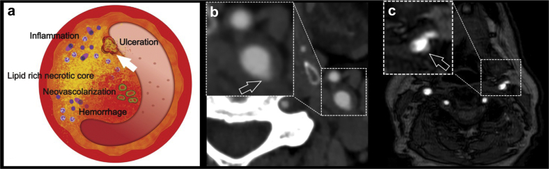
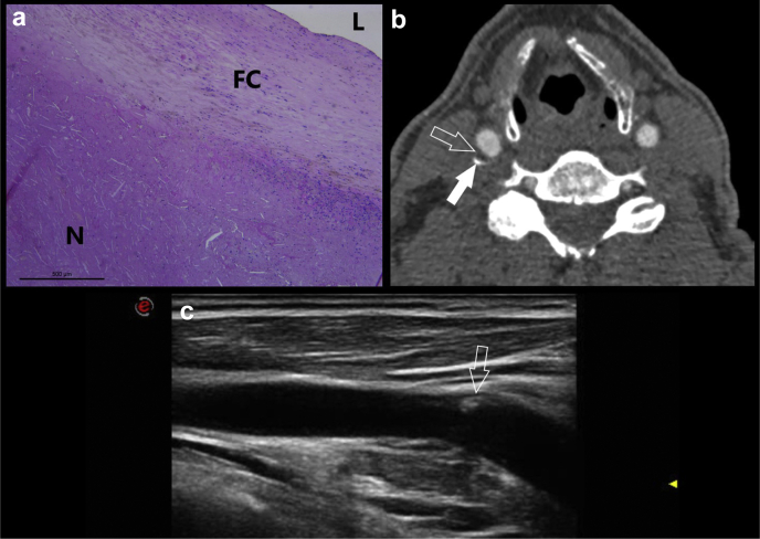
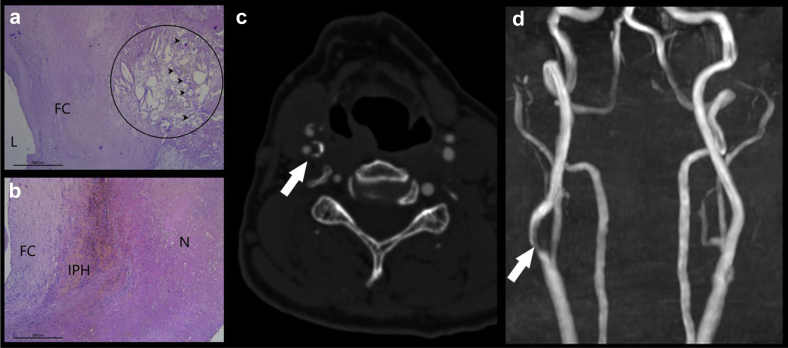
Identification of carotid artery atherosclerosis is conventionally based on measurements of luminal stenosis. However, histopathologic studies demonstrate considerable differences between plaques with identical degrees of stenosis and indicate that certain plaque features are associated with increased risk for ischemic events. As a result of the rapid technological evolution in medical imaging, several important steps have been taken in the field of carotid plaque imaging allowing us to visualize the carotid atherosclerotic plaque and its composition in great detail. For computed tomography, magnetic resonance imaging, positron emission tomography, and ultrasound scan, evidence has accumulated on novel imaging-based markers that confer information on carotid plaque vulnerability, such as intraplaque hemorrhage and lipid-rich necrotic cores. In terms of the imaging-based identification of individuals at high risk of stroke, routine assessments of such imaging markers are the way forward for improving current clinical practice. The current review highlights the main characteristics of the vulnerable plaque indicating their role in the etiology of ischemic stroke as identified by intensive plaque imaging.
Keywords: Carotid; Plaque; Stenosis.
© 2021 by the Society for Vascular Surgery. Published by Elsevier Inc.
Figures
References
-
- Katan M., Luft A. Global burden of stroke. Semin Neurol. 2018;38:208–211. - PubMed
-
- Kumamaru H., Jalbert J.J., Nguyen L.L., Gerhard-Herman M.D., Williams L.A., Chen C.-Y. Surgeon case volume and 30-day mortality after carotid endarterectomy among contemporary Medicare beneficiaries: before and after national coverage determination for carotid artery stenting. Stroke. 2015;46:1288–1294. - PubMed
-
- Abbott A.L., Adelman M.A., Alexandrov A.V., Barber P.A., Barnett H.J.M., Beard J. Why calls for more routine carotid stenting are currently inappropriate: an international, multispecialty, expert review and position statement. Stroke. 2013;44:1186–1190. - PubMed
Publication types
LinkOut - more resources
Full Text Sources
Research Materials
