. 2021 Nov 16;50(44):16053-16066.
doi: 10.1039/d1dt02763h.

Liposomal formulations of anticancer copper(II) thiosemicarbazone complexes

Affiliations

Liposomal formulations of anticancer copper(II) thiosemicarbazone complexes

Marlene Mathuber et al. Dalton Trans. .

Abstract

α-N-Heterocyclic thiosemicarbazones such as triapine and COTI-2 are currently investigated as anticancer therapeutics in clinical trials. However, triapine was widely inactive against solid tumor types. A likely explanation is the short plasma half-life time and fast metabolism. One promising approach to overcome these drawbacks is the encapsulation of the drug into nanoparticles (passive drug-targeting). In a previous work we showed that it was not possible to stably encapsulate free triapine into liposomes. Hence, in this manuscript we present the successful preparation of liposomal formulations of the copper(II) complexes of triapine and COTI-2. To this end, various drug-loading strategies were examined and the resulting liposomes were physico-chemically characterized. Especially for liposomal Cu-triapine, a decent encapsulation efficacy and a slow drug release behavior could be observed. In contrast, for COTI-2 and its copper(II) complex no stable loading could be achieved. Subsequent in vitro studies in different cell lines with liposomal Cu-triapine showed the expected strongly reduced cytotoxicity and DNA damage induction. Also in vivo distinctly higher copper plasma levels and a continuous release could be observed for the liposomal formulation compared to free Cu-triapine. Taken together, the here presented nanoformulation of Cu-triapine is an important step further to increase the plasma half-life time and tumor targeting properties of anticancer thiosemicarbazones.


Conflict of interest statement

The authors declare no competing interest.

Figures

Fig. 1
Fig. 1. Chemical structures of the clinically investigated thiosemicarbazones triapine and COTI-2 as well as their copper(ii) complexes.
Scheme 1
Scheme 1. Preparation of the liposomal formulations by addition of the drug at the beginning of the synthetic procedure.
Scheme 2
Scheme 2. Preparation of the liposomal formulations by the remote-loading approach.
Fig. 2
Fig. 2. (A) Size distribution of l-Cu–Tria (by intensity) measured by DLS (each line represents measurements in triplicate). (B) Transmission electron microscopy (TEM) image of l-Cu–Tria; samples were prepared by negative staining with Uranyless.
Fig. 3
Fig. 3. Cellular copper levels of SW480 cells measured by ICP-MS after treatment with empty liposomes, CuSO4, triapine, Cu–Tria or l-Cu–Tria at the indicated concentrations for 3 h. Values given are the mean ± standard deviation (SD) of triplicates. Significance to control (stars above bars) and to other treatments (stars above brackets between bars) was calculated by one-way ANOVA and Tukey's multiple comparison test (* p < 0.05; ** p < 0.01; **** p < 0.0001).
Fig. 4
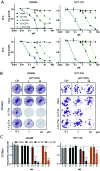
Fig. 4. (A) Cell viability of SW480 and HCT-116 cells treated with free complexes, drug-free or -loaded liposomes measured by MTT assay after 48 and 72 h. Values given in the graph are the mean ± SD of triplicates from one representative experiment out of three. (B) Cell viability after long-term treatment of SW480 and HCT-116 cells with CuSO4, free complexes, drug-free or -loaded liposomes. After 10 days, cells were fixed and stained with crystal violet. Representative images of stained cells (violet) are shown. Fold growth to untreated cells was calculated from integrated density analyzed with Image J and shown in (C). Values given are the mean ± SD of duplicates from three experiment. Significance to control was calculated in with two-way ANOVA and Dunnett's multiple comparison test (**** p < 0.0001, * p ≤ 0.05).
Fig. 5
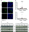
Fig. 5. (A) Representative images of pH2AX immunofluorescence staining (green) in nuclei (DAPI, blue) of SW480 cells treated as indicated. (B) Quantification of immunofluorescence intensities in the nucleus of the DNA damage marker pH2AX in SW480 and HCT-116 cells treated with the indicated drugs and concentrations for 24 h. Values given are the mean fold intensities ± SD per nucleus. Significance to control was calculated with two-way ANOVA and Dunnett's multiple comparison test (**** p < 0.0001, ** p ≤ 0.01). (C) Western blot analysis of pH2AX expressed by SW480 and HCT-116 cells treated with indicated drugs and concentrations 24 h and 7 days. β-Actin was used as a loading control. Normalized pH2AX values to β-actin and compared to control are given below the bands.
Fig. 6
Fig. 6. Serum copper levels of female Balb/c mice treated i.v either with 1.75 mg kg−1 Cu–Tria or l-Cu–Tria and blood drawn via the facial vein at the indicated time points. Samples for baseline were collected from the same mice 7 days prior drug treatment. Values given are mean ± standard error of the mean (SEM) and significance was calculated by Kruskal–Wallis test against the baseline with false discovery rate by Benjamini and Hochberg (* p < 0.05; ** p < 0.01).
Fig. 7
Fig. 7. Time resolved UV-Vis spectroscopy of the reaction between human HbO2 and (A) triapine and (B) l-Cu–Tria in the presence of iron(iii) during 21 min (inset zoom of the range 500–650 nm).

References

    1. Lobana T. S. Sharma R. Bawa G. Khanna S. Bonding and structure trends of thiosemicarbazone derivatives of metals—an overview. Coord. Chem. Rev. 2009;253(7–8):977–1055. doi: 10.1016/j.ccr.2008.07.004. - DOI
    1. Beraldo H. Gambino D. The wide pharmacological versatility of semicarbazones, thiosemicarba-zones and their metal complexes. Mini-Rev. Med. Chem. 2004;4(1):31. doi: 10.2174/1389557043487484. - DOI - PubMed
    1. Yu Y. Kalinowski D. S. Kovacevic Z. Siafakas A. R. Jansson P. J. Stefani C. Lovejoy D. B. Sharpe P. C. Bernhardt P. V. Richardson D. R. Thiosemicarbazones from the old to new: iron chelators that are more than just ribonucleotide reductase inhibitors. J. Med. Chem. 2009;52(17):5271–5294. doi: 10.1021/jm900552r. - DOI - PubMed
    1. Heffeter P. Pape V. F. Enyedy É. A. Keppler B. K. Szakacs G. Kowol C. R. Anticancer thiosemicarbazones: chemical properties, interaction with iron metabolism, and resistance development. Antioxid. Redox Signal. 2019;30(8):1062–1082. doi: 10.1089/ars.2017.7487. - DOI - PubMed
    1. Hager S. Pape V. F. Pósa V. Montsch B. Uhlik L. Szakács G. Tóth S. Jabronka N. Keppler B. K. Kowol C. R. High copper complex stability and slow reduction kinetics as key parameters for improved activity, paraptosis induction and impact on drug-resistant cells of anticancer thiosemicarbazones. Antioxid. Redox Signal. 2020;33(6):395–414. doi: 10.1089/ars.2019.7854. - DOI - PubMed

MeSH terms