. 2021 Dec;232(6):2295-2307.
doi: 10.1111/nph.17765. Epub 2021 Oct 21.

Physiological roles of Casparian strips and suberin in the transport of water and solutes

Affiliations

Physiological roles of Casparian strips and suberin in the transport of water and solutes

Monica Calvo-Polanco et al. New Phytol. 2021 Dec.

Abstract

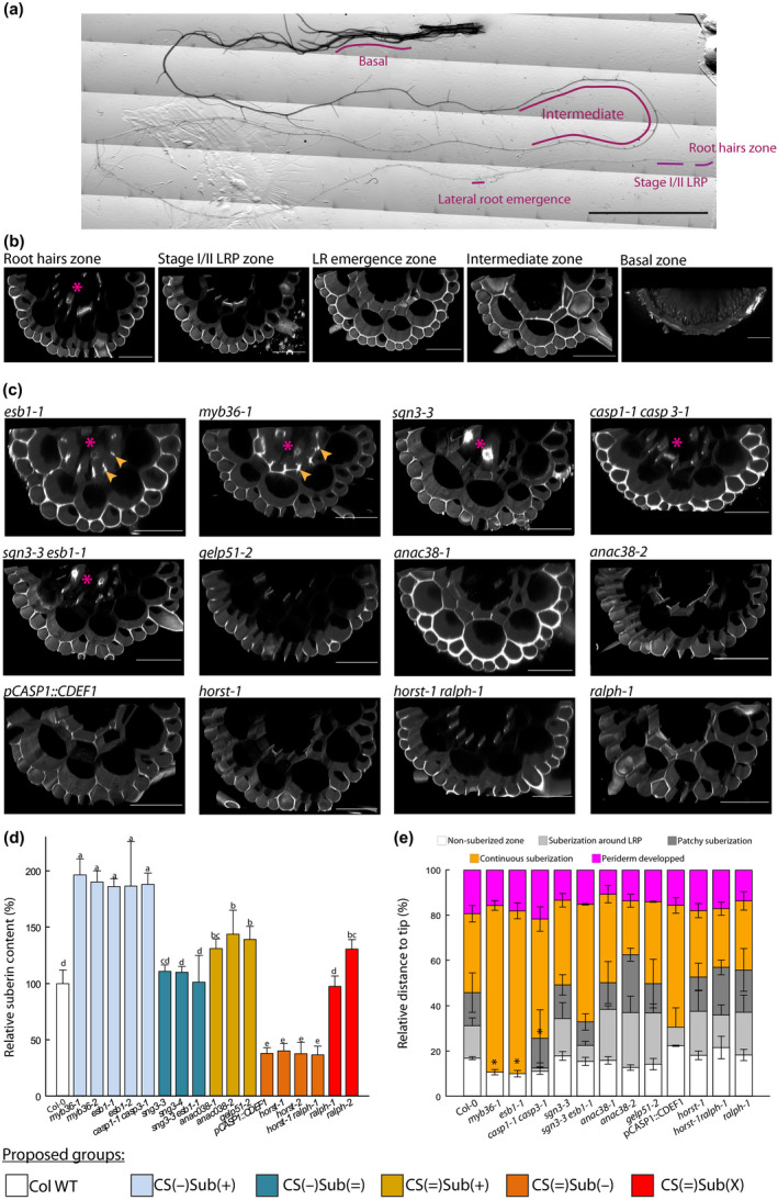
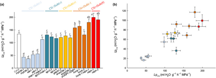
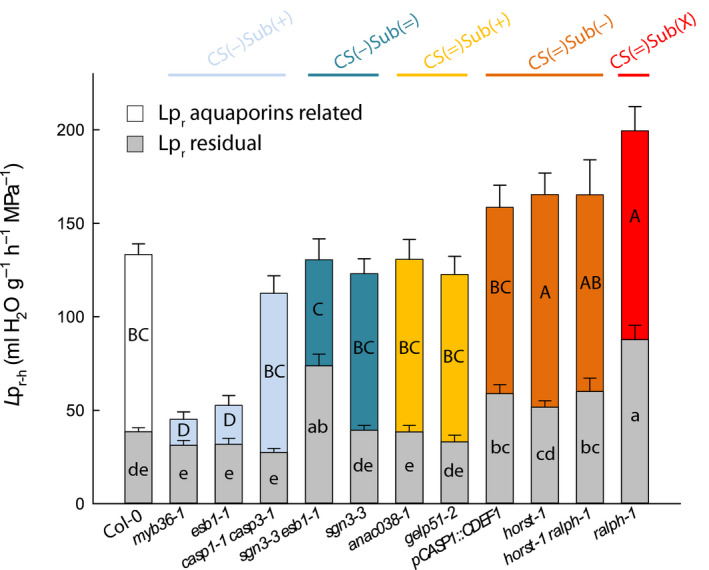
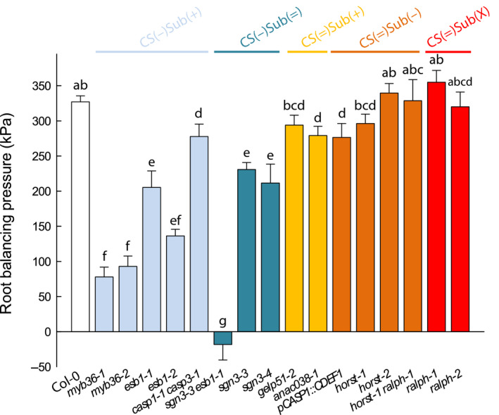
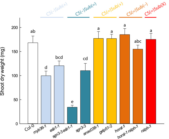
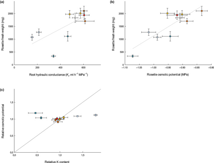
The formation of Casparian strips (CS) and the deposition of suberin at the endodermis of plant roots are thought to limit the apoplastic transport of water and ions. We investigated the specific role of each of these apoplastic barriers in the control of hydro-mineral transport by roots and the consequences on shoot growth. A collection of Arabidopsis thaliana mutants defective in suberin deposition and/or CS development was characterized under standard conditions using a hydroponic system and the Phenopsis platform. Mutants altered in suberin deposition had enhanced root hydraulic conductivity, indicating a restrictive role for this compound in water transport. In contrast, defective CS directly increased solute leakage and indirectly reduced root hydraulic conductivity. Defective CS also led to a reduction in rosette growth, which was partly dependent on the hydro-mineral status of the plant. Ectopic suberin was shown to partially compensate for defective CS phenotypes. Altogether, our work shows that the functionality of the root apoplastic diffusion barriers greatly influences the plant physiology, and that their integrity is tightly surveyed.

Keywords: Arabidopsis thaliana; Casparian strips; apoplastic barriers; aquaporins; root hydraulic conductivity; solutes diffusion; suberin; water transport.


Figures

Fig. 1
Fig. 1
Characterization of Casparian strip permeability and suberin development. (a) Reconstituted picture of a 21 d‐old primary root, and the zones that were monitored for propidium iodide (PI) permeability. Bar, 1 cm. (b) Confocal cross‐sections of a 21 d‐old plants, PI stained, Arabidopsis root from Col‐0 at various zones: root hairs, stages I and II lateral root primordium (LRP), first lateral root (LR) emergence, intermediate, and basal. Bars, 50 µm. (c) Confocal cross‐section of PI staining in roots of 21 d‐old plant at stages I and II LRP development. Arrows highlight the staining related to ectopic deposition of cell wall polymers at the cell corners, stars indicate when the vessels are stained and hence, PI was able to penetrate through the stele. Bars, 50 µm. (d) Relative suberin content related to wild‐type Arabidopsis plants (Col‐0) of 17 Casparian strips (CS) and/or suberin mutants of 21 d‐old plants. Suberin was analyzed using gas chromatography after release by transesterification using boron trifluoride in methanol from solvent extracted root cell walls. Bars represent mean values in μg per mg dry weight ± SE (n = 3–5). *Suberin content taken from literature esb1‐2 (Baxter et al., 2009), pCASP1::CDEF1 (Barberon et al., 2016), horst‐1, horst‐2 (Hofer et al., 2008), ralph‐1, ralph‐2 (Compagnon et al., 2009). (e) Scoring of the suberin stages along the root, as a relative position from the tip ± SE, after staining with the lignin/suberin dye Auramine‐O (n = 3–5). Method detailed in Supporting Information Fig. S2. Asterisks indicate significant difference (P < 0.05) to Col‐0 plants. Colors patterns of (c) allow to visually identify the groups that are defined in the first section of the results. They are reproduced similarly over all the figures.
Fig. 2
Fig. 2
Hydrostatic root hydraulic conductivity (Lpr–h) (a), and its relation with the osmotically root hydraulic conductivity (Lpr–o) (b) in Col‐0, and in a collection of 16 Casparian strips (CS) and/or suberin mutants in Arabidopsis. The plants that were grown hydroponically for 19 to 21 d under environmental controlled conditions, and measured using pressure chambers (Lpr–h) (means ± SE, n = 15–20, n = 3) or by the exudation method (Lpr–o) (means ± SE, n = 20–25, n = 3). In (a), anac038‐2 is presented at a ‘virtual Lpr’ of 119.38 with respect to a wild‐type (WT) value of 134.08 ml g−1 h−1 MPa−1, when ‘real values’ obtained during a dedicated experiment were of 205.0 and 230.2 ml g−1 h−1 MPa−1, respectively. One‐way ANOVA and Tukey’s test were used to determine significant differences (α = 0.05). Data of Lpr–h for pCASP1::CDEF1 are the same as in Wang et al. (2019).
Fig. 3
Fig. 3
Effects of sodium azide (NaN3) on root hydraulic conductivity and effects of root barrier mutations on residual water transport. Measurements were performed in a collection of 19 to 21 d‐old Arabidopsis Col‐0 and 11 Casparian strips (CS) and/or suberin mutants (means ± SE, n = 15–20, n = 3). The aquaporin‐dependent pathway (colored bar) was derived from the substraction of xylem sap flow before (full bar) and after 40 min (residual‐Lpr, gray bars) of a NaN3 treatment. Uppercase and lowercase letters inside the bars indicate significant differences. Data were analyzed using one‐way ANOVA and Tukey’s test at α = 0.05.
Fig. 4
Fig. 4
Correlations between root hydraulic conductivity (Lpr–h) and aquaporins AtPIP1;5 (a) or AtPIP2;1 (b) expression levels in Col‐0 and a collection of seven Arabidopsis mutants with alterations in endodermal Casparian strips (CS) and/or suberin. Spearman’s correlations are statistically significant for each gene (P < 3 × 10–2 and 2 × 10–7, respectively). Plants were grown hydroponically for 19 to 21 d (means ± SE, n = 3).
Fig. 5
Fig. 5
Root balancing pressure (P Jv0) in Col‐0 and in a collection of 16 Casparian strips (CS) and/or suberin mutants in Arabidopsis that were grown hydroponically for 19 to 21 d (means ± SE, n = 15–20, n = 3). The measurements were performed using pressure chambers at a constant pressure (320 kPa) and after subjecting the roots to 100 mM sodium chloride (NaCl) for 1 h. Letters above the bars indicate significant differences among means after one‐way ANOVA and post hoc Tukey's test (α = 0.05).
Fig. 6
Fig. 6
Relative shoot growth in Col‐0 and a collection of nine Casparian strips (CS) and/or suberin Arabidopsis mutants in control conditions. Plants were grown for 5 wk in a Phenopsis phenotyping platform under controlled environmental conditions (means ± SE, n = 7). Letters above the bars indicate significant differences among means after one‐way ANOVA and post hoc Tukey's test (α = 0.05).
Fig. 7
Fig. 7
Relationship between rosette fresh weight (FW) and root hydraulic conductance (K r) (a) and rosette osmotic potential (± SEM) from elongating leaves (b) in a collection of nine Casparian strips (CS) and/or suberin Arabidopsis mutants and Col‐0 grown in soil for 5 wk in a Phenopsis phenotyping platform under controlled environmental conditions (n = 7). Pearson correlation coefficients are 0.69 (P‐value < 0.05) and 0.84 (P‐value < 0.01), for FW vs K r and FW vs shoot osmotic potential, respectively. (c) Rosette osmotic potential vs potassium (K) content for mutants grown under control conditions, relatively to Col‐0.

References

    1. Alassimone J, Naseer S, Geldner N. 2010. A developmental framework for endodermal differentiation and polarity. Proceedings of the National Academy of Sciences, USA 107: 5214–5219. - PMC - PubMed
    1. Andersen TG, Naseer S, Ursache R, Wybouw B, Smet W, De Rybel B, Vermeer JEM, Geldner N. 2018. Diffusible repression of cytokinin signalling produces endodermal symmetry and passage cells. Nature 555: 529–533. - PMC - PubMed
    1. Barberon M, Vermeer J, De Bellis D, Wang P, Naseer S, Andersen T, Humbel B, Nawrath C, Takano J, Salt D et al. 2016. Adaptation of root function by nutrient‐induced plasticity of endodermal differentiation. Cell 164: 447–459. - PubMed
    1. Baxter I, Hosmani PS, Rus A, Lahner B, Borevitz JO, Muthukumar B, Mickelbart MV, Schreiber L, Franke RB, Salt DE. 2009. Root suberin forms an extracellular barrier that affects water relations and mineral nutrition in Arabidopsis. PLoS Genetics 5: e1000492. - PMC - PubMed
    1. Beisson F, Li Y, Bonaventure G, Pollard M, Ohlrogge JB. 2007. The acyltransferase GPAT5 is required for the synthesis of suberin in seed coat and root of Arabidopsis. Plant Cell 19: 351–368. - PMC - PubMed

Publication types

LinkOut - more resources