. 2021 Oct 26;9(21):7297-7310.
doi: 10.1039/d1bm01262b.

Decellularised extracellular matrix decorated PCL PolyHIPE scaffolds for enhanced cellular activity, integration and angiogenesis

Affiliations

Decellularised extracellular matrix decorated PCL PolyHIPE scaffolds for enhanced cellular activity, integration and angiogenesis

Serkan Dikici et al. Biomater Sci. .

Abstract

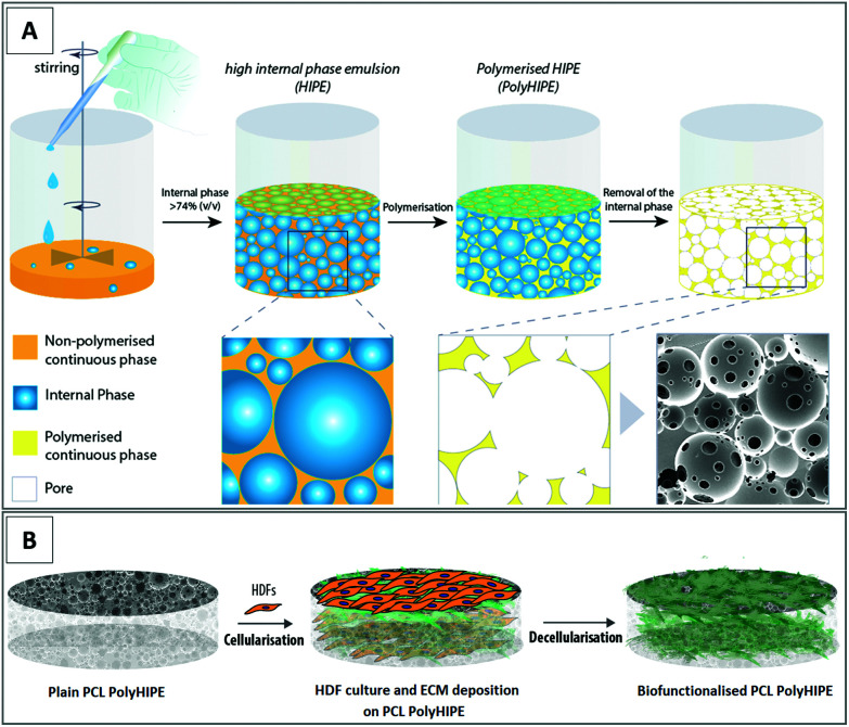
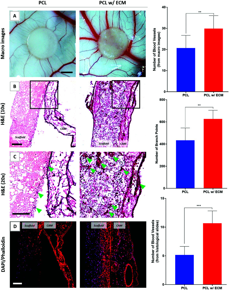
Wound healing involves a complex series of events where cell-cell and cell-extracellular matrix (ECM) interactions play a key role. Wounding can be simple, such as the loss of the epithelial integrity, or deeper and more complex, reaching to subcutaneous tissues, including blood vessels, muscles and nerves. Rapid neovascularisation of the wounded area is crucial for wound healing as it has a key role in supplying oxygen and nutrients during the highly demanding proliferative phase and transmigration of inflammatory cells to the wound area. One approach to circumvent delayed neovascularisation is the exogenous use of pro-angiogenic factors, which is expensive, highly dose-dependent, and the delivery of them requires a very well-controlled system to avoid leaky, highly permeable and haemorrhagic blood vessel formation. In this study, we decorated polycaprolactone (PCL)-based polymerised high internal phase emulsion (PolyHIPE) scaffolds with fibroblast-derived ECM to assess fibroblast, endothelial cell and keratinocyte activity in vitro and angiogenesis in ex ovo chick chorioallantoic membrane (CAM) assays. Our results showed that the inclusion of ECM in the scaffolds increased the metabolic activity of three types of cells that play a key role in wound healing and stimulated angiogenesis in ex ovo CAM assays over 7 days. Herein, we demonstrated that fibroblast-ECM functionalised PCL PolyHIPE scaffolds appear to have great potential to be used as an active wound dressing to promote angiogenesis and wound healing.


Conflict of interest statement

There are no conflicts to declare.

Figures

Fig. 1
Fig. 1. Fabrication and bio-functionalisation of the constructs. (A) Preparation of the emulsions made of photocurable PCL and water and polymerisation to form PCL PolyHIPE structure and (B) culture of HDFs and ECM deposition on PCL PolyHIPE scaffolds and decellularisation to prepare the bio-functionalised PCL PolyHIPE scaffolds.
Fig. 2
Fig. 2. (A) Synthesis of 4PCL from the monomers via ring-opening polymerization and methacrylate functionalization of the hydroxyl end groups (4PCLMA), (B) chemical structure diagram and proton NMR spectrum of 4PCL and 4PCLMA and the relative assignments.
Fig. 3
Fig. 3. (A) The SEM images of the PCL PolyHIPE showing the microporous and interconnected structure of the constructs. (B) Pore size and (C) window size distribution of PCL PolyHIPE. (D) Liquid absorption capability of the plasma-treated and non-treated PCL PolyHIPE scaffolds. ** p < 0.01, * p < 0.05, ns not significant, p > 0.05, n = 3 ± SD. Scale bar represents 200 µm.
Fig. 4
Fig. 4. (A) H&E and Sirius Red-stained sections of the recellularised PCL PolyHIPE scaffolds with HDFs over 21 days. Circular images show the macro images of the Sirius Red-stained scaffolds. (B) AlamarBlue assay and (C) Sirius Red assay results showing cell viability and collagen production, respectively, over 21 days. AA2P was observed to increase both metabolic activity and collagen production compared with non-supplemented controls. Scale bars represent 100 µm. *** p < 0.001, * p < 0.05, ns not significant, p > 0.05, n = 3 ± SD.
Fig. 5
Fig. 5. The SEM images of the HDFs cultured on PCL PolyHIPE scaffolds over 21 days either in the presence or absence of AA2P.
Fig. 6
Fig. 6. SEM images of the (A) surface and (B) cross-sections of the decellularised PCL PolyHIPE scaffolds. DAPI (blue) and phalloidin-TRITC (red) stained sections of the (C) cellular and (D) decellularised scaffolds. Graphs show the confirmation of (E) the DNA removal with Picogreen assay and (F) the preservation of the deposited collagen with Sirius Red assay. Scale bars represent 100 µm for fluorescent images. *** p < 0.001, ns not significant, p > 0.05, n = 3 ± SD.
Fig. 7
Fig. 7. The mechanical testing results (Young's modulus, UTS, and elongation at yield) of plain and ECM containing PCL PolyHIPE scaffolds. ns not significant, p > 0.05, n = 3 ± SD.
Fig. 8
Fig. 8. (A) H&E-stained sections of the HDFs cultured for 7 days on bio-functionalised scaffolds and (B) the metabolic activity of HDFs over 7 days. (C) H&E-stained sections of the HAECs cultured for 7 days on bio-functionalised scaffolds and (D) the metabolic activity of HAECs over 7 days. (E) H&E stained sections of the HEKs cultured for 7 days on bio-functionalised scaffolds and (F) the metabolic activity of HEKs over 7 days. Scale bars represent 100 µm. *** p < 0.001, ** p < 0.01, * p < 0.05, n = 3 ± SD.
Fig. 9
Fig. 9. Evaluation of the angiogenic performance of the plain PCL and bio-functionalised scaffolds. (A) Macro images, (B) lower and (C) higher magnifications of the H&E-stained sections. Blood vessels are shown with green arrows. (D) Phalloidin-TRITC (red)/DAPI (blue) stained sections of the scaffolds. Graphs show the quantified results. Scale bars represent 2 mm for macro images of the CAM (A) and 100 μm for H&E (B and C) and fluorescent stained (D) sections. ***p < 0.001, **p < 0.01, n = 3 ± SD.

Similar articles

Cited by

References

    1. Lee S. H. Jeong S. K. Ahn S. K. Yonsei Med. J. 2006;47:293–306. doi: 10.3349/ymj.2006.47.3.293. - DOI - PMC - PubMed
    1. Forslind B. Engström S. Engblom J. Norlén L. J. Dermatol. Sci. 1997;14:115–125. doi: 10.1016/S0923-1811(96)00559-2. - DOI - PubMed
    1. Rees J. Lancet. 1999;354:1491–1492. doi: 10.1016/S0140-6736(99)00281-0. - DOI - PubMed
    1. Dong R. H. Jia Y. X. Qin C. C. Zhan L. Yan X. Cui L. Zhou Y. Jiang X. Long Y. Z. Nanoscale. 2016;8:3482–3488. doi: 10.1039/C5NR08367B. - DOI - PubMed
    1. Stroncek J. D. and Reichert W. M., in Indwelling Neural Implants: Strategies for Contending with the in Vivo Environment, 2007, pp. 3–38 - PubMed