. 2021 Jan-Dec:25:23312165211045306.
doi: 10.1177/23312165211045306.

Auditory and Visual Integration for Emotion Recognition and Compensation for Degraded Signals are Preserved With Age

Affiliations

Auditory and Visual Integration for Emotion Recognition and Compensation for Degraded Signals are Preserved With Age

Minke J de Boer et al. Trends Hear. 2021 Jan-Dec.

Abstract

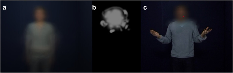
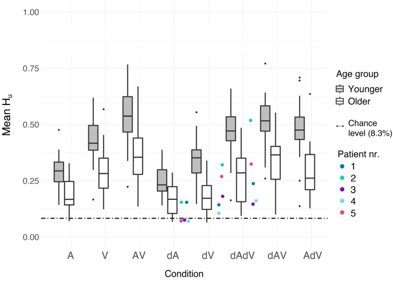
Since emotion recognition involves integration of the visual and auditory signals, it is likely that sensory impairments worsen emotion recognition. In emotion recognition, young adults can compensate for unimodal sensory degradations if the other modality is intact. However, most sensory impairments occur in the elderly population and it is unknown whether older adults are similarly capable of compensating for signal degradations. As a step towards studying potential effects of real sensory impairments, this study examined how degraded signals affect emotion recognition in older adults with normal hearing and vision. The degradations were designed to approximate some aspects of sensory impairments. Besides emotion recognition accuracy, we recorded eye movements to capture perceptual strategies for emotion recognition. Overall, older adults were as good as younger adults at integrating auditory and visual information and at compensating for degraded signals. However, accuracy was lower overall for older adults, indicating that aging leads to a general decrease in emotion recognition. In addition to decreased accuracy, older adults showed smaller adaptations of perceptual strategies in response to video degradations. Concluding, this study showed that emotion recognition declines with age, but that integration and compensation abilities are retained. In addition, we speculate that the reduced ability of older adults to adapt their perceptual strategies may be related to the increased time it takes them to direct their attention to scene aspects that are relatively far away from fixation.

Keywords: aging; audiovisual; emotion recognition; eye-tracking; sensory impairments.


Conflict of interest statement

Declaration of Conflicting Interests: The authors declared no potential conflicts of interest with respect to the research, authorship, and/or publication of this article.

Figures

Figure 1.
Figure 1.
Individual levels of visual acuity (left) and contrast sensitivity (middle, in logCS), measured binocularly, for younger, older, and patient participants. Left: individual hearing thresholds in dB HL for the better ear for younger, older, and patient participants. Note: one patient participant (4) did not respond when the frequencies  ≥ 3,000 Hz were presented at 90 dB HL, at which point testing stopped to not further damage hearing. The thresholds in the figure were set at 95 dB HL to indicate this, the actual hearing thresholds for those audiometric test frequencies are unknown.
Figure 2.
Figure 2.
(a) Still image created by averaging together all frames of all videos. This image preceded stimulus presentation in all conditions with video. (b) Shape and approximate size of the scotoma mask. The scotoma was gaze-contingent with its center positioned on the point of gaze. Four different orientations were used during the experiment (randomly intermixed): as shown in this figure, left-right flipped, up-down flipped, and left-right and up-down flipped. (c) Scotoma overlaid on a still image of one video. The red dot indicates the point of gaze, this dot was not visible to participants. The still image in (c) is retrieved from one of the video's of the GEMEP core set from Bänziger et al. (2012). Published with permission from the Swiss Center for Affective Sciences.
Figure 3.
Figure 3.
Task performance for each condition and age group, shown as unbiased hit-rates. Performance is averaged across emotions and blocks. Each box shows the data between the first and third quartiles. The horizontal solid line in each box denotes the median. The whiskers extend to the lowest/highest value still within 1.5*inter-quartile range (IQR), data outside the 1.5*IQR are plotted as dots. Performance for young participants is shown in light grey boxes, performance for older participants is shown in white boxes. Performance for individual patient participants is shown in the colored dots. Note that these participants did not receive degraded stimuli, but their hearing and visual acuity tests indicate that their perception is degraded. Thus, for patient participants, dA corresponds to stimuli presented in A, likewise for dV and dAdV. The dashed line indicates chance level performance.
Figure 4.
Figure 4.
Fixation durations in ms for all conditions and age groups. As for Figure 3, fixation durations are averaged across emotions and blocks. Each box shows the data between the first and third quartiles. The horizontal solid line in each box denotes the median. The whiskers extend to the lowest/highest value still within 1.5*IQR, data outside the 1.5*IQR are plotted as dots. Performance for young participants is shown in light grey boxes, performance for older participants is shown in white boxes.
Figure 5.
Figure 5.
Saccadic amplitudes in degree of visual angle for all conditions and age groups. Amplitudes are averaged across emotions and blocks. Each box shows the data between the first and third quartiles. The horizontal solid line in each box denotes the median. The whiskers extend to the lowest/highest value still within 1.5*IQR, data outside the 1.5*IQR are plotted as dots. Performance for young participants is shown in light grey boxes, performance for older participants is shown in white boxes. The dashed line indicates the minimal radius of the scotoma.
Figure 6.
Figure 6.
Mean fixation proportions on the face and hand AOIs (areas of interest) for all conditions and both age groups, and averaged over emotions and blocks. Error bars denote the standard error of the mean (SEM). Intact conditions are indicated by a black outline.

References

    1. Bach M. (1996). The Freiburg visual acuity test-automatic measurement of visual acuity. Optometry & Vision Science, 73(1), 49–53. 10.1097/00006324-199601000-00008 - DOI - PubMed
    1. Bach M. (2006). The Freiburg visual acuity test-variability unchanged by post-hoc re-analysis. Graefe's Archive for Clinical and Experimental Ophthalmology, 245(7), 965–971. 10.1007/s00417-006-0474-4 - DOI - PubMed
    1. Bassili J. N. (1979). Emotion recognition: The role of facial movement and the relative importance of upper and lower areas of the face. Journal of Personality and Social Psychology, 37(11), 2049. 10.1037/0022-3514.37.11.2049 - DOI - PubMed
    1. Bänziger T., Mortillaro M., Scherer K. R. (2012). Introducing the Geneva multimodal expression corpus for experimental research on emotion perception. Emotion (Washington, DC), 12(5), 1161–1179. 10.1037/a0025827 - DOI - PubMed
    1. Bennett R. M. C., Hohmann V. (2012). Simulation of reduced frequency selectivity found with cochlear hearing loss using a model based procedure. Annual Meeting of the German Society of Audiology.

Publication types