. 2021 Oct 26;15(10):16642-16653.
doi: 10.1021/acsnano.1c06488. Epub 2021 Oct 7.

Direct Nanopore Sequencing of Individual Full Length tRNA Strands

Affiliations

Direct Nanopore Sequencing of Individual Full Length tRNA Strands

Niki K Thomas et al. ACS Nano. .

Abstract

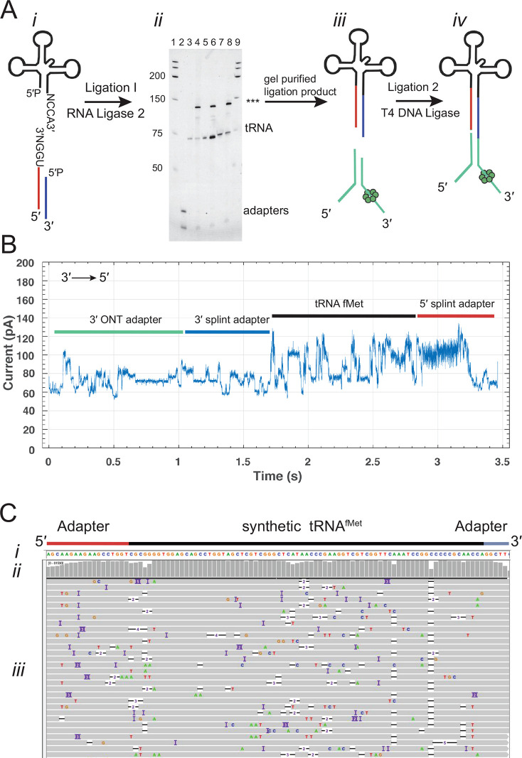
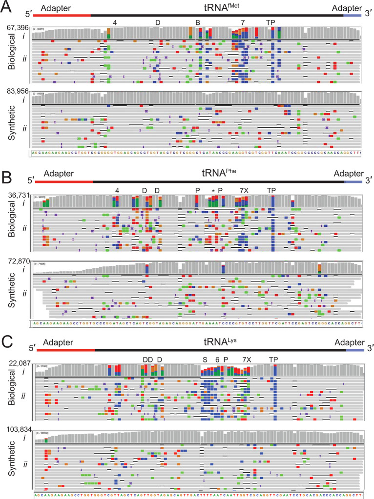
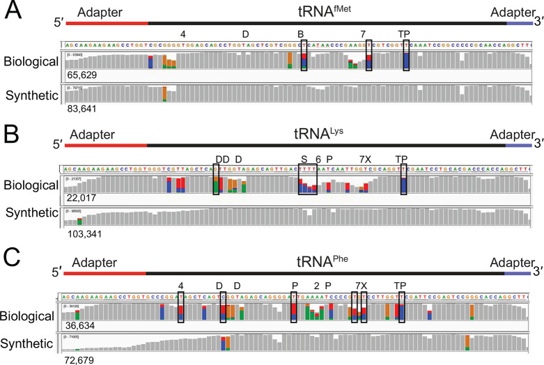
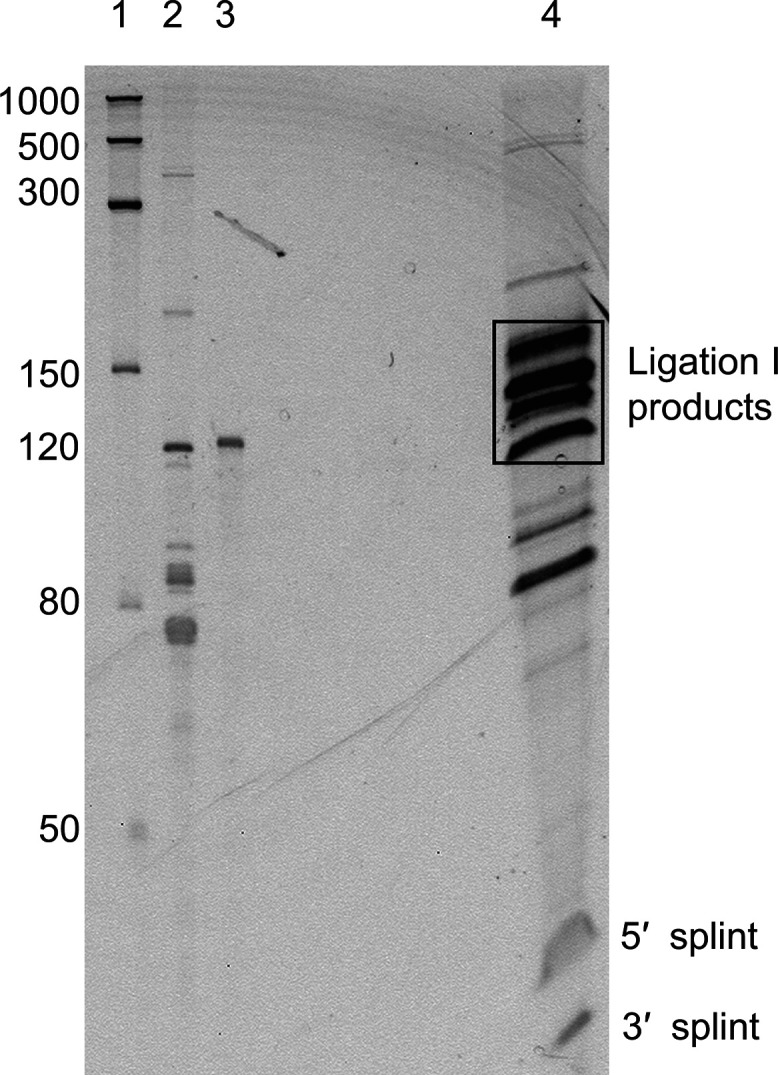
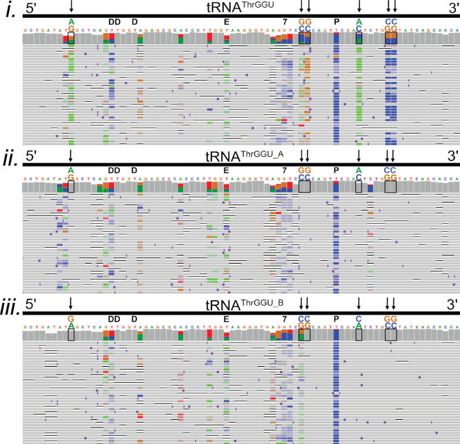
We describe a method for direct tRNA sequencing using the Oxford Nanopore MinION. The principal technical advance is custom adapters that facilitate end-to-end sequencing of individual transfer RNA (tRNA) molecules at subnanometer precision. A second advance is a nanopore sequencing pipeline optimized for tRNA. We tested this method using purified E. coli tRNAfMet, tRNALys, and tRNAPhe samples. 76-92% of individual aligned tRNA sequence reads were full length. As a proof of concept, we showed that nanopore sequencing detected all 43 expected isoacceptors in total E. coli MRE600 tRNA as well as isodecoders that further define that tRNA population. Alignment-based comparisons between the three purified tRNAs and their synthetic controls revealed systematic nucleotide miscalls that were diagnostic of known modifications. Systematic miscalls were also observed proximal to known modifications in total E. coli tRNA alignments, including a highly conserved pseudouridine in the T loop. This work highlights the potential of nanopore direct tRNA sequencing as well as improvements needed to implement tRNA sequencing for human healthcare applications.

Keywords: E. coli; isoacceptor; isodecoder; modifications; nanopore; pseudouridine; tRNA.


Conflict of interest statement

The authors declare the following competing financial interest(s): M.A. holds shares in Oxford Nanopore Technologies (ONT). M.A. is a paid consultant to ONT. M.A. and M.J. received reimbursement for travel, accommodations, and conference fees to speak at events organized by ONT. M.A. is an inventor on 11 UC patents licensed to ONT (6,267,872, 6,465,193, 6,746,594, 6,936,433, 7,060,50, 8,500,982, 8,679,747, 9,481,908, 9,797,013, 10,059,988, and 10,081,835). M.A. received research funding from ONT.

Figures

Figure 1
Figure 1
Overview of the tRNA sequencing strategy using synthetic canonical tRNAs. (A) tRNA adaptation for nanopore sequencing. From left to right: (i) The tRNA is ligated to a double-stranded splint adapter using RNA ligase 2. (ii) Gel purification of the ligation I product for synthetic tRNAs. The denaturing PAGE gel shows the first ligation of three synthetic tRNAs to the splint adapter. The lanes are as follows: 1, RNA ladder; 2, splint adapter; 3, synthetic tRNAfMet; 4, synthetic tRNAfMet ligation reaction; 5, synthetic tRNALys; 6, synthetic tRNALys ligation reaction; 7, synthetic tRNAPhe; 8, synthetic tRNAPhe ligation reaction; and 9, RNA ladder. The full length product (***) is excised and purified (see Figure S1 for biological tRNA). (iii) The purified product is ligated to the ONT sequencing adapters using T4 DNA Ligase. (iv) Adapted, nanopore sequencing-ready tRNA product. In the line drawing, the adapters and tRNA are not to scale. (B) An example ionic current trace of adapter-ligated synthetic tRNAfMet. Regions of the trace are indicated with colored bars corresponding to structures in (A): The 3′ strand of the ONT RMX adapter (teal); the 3′ strand of the splint adapter (blue); the tRNA (black); and the 5′ strand of the splint adapter (red). (C) Primary alignments of synthetic tRNAfMet to the reference sequence visualized using IGV. The reference sequence and its components are labeled as (i). The coverage at each position (coverage plot) is indicated by gray columns, where a maximally tall bar means every aligned read is covered at that position (ii). Beneath the coverage plot is a diagram of a randomly downsized sample of the aligned reads (iii). Gray rows denote continuous alignment and agreement with the reference nucleotide. Within each read, positions that do not match the reference (U(T) = red, A = green, C = blue, G = gold) are shown. White spaces bisected with a black bar within an aligned read indicate a deletion. Insertions are indicated in purple. The rows of aligned reads are presented as they were displayed on IGV.
Figure 2
Figure 2
Biological and canonical tRNA strand reads aligned against reference sequences. (A) tRNAfMet, (B) tRNAPhe, and (C) tRNALys. In each panel (i) is base coverage along the reference sequence at each position (coverage plot) and (ii) is a randomly selected subset of individual aligned nanopore reads. The total numbers of aligned reads are shown to the left of the coverage plots. The positions of expected modifications on biological tRNA3 are indicated above the coverage plots and are abbreviated: 4 = 4-thiouridine; D = Dihydrouridine; B = 2′-O-methylcytidine; 7 = 7-methylguanosine; T = 5-methyluridine; P = pseudouridine; X = 3-(3-amino-3-carboxypropyl)uridine; * = 2-methylthio-N6-isopentenyladenosine; S = 5-methyl-aminomethyl-2-thiouridine; and 6 = N6-threonylcarbamoyl-adenosine. Gray columns in the coverage plots indicate positions along the reference where 80% or more of the quality weighted reads are the expected canonical nucleotide. At positions where the value is under the 80% threshold, the proportion of each nucleotide call is shown in color where U(T) = red, A = green, C = blue, and G = gold. Similarly, the rows of individual aligned reads (A–C, ii) are gray at positions matching the reference and colored (using the previously mentioned convention) at positions with mismatches. The black horizontal bars in the aligned reads indicate a deletion, and purple bars indicate an insertion. The rows of aligned reads are presented as they were displayed on IGV.
Figure 3
Figure 3
Systematic base miscalls in purified biological and canonical tRNAfMet, tRNALys, and tRNAPhe. The coverage plots (A–C) for biological and canonical synthetic tRNAs were generated from alignments using marginAlign. The number of aligned reads for each tRNA is shown under each coverage plot. Boxes surrounding base positions denote systematic miscalls (posterior probability of ≥30%). No systematic miscalls were identified in the synthetic canonical tRNAs. Colored bars are at positions where <80% of the quality weighted alignments match the reference. At these positions, the proportion of individual bases called are shown in color (U(T) = red, A = green, C = blue, and G = gold). The known modifications for the biological tRNAs3 are indicated above the coverage plots. Modified nucleotides are indicated above the reference sequence with abbreviations (4 = 4-thiouridine, D = dihydrouridine, B = 2′-O-methylcytidine, 7 = 7-methylguanosine, T = 5-methyluridine, P = pseudouridine, X = 3-(3-amino-3-carboxy-propyl)uridine, * = 2-methylthio-N6- isopentenyladenosine, S = 5-methylaminomethyl-2-thiouridine, and 6 = N6-threonylcarbamoyl-adenosine). The falloff in 5′ coverage of synthetic tRNALys (B, lower panel) and tRNAPhe (C, lower panel) is likely due to incomplete 5′ phosphorylation of these substrates.
Figure 4
Figure 4
PAGE gel of total tRNA ligated to splint adapters. Lane 1: The ssRNA ladder with sizes in nucleotides indicated to the left. Lane 2: E. coli MRE600 total tRNA. Lane 3: A 121 nt IVT human 5S rRNA used as a size marker. Lane 4: The products of the ligation reaction of total E. coli tRNA and the four types of splint adapters. The two bands under 50 nt are the 30 nt and 24 nt strands of the splint adapters that did not ligate to the tRNA. Successful ligation of the double-stranded splint will add 54 nt to the tRNA. As tRNA ranges from 75 to 93 nt, the expected ligation products are 129–140 nt. A block of gel encompassing fragments of approximately 110–180 nt, indicated on the gel as a black rectangle, was excised, purified, and carried forward for the library preparation.
Figure 5
Figure 5
Confirmation of two known isodecoders in nanopore tRNAThrGGU sequence alignments. The isoacceptor tRNAThrGGU has two isodecoder forms which have canonical sequence variations at positions 9 (G/A), 49 (C/G), 50 (C/G), 59 (C/A), 64 (G/C), and 65 (G/C). (i–iii) Black boxes on the IGV coverage plots surround the positions of these variations. Black arrows also point to variant positions for clarity. Above each black box, the reference nucleotide is enlarged, and the alternative nucleotide is above it, labeled and colored in accordance with IGV schema (A = green, G = gold, C = blue, and T(U) = red). (i) Alignments of total E. coli tRNA reads to the tRNAThrGGU isoacceptor. At positions that vary between tRNAThrGGU isodecoders, the colors representing the reference and alternative nucleotides are seen in the coverage plot. This can be interpreted as both isodecoder forms being present in the data. (ii) Alignments of total E. coli tRNA read to the tRNAThrGGU_A isodecoder. Gray in the coverage plot and the rows of aligned reads indicate agreement with the tRNAThrGGU_A reference. (iii) Alignments of total E. coli tRNA read to the tRNAThrGGU_B isodecoder. Gray in the coverage plot and the rows of aligned reads indicate agreement with the tRNAThrGGU_B reference. The known modifications D (dihydrouridine), E (N6-methyl-N6-threonylcarbomoyladenosine), 7 (7-methylguanosine), and P (pseudouridine) are denoted in black above the reference sequence. Adapter sequences were included in the alignments but are not shown.

References

    1. Raina M.; Ibba M. tRNAs as Regulators of Biological Processes. Front. Genet. 2014, 5, 1–14. 10.3389/fgene.2014.00171. - DOI - PMC - PubMed
    1. Zhang J.; Ferré-D’amaré A. R. The tRNA Elbow in Structure, Recognition and Evolution. Life 2016, 6, 3.10.3390/life6010003. - DOI - PMC - PubMed
    1. Boccaletto P.; MacHnicka M. A.; Purta E.; Pitkowski P.; Baginski B.; Wirecki T. K.; De Crécy-Lagard V.; Ross R.; Limbach P. A.; Kotter A.; Helm M.; Bujnicki J. M. MODOMICS: A Database of RNA Modification Pathways. 2017 Update. Nucleic Acids Res. 2018, 46, D303–D307. 10.1093/nar/gkx1030. - DOI - PMC - PubMed
    1. Suzuki T. The Expanding World of tRNA Modifications and Their Disease Relevance. Nat. Rev. Mol. Cell Biol. 2021, 22, 375–392. 10.1038/s41580-021-00342-0. - DOI - PubMed
    1. Zheng G.; Qin Y.; Clark W. C.; Dai Q.; Yi C.; He C.; Lambowitz A. M.; Pan T. Efficient and Quantitative High-Throughput tRNA Sequencing. Nat. Methods 2015, 12, 835–837. 10.1038/nmeth.3478. - DOI - PMC - PubMed

Publication types