. 2021 Nov;297(5):101275.
doi: 10.1016/j.jbc.2021.101275. Epub 2021 Oct 4.

Coordinated regulation of the ribosome and proteasome by PRMT1 in the maintenance of neural stemness in cancer cells and neural stem cells

Affiliations

Coordinated regulation of the ribosome and proteasome by PRMT1 in the maintenance of neural stemness in cancer cells and neural stem cells

Lu Chen et al. J Biol Chem. 2021 Nov.

Abstract

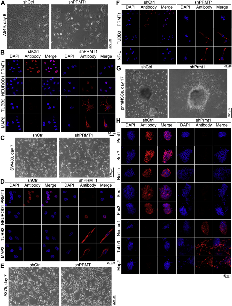
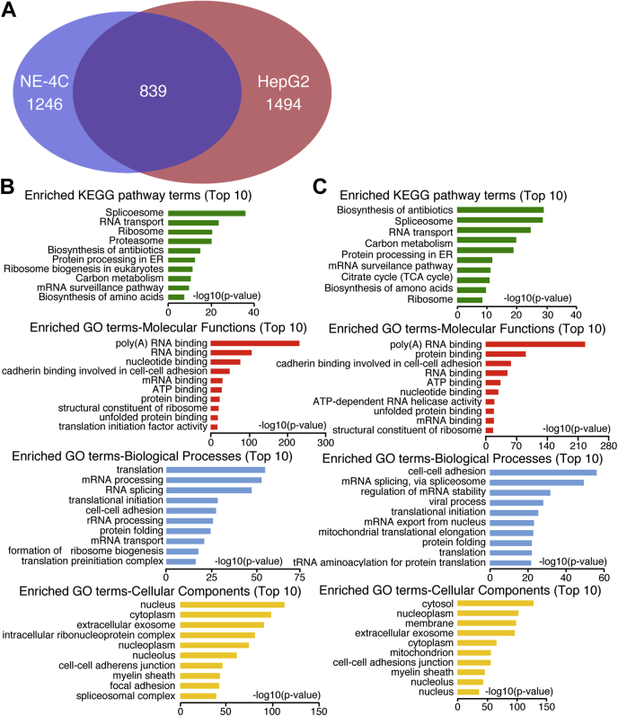
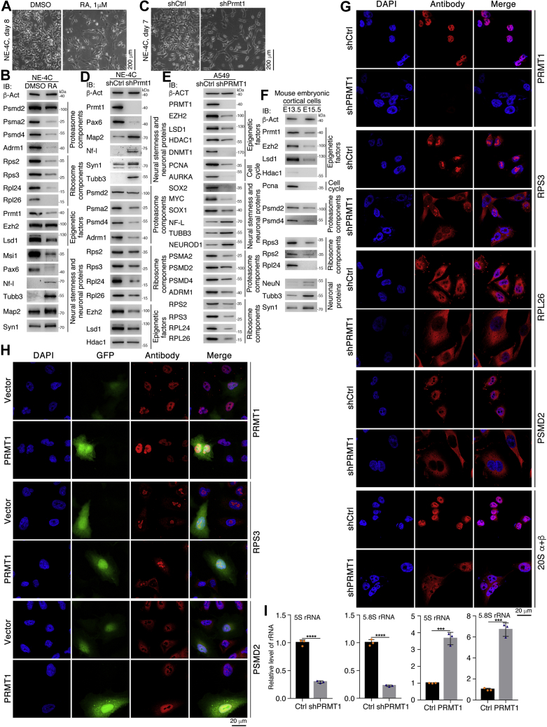
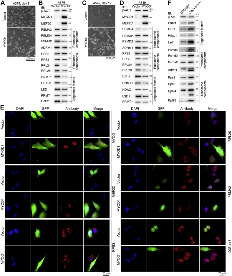
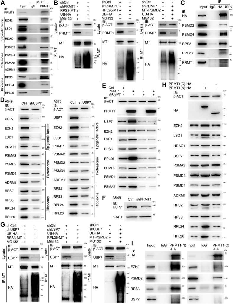
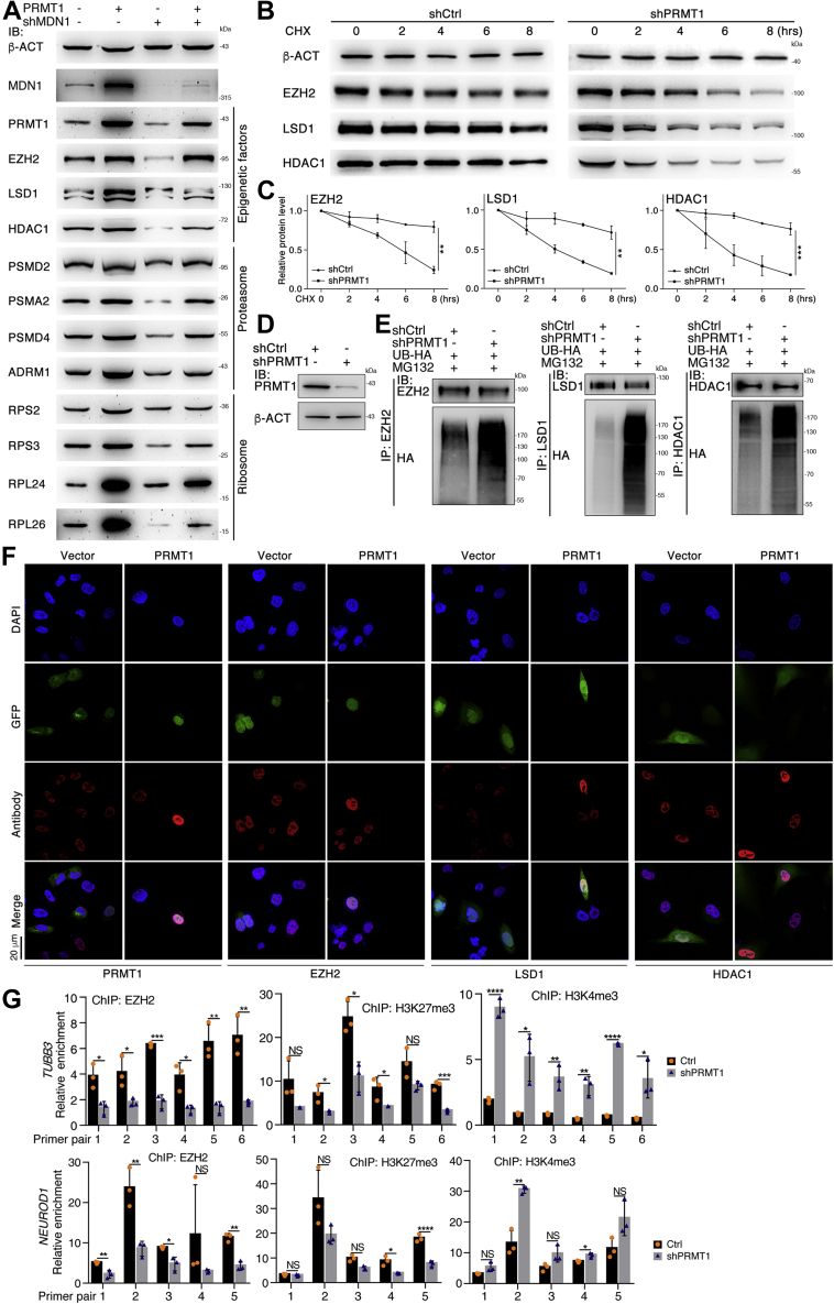
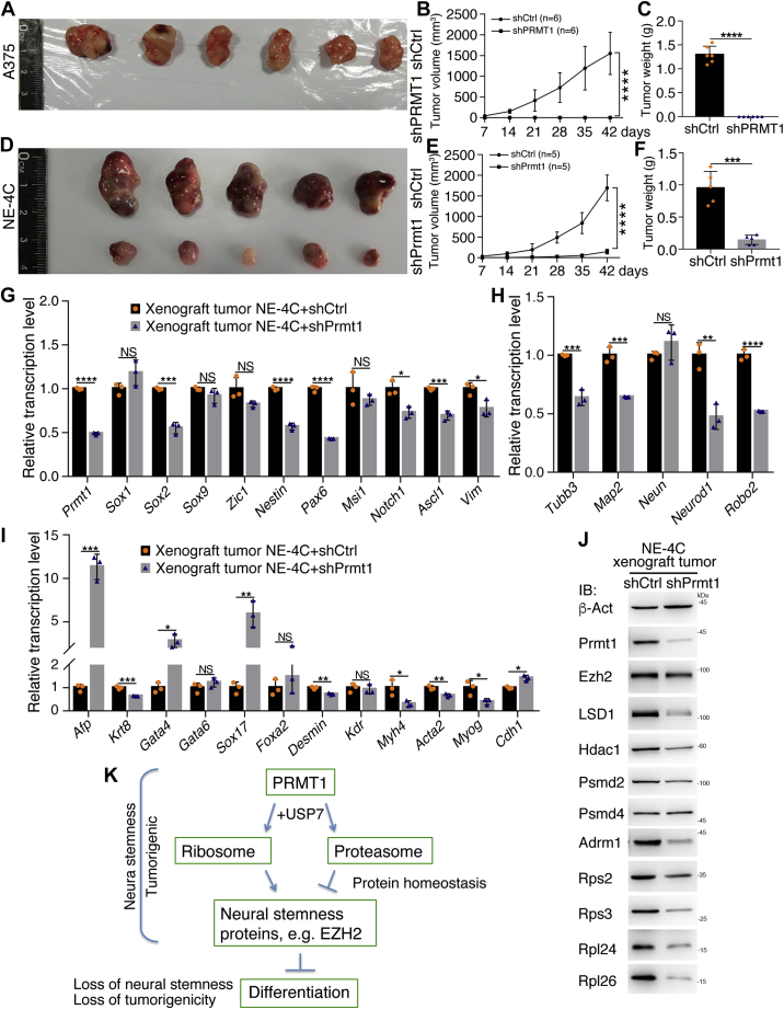
Previous studies suggested that cancer cells resemble neural stem/progenitor cells in regulatory network, tumorigenicity, and differentiation potential, and that neural stemness might represent the ground or basal state of differentiation and tumorigenicity. The neural ground state is reflected in the upregulation and enrichment of basic cell machineries and developmental programs, such as cell cycle, ribosomes, proteasomes, and epigenetic factors, in cancers and in embryonic neural or neural stem cells. However, how these machineries are concertedly regulated is unclear. Here, we show that loss of neural stemness in cancer or neural stem cells via muscle-like differentiation or neuronal differentiation, respectively, caused downregulation of ribosome and proteasome components and major epigenetic factors, including PRMT1, EZH2, and LSD1. Furthermore, inhibition of PRMT1, an oncoprotein that is enriched in neural cells during embryogenesis, caused neuronal-like differentiation, downregulation of a similar set of proteins downregulated by differentiation, and alteration of subcellular distribution of ribosome and proteasome components. By contrast, PRMT1 overexpression led to an upregulation of these proteins. PRMT1 interacted with these components and protected them from degradation via recruitment of the deubiquitinase USP7, also known to promote cancer and enriched in embryonic neural cells, thereby maintaining a high level of epigenetic factors that maintain neural stemness, such as EZH2 and LSD1. Taken together, our data indicate that PRMT1 inhibition resulted in repression of cell tumorigenicity. We conclude that PRMT1 coordinates ribosome and proteasome activity to match the needs for high production and homeostasis of proteins that maintain stemness in cancer and neural stem cells.

Keywords: PRMT1; USP7; cancer cell; cell differentiation; deubiquitination; neural stem cell (NSC); neural stemness; proteasome; ribosome; tumorigenicity.


Conflict of interest statement

Conflicts of interest The authors declare that they have no conflicts of interest with the contents of this article.

Figures

Figure 1
Figure 1
Neuronal differentiation effect induced by knockdown of PRMT1/Prmt1 in cancer cells or NSCs.A and B, neuronal-like differentiation phenotype by knockdown of PRMT1 (shPRMT1) in A549 cells (A), and validation of knockdown effect of PRMT1 and detection of neuronal protein expression (NEUROD1, TUBB3, MAP2) in cells using immunofluorescence (IF) (B). Cells infected with lentivirus containing empty vector (shCtrl) were used as control. C and D, neuronal-like differentiation phenotype by knockdown of PRMT1 in SW480 cells (C), and validation of knockdown effect of PRMT1 and detection of neuronal proteins in cells using IF (D). E and F, neuronal-like differentiation effect in A375 cells in response to PRMT1 knockdown (E), and validation of knockdown effect of PRMT1 and detection of neuronal markers in cells using IF (F). G, neurosphere formation of primNSCs derived from mouse ESCs in NSC serum-free medium (left) and neuronal differentiation phenotype induced by knockdown of Prmt1 in primNSCs (right). H, analysis of Prmt1 knockdown effect and detection of neural stemness markers (Sox2, Nestin, Sox1, Pax3) and neuronal proteins (Neurod1, Tubb3, Map2) in neurospheres and in cells with Prmt1 knockdown using IF. In all IF assays, nuclei were counterstained with DAPI.
Figure 2
Figure 2
Identification of putative interaction proteins of PRMT1/Prmt1 using mass spectrometry, and bioinformatic analysis on the identified proteins.A, a diagram summarizing the number of identified interaction partners of Prmt1/PRMT1 in NE-4C cells and HepG2 cells, and the shared interaction partners between the two cells. B and C, bioinformatic analysis on enriched pathway and GO terms in the interaction partners in NE-4C (B) or HepG2 (C) cells.
Figure 3
Figure 3
Coordinated regulation of ribosome and proteasome component proteins during neuronal differentiation.A and B, RA-induced neuronal differentiation phenotype in NE-4C cells shown at the time indicated after treatment (A), and immunoblotting (IB) detection of a series of proteasome, ribosome components, epigenetic factors, and neural stemness and neuronal proteins in control cells treated with vehicle (DMSO) and cells treated with RA, as indicated (B). C and D, neuronal differentiation phenotype in NE-4C cells induced by Prmt1 knockdown at the indicated time (C), and IB detection of expression of proteins, as indicated, in control (shCtrl) and knockdown (shPrmt1) cells (D). E, IB detection of various proteins, as indicated, in control A549 (shCtrl) cells and cells with knockdown of PRMT1 (shPRMT1). F, IB detection of various proteins, as indicated, in mouse embryonic cortical cells at two developmental stages. G, IF detection of the effect of PRMT1 knockdown on the subcellular distribution of ribosome components RPS3 and RPL26, proteasome protein PSMD2 and 20S subunit (20S α + β) in A375 cells. H, IF detection of the effect of PRMT1 overexpression on the expression of ribosome and proteasome proteins in A549 cells. Cells infected with the virus containing only the vector (Vector) as a control. I, detection of the change in 5S and 5.8S rRNA in A549 cells with PRMT1 knockdown or overexpression using RT-qPCR. Significance in change of rRNA levels was calculated for experiments in triplicate using unpaired Student’s t test. Data are shown as mean ± SD. ∗∗∗p < 0.001, ∗∗∗∗p < 0.0001.
Figure 4
Figure 4
Effect of MYOD1-induced differentiation on the expression of ribosome and proteasome components and epigenetic factors.A, phenotypic change in A375 cells infected with virus containing MYOD1. Cells infected with virus containing the empty vector were used as a control. B, IB detection of expression change of a series of proteins, as indicated, in control A375 cells and cells with forced MYOD1 expression. C, phenotypic change in A549 cells in response to MYOD1 expression. D, detection of protein expression, as indicated, in control cells and cells with MYOD1 expression using IB. E, IF detection of expression and subcellular distribution of a muscle cell marker, ribosome and proteasome proteins, and the proteasome 20S subunit in control cells and cells with MYOD1 expression. F, expression alteration in a series of proteins, as indicated, in wild-type C2C12 (C2C12WT) cells and the cells with knockout of Myod1 gene (C2C12Myod1−/−).
Figure 5
Figure 5
Regulation of ribosome, proteasome component proteins, and epigenetic factors by PRMT1.A, Co-IP confirmation of interaction between PRMT1 and a series of epigenetic factors, ribosome and proteasome proteins and a neural stemness protein, which were identified in mass spectrometry. B, ubiquitination of overexpressed RPS3, RPL26, and PSMD2, and the effect of PRMT1 knockdown on the ubiquitination of these proteins. C, interaction of USP7 with ribosome and proteasome proteins and PRMT1. D, IB detection of USP7 knockdown on the expression of proteins as indicated, in A549 and A375 cells. E, dependence of PRMT1-mediated protein expression on USP7. F, effect of PRMT1 knockdown on USP7 expression. G, the effect of USP7 knockdown on the ubiquitination of overexpressed RPS3, RPL26, and PSMD2. H, forced expression of PRMT1(N) and PRMT1(C) on the expression of proteins. I, differential interaction of PRMT1(N) and PRMT1(C) with proteins.
Figure 6
Figure 6
Regulatory effect of ribosome and proteasome on expression of proteins that maintain neural stemness.A, IB detection of the repressed expression of proteins by specific disruption of ribosome activity, and the rescue effect by PRMT1 in A549 cells. B, analysis of the effect of protein degradation in response to inhibition of de novo protein synthesis via CHX treatment of HEK293T cells in a time series and PRMT1 knockdown. C, quantification of relative protein levels in triplicate experiments as in (B). Significance in difference of protein level was calculated using unpaired Student’s t test. Data are shown as the mean ± SD. ∗∗p < 0.01, ∗∗∗p < 0.001. D and E, analysis of the effect of protein ubiquitination in response to PRMT1 knockdown. Control cells or cells with PRMT1 knockdown were overexpressed with HA-tagged Ubiquitin (UB-HA) and treated with MG132. Ubiquitination of EZH2, LSD1, and HDAC1 in control cells and knockdown cells was detected with immunoprecipitated proteins with their respective antibodies. D, displays PRMT1 knockdown efficiency in the cells. F, IF detection of the effect of PRMT1 overexpression on the expression of EZH2, LSD1, and HDAC1 in A549 cells. Nuclei were counterstained with DAPI. G, influence of PRMT1 knockdown on the binding of EZH2 to TUBB3 and NEUROD1 promoters, and on the change in H3K27me3 and H3K4me3 in these promoters. In control (Ctrl) or knockdown A375 cells, chromatin fragments were precipitated with antibodies against EZH2, H3K27me3, and H3K4me3, respectively, and detected with qPCR using primers amplifying different regions of promoters. Significance in difference in amplified promoter fragments was calculated using unpaired Student’s t test based on experiments in triplicate. Data are shown as the mean ± SD. ∗p < 0.05, ∗∗p < 0.01, ∗∗∗p < 0.001, ∗∗∗∗p < 0.0001. NS, not significant.
Figure 7
Figure 7
Suppression effect of PRMT1/Prmt1 knockdown on cell tumorigenicity.AC, tumor formation of control (shCtrl) and knockdown (shPRMT1) A375 cells in each six injected nude mice (A), and difference in tumor volume (B) and weight (C) between the two groups. DF, tumor formation of control (shCtrl) and knockdown (shPrmt1) NE-4C cells in each five injected nude mice (D), and difference in tumor volume (E) and weight (F) between the two groups. In (B) and (E), significance of difference in tumor volume between two groups of mice was calculated using two-way ANOVA-Bonferroni/Dunn test. In (C) and (F), significance of difference in tumor weight was calculated using unpaired Student’s t test. Data are shown as the mean ± SD. ∗∗∗p < 0.001, ∗∗∗∗p < 0.0001. GI, comparison of expression of genes representing neural stemness (G), neuronal differentiation (H), and mesodermal and endodermal differentiation (I) between xenograft tumors derived from control NE-4C cells (NE-4C+shCtrl) and tumors from NE-4C knockdown cells (NE-4C+shPrmt1), as detected with RT-qPCR. Significance in expression change was calculated for experiments in triplicate using unpaired Student’s t test. Data are shown as the mean ± SD. ∗p < 0.05, ∗∗p < 0.01, ∗∗∗p < 0.001, ∗∗∗∗p < 0.0001. J, protein expression difference between tumors from control NE-4C cells and tumors from knockdown cells. Pooled protein samples of the control and knockdown groups, respectively, were used for IB. K, a model depicting the coordinated regulation of ribosome and proteasome by PRMT1 in the maintenance of neural stemness, the basic cell state of fast cell cycle, and proliferation. NS, not significant.

Similar articles

Cited by

References

    1. de la Cruz J., Karbstein K., Woolford J.L., Jr. Functions of ribosomal proteins in assembly of eukaryotic ribosomes in vivo. Annu. Rev. Biochem. 2015;84:93–129. - PMC - PubMed
    1. Pelletier J., Thomas G., Volarević S. Ribosome biogenesis in cancer: New players and therapeutic avenues. Nat. Rev. Cancer. 2018;18:51–63. - PubMed
    1. Konikkat S., Woolford J.L., Jr. Principles of 60S ribosomal subunit assembly emerging from recent studies in yeast. Biochem. J. 2017;474:195–214. - PMC - PubMed
    1. Kressler D., Hurt E., Bergler H., Bassler J. The power of AAA-ATPases on the road of pre-60S ribosome maturation-molecular machines that strip pre-ribosomal particles. Biochim. Biophys. Acta. 2012;1823:92–100. - PMC - PubMed
    1. Baserga R. Is cell size important? Cell Cycle. 2007;6:814–816. - PubMed

Publication types

MeSH terms