. 2021 Nov;599(7886):692-696.
doi: 10.1038/s41586-021-04058-1. Epub 2021 Oct 7.

Transposon-associated TnpB is a programmable RNA-guided DNA endonuclease

Affiliations

Transposon-associated TnpB is a programmable RNA-guided DNA endonuclease

Tautvydas Karvelis et al. Nature. 2021 Nov.

Abstract

Transposition has a key role in reshaping genomes of all living organisms1. Insertion sequences of IS200/IS605 and IS607 families2 are among the simplest mobile genetic elements and contain only the genes that are required for their transposition and its regulation. These elements encode tnpA transposase, which is essential for mobilization, and often carry an accessory tnpB gene, which is dispensable for transposition. Although the role of TnpA in transposon mobilization of IS200/IS605 is well documented, the function of TnpB has remained largely unknown. It had been suggested that TnpB has a role in the regulation of transposition, although no mechanism for this has been established3-5. A bioinformatic analysis indicated that TnpB might be a predecessor of the CRISPR-Cas9/Cas12 nucleases6-8. However, no biochemical activities have been ascribed to TnpB. Here we show that TnpB of Deinococcus radiodurans ISDra2 is an RNA-directed nuclease that is guided by an RNA, derived from the right-end element of a transposon, to cleave DNA next to the 5'-TTGAT transposon-associated motif. We also show that TnpB could be reprogrammed to cleave DNA target sites in human cells. Together, this study expands our understanding of transposition mechanisms by highlighting the role of TnpB in transposition, experimentally confirms that TnpB is a functional progenitor of CRISPR-Cas nucleases and establishes TnpB as a prototype of a new system for genome editing.


Conflict of interest statement

T.K. and V.S. are co-inventors on a patent application (PCT/IB2021/055958) filed by Vilnius University relating to the work described in this paper. V.S. is a chairman of and has financial interest in CasZyme.

Figures

Fig. 1
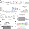
Fig. 1. ISDra2 MGE of the IS200/IS605 family.
a, Schematic representation of the D. radiodurans ISDra2 locus. The MGE consists of the tnpA and tnpB genes flanked by left end (LE) and right end (RE) partially palindromic elements (shown in red and blue, respectively). Amino acid residues at the predicted RuvC nuclease active site are indicated above the tnpB gene. b, TnpA-mediated ‘peel and paste’ transposition mechanism for ISDra2. The TnpA dimer catalyses transposon excision from the lagging strand during DNA replication forming a circular single-stranded DNA intermediate and a donor joint. The excised transposon circle inserts at the acceptor joint into the lagging DNA strand 3′ to the TTGAT sequence, completing the transposition cycle. Transposon excision/insertion sites are marked by red triangles. c, Experimental workflow of the expression and purification of the TnpB complex from E. coli cells and bound RNA extraction. sRNA-seq, small RNA sequencing. d, Alignment of sRNA sequenced reads to the ISDra2 locus. The blue colour shows the RNA sequences derived from the RE element, and the green marks the last 16 nt at the sequenced RNA 3′ ends, which align with the transposon flanking DNA.
Fig. 2
Fig. 2. TnpB protein is an RNA-guided dsDNA nuclease.
a, Experimental workflow for the establishment of dsDNA cleavage requirements by the TnpB–reRNA complex. E. coli cells were transformed with a plasmid expressing TnpB and HDV ribozyme-terminated reRNA, containing the 16-nt sequence that matched the target in the plasmid DNA library, flanked by the randomized 7-nt sequence (7N). Cell lysate was used for library digestion followed by double-stranded break (DSB) capture. F, forward primer, annealing to the ligated adapter; R1 and R2, reverse primers, annealing to the target plasmid backbone. b, Determination of adapter ligation positions indicate the formation of DSBs in the targeted sequence. ‘–TnpB’ represents the cleavage reactions using lysates obtained from the cells that do not express TnpB. The blue and red triangles indicate the positions of F + R1-enriched and F + R2-enriched adapter ligated reads, respectively. c, WebLogo representation of motifs identified in the 7N randomized region at 20–21-bp F + R1-enriched adapter ligated reads. d, Experimental workflow of the expression and purification of the TnpB RNP complex. E. coli cells were transformed with a plasmid expressing TnpB and a separate plasmid expressing HDV ribozyme-terminated reRNA. The reRNA-encoding construct contained the 16-nt guide sequence, which was different from the guide sequence used in the plasmid library cleavage experiment. e, The TnpB RNP complex cleaves supercoiled and linearized target plasmid in vitro. Cleavage is blocked by the D191A mutation at the RuvC-like active site. f, Target plasmid cleavage (TAM+/Target+, TAM−/Target+ and TAM+/Target−) by the TnpB RNP complex in vitro. TAM and the target complementary to the reRNA 3′-end sequence are required for plasmid DNA cleavage. g, Sanger sequencing of the TnpB-cleaved plasmid products reveals multiple cleavage positions at the non-targeted strand (NTS) and a single cleavage site at the target strand (TS) (marked with red triangles). For uncropped gel images, see Supplementary Fig. 1.
Fig. 3
Fig. 3. TnpB-mediated plasmid interference in vivo.
a, Experimental workflow of the plasmid interference assay in E. coli. The cleavage of a target plasmid results in loss of kanamycin (Kn) resistance. The reRNA-encoding construct contained the 16-nt guide sequence. AmpR, ampicillin/carbenicillin (Ap/Cb) resistance gene; KanR, kanamycin resistance gene. b, Plasmid interference assay. E. coli culture samples were serially diluted (10×) and the E. coli transformants were grown on the media supplemented with Cb and Kn at 25 °C for 44 h. Interference is compromised for the catalytically dead D191A and E278A TnpB variants. Target ‘+’ or ‘−’ indicates the plasmids with or without the target, respectively. For the uncropped plate image, see Supplementary Fig. 1. c, Proposed role of TnpB in transposition. The IS200/IS605 transposon circle is excised from the lagging strand during DNA replication resulting in two DNA copies: one copy that originates from the leading strand and carries an intact transposon, and another copy that originates from the lagging strand and lacks the transposon at the original site due to the strand-specific transposon excision. However, the latter DNA copy still carries the transposon ‘footprint’ in the form of the donor joint, comprised of the 5′-TTGAT sequence and the 3′-flanking DNA sequence that becomes a target to the TnpB–reRNA complex. In this case, the 5′-TTGAT sequence serves as a TAM that initiates the binding of the reRNA sequence to the matching DNA sequence followed by dsDNA cleavage. TnpB-induced DSB could facilitate homology-directed repair to reinstate the transposon at the donor joint using its intact copy on the sister chromatid, ensuring that both DNA copies have a transposon-coding gene before cell division. Red triangles indicate DNA cleavage sites.
Fig. 4
Fig. 4. TnpB nuclease is a novel genome editor.
a, The experimental workflow of the human cell line (HEK293T) genome-editing experiment. NLS, nuclear localization sequence. b, Detection of indel activity in the five tested targets of 20 nt in length in human gDNA (represented as the mean of three biologically independent experiments (shown in dots) ± standard deviation). The TnpB (non-targeting) expression plasmid used as a negative control encodes the reRNA-containing guide sequence that does not match any target in the human gDNA. c, Indel profile distribution within the target sequence in the EMX1-1 site showing the distribution of deletions (blue line) and insertions (red line) across the cleavage site (dotted line). The profile was obtained by aligning all reads at the EMX1-1 site and counting deletions and insertions at each position.
Extended Data Fig. 1
Extended Data Fig. 1. Purification of ISDra2 TnpB.
a, b, Experimental workflow for expression and purification of a single TnpB (a) and TnpB co-expressed with ISDra2 (ΔTnpA) (b) in E. coli cells. SDS-PAGE gels show elution fractions of proteins bound to HisTrap chelating column. Red boxes denote bands corresponding to an intact 10×MBP-TnpB protein (95.4 kDa). c, SDS-PAGE gel of pooled fractions indicated in a and b. d, Detection and analysis of nucleic acids co-purifying with TnpB protein. For uncropped gel images, see Supplementary Fig. 2.
Extended Data Fig. 2
Extended Data Fig. 2. TnpB RNP complex cleaves dsDNA in a TAM dependent manner.
a, Experimental workflow of double-stranded (ds) DNA cleavage activity detection. The reRNA encoding construct contained 20 nt guide sequence. F – forward primer, annealing to ligated adapter. R1 and R2 – reverse primers, annealing to the plasmid backbone. 7N represents the randomized region in the plasmid library next to the targeted sequence. b, Adapter ligation position determination indicating double-stranded break (DSB) formation in the targeted sequence. “–TnpB” represents the cleavage reactions using lysates obtained from the cells that do not express TnpB. c, d, WebLogo representation of motifs identified in 7N randomized region at 20–21 bp F + R1 and F + R1 (-TnpB) enriched adapter ligated reads, respectively. No substantial enrichment is observed at 7N region for “-TnpB” reactions.
Extended Data Fig. 3
Extended Data Fig. 3. Position frequency matrix (PFM) of the nucleotides at 7N region obtained after plasmid library cleavage using E. coli lysates.
a, b, PFM representation of nucleotides distribution identified in 7N randomized region at 20–21 bp F + R1 enriched adapter ligated reads obtained from E. coli cells expressing TnpB and reRNA constructs with 16 nt and 20 nt guide sequences, respectively. c, the cleavage reaction using lysate obtained from the cells that do not express TnpB demonstrates no substantial enrichment of motifs at 7N region.
Extended Data Fig. 4
Extended Data Fig. 4. TnpB RNP complex purification.
a, Experimental workflow of TnpB RNP complex expression and multi-step purification. The reRNA encoding construct contained a 16 nt guide sequence and carried an HDV ribozyme sequence at the 3’-end, which enabled production of the reRNA with fixed 16 nt length guide sequence. b, SDS-PAGE analysis of the purified TnpB and TnpB (D191A) RNP complexes. For uncropped gel image, see Supplementary Fig. 2. c, Molecular mass of TnpB and reRNA RNP complex determined by mass photometry. Experimentally established molecular mass corresponds to TnpB RNP complex consisting of a TnpB protein monomer bound to a ~150 nt long reRNA in a 1:1 molar ratio.
Extended Data Fig. 5
Extended Data Fig. 5. Synthetic dsDNA cleavage by TnpB RNP complex.
a, b, Cleavage of dsDNA substrates containing a target (represented in green colour) with TAM (red colour) (a) or without TAM sequence (b). TnpB RNP complex cleaves dsDNA in a TAM-dependent manner. Cleavage is blocked by D191A mutation at the RuvC-like active site (lane D). NTS and TS represent non-target and target strand, respectively. M – DNA marker lane. For uncropped gel images, see Supplementary Fig. 2.
Extended Data Fig. 6
Extended Data Fig. 6. Synthetic ssDNA cleavage by TnpB RNP complex.
a, b, Cleavage of ssDNA substrates containing a target (represented in green colour) with TAM (red colour) (a) or without TAM sequence (b). TnpB RNP complex cleaves ssDNA in a TAM-independent manner. Cleavage is blocked by D191A mutation at the RuvC-like active site (lane D). NTS and TS represent non-target and target strand, respectively. M – DNA marker lane. For uncropped gel images, see Supplementary Fig. 2.
Extended Data Fig. 7
Extended Data Fig. 7. Comparison of a TnpB model with experimentally determined representative structures of different Cas12 groups.
a, Schematic representation of common structural domains/motifs (coloured) and unique structural regions (grey) along the sequence; WED – wedge domain, corresponding to the β-barrel, REC – helical bundle, RuvC – RuvC domain with the inserted helical hairpin (HH) and either the zinc-finger domain (ZnF), or analogous domain inserted in the same relative position. Length of all sequences are approximately to scale. TnpB represents a minimal domain organization also present in Cas12 groups. b, Comparison of N-terminal regions between TnpB and other Cas12 proteins; the N-terminal WED and REC domains are involved in PAM recognition in Cas12f and are expected to participate in TAM recognition in TnpB. c, Comparison of corresponding RuvC regions.

Comment in

References

    1. Koonin EV. Viruses and mobile elements as drivers of evolutionary transitions. Phil. Trans. R. Soc. B. 2016;371:20150442. - PMC - PubMed
    1. Siguier P, Gourbeyre E, Chandler M. Bacterial insertion sequences: their genomic impact and diversity. FEMS Microbiol. Rev. 2014;38:865–891. - PMC - PubMed
    1. Kersulyte D, Mukhopadhyay AK, Shirai M, Nakazawa T, Berg DE. Functional organization and insertion specificity of IS607, a chimeric element of Helicobacter pylori. J. Bacteriol. 2000;182:5300–5308. - PMC - PubMed
    1. Kersulyte D, et al. Transposable element ISHp608 of Helicobacter pylori: nonrandom geographic distribution, functional organization, and insertion specificity. J. Bacteriol. 2002;184:992–1002. - PMC - PubMed
    1. Pasternak C, et al. ISDra2 transposition in Deinococcus radiodurans is downregulated by TnpB. Mol. Microbiol. 2013;88:443–455. - PubMed

Publication types