. 2021 Nov 23;5(22):4578-4589.
doi: 10.1182/bloodadvances.2020001514.

Third-party type 2 innate lymphoid cells prevent and treat GI tract GvHD

Affiliations

Third-party type 2 innate lymphoid cells prevent and treat GI tract GvHD

Danny W Bruce et al. Blood Adv. .

Abstract

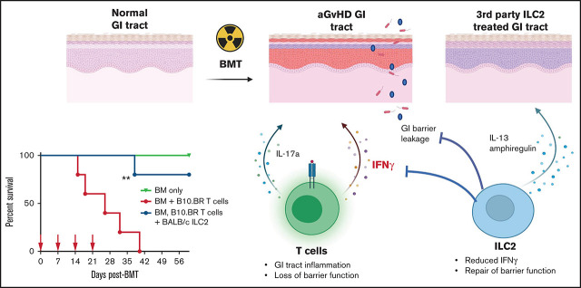
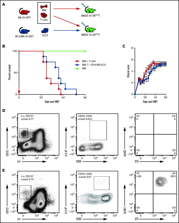
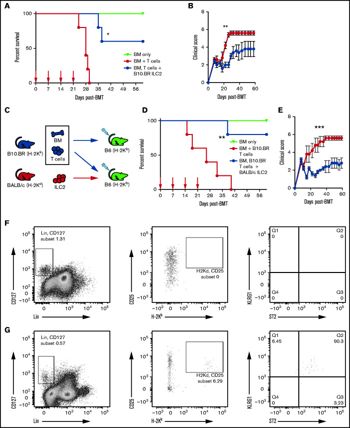
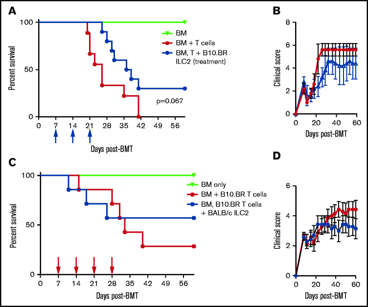
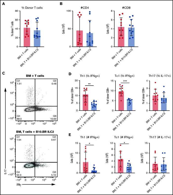
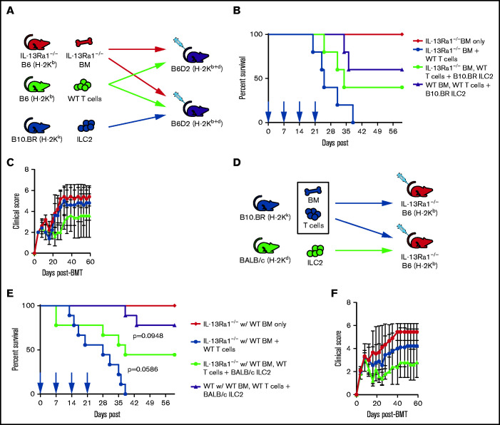
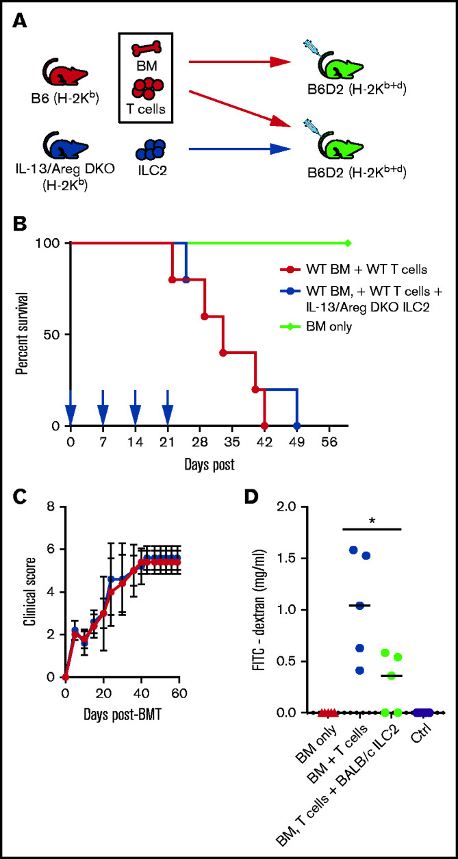
Acute graft-versus-host disease (aGVHD), mediated by the recognition of host major histocompatibility complex/peptide polymorphisms by donor T cells, remains a significant complication of allogeneic hematopoietic stem cell transplantation (allo-HSCT). aGVHD most commonly involves the gastrointestinal tract, liver, and skin; symptomatic aGVHD is treated with corticosteroids. Steroid-nonresponsive aGVHD is a significant problem for patients undergoing allo-HSCT, with <15% of these patients alive 1 year after diagnosis. Previously, we found that the infusion of donor innate lymphoid type 2 (ILC2) cells could prevent and treat aGVHD of the lower gastrointestinal tract with no effect on the graft-versus-leukemia response. This approach for clinical translation is cumbersome, as it would require the generation of donor-derived ILC2 cells for each recipient. Thus, the ability to use third-party ILC2 cells would provide an "off-the-shelf" reagent that could be used to treat and/or prevent aGVHD. Here, we show that third-party ILC2 cells enhance the survival of allo-HSCT recipients. Treatment required at least 4 weekly infusions of ILC2 cells. Mechanistically, we show that ILC2 cell function was completely lost if the cells could not express both interleukin-13 (IL-13) and amphiregulin. Finally, we show that the activity of IL-13 has a greater dependence on the expression of the IL-13R on host rather than donor bone marrow cells. The ability to generate third-party ILC2 cells offers a new avenue for the prevention of aGVHD.


Figures

None
Graphical abstract
Figure 1.
Figure 1.
Third-party ILC2s improve survival but do not persist. (A) Diagram for B6 into B6D2 transplantation. Each B6D2 recipient received B6 BM and B6 splenic T cells, with one group of B6D2 recipients also being given B10.BR ILC2s. Lethally irradiated B6D2 mice received TCD BM (BM only), BM plus total splenic T cells (BM + T cells), or BM plus T cells with activated B10.BR LC2s (BM, T cells + B10.BR ILC2). (B) Kaplan-Meier plot of survival following allo-HSCT; 1 representative of 2 experiments shown (n = 5 each experiment), log-rank (Mantel-Cox) test. (C) Clinical score posttransplantation, analyzed by using a two-way analysis of variance, with Bonferroni correction for repeated measures of multiple comparisons. Flow cytometry analysis of donor B10.BR (H-2Kk) ILC2 7 days’ posttransplant in the small intestine (D) and lung (E). BMT, BM transplantation.
Figure 2.
Figure 2.
Multiple infusions of third-party ILC2s reduce aGVHD incidence. Lethally irradiated B6D2 mice received B6 TCD BM (BM only), B6 BM plus total splenic B6 T cells (BM + T cells), or BM plus T cells with activated ILC2s (BM, T cells + B10.BR ILC2). (A) Kaplan-Meier plot of male recipient survival following allo-HSCT; red arrows indicate time points for B10.BR ILC2 injections, 1 representative of 2 experiments shown (n = 5 each experiment), log-rank (Mantel-Cox) test. (B) Clinical score of recipients from panel A, analyzed by using a two-way analysis of variance, with Bonferroni correction for repeated measures of multiple comparisons. (C) Diagram for B10.BR into B6 transplantation. Each B6 recipient received WT B10.BR BM and B10.BR splenic T cells, with one group of B6 recipients also being given BALB/c ILC2s. Lethally irradiated B6 mice received TCD BM (BM only), BM plus total splenic T cells (BM + B10.BR T cells), or BM plus T cells with activated B10.BR ILC2s (BM, B10.BR T cells + BALB/c ILC2s). (D) Kaplan-Meier plot of survival after allo-HSCT; white arrows indicate time points for BALB/c ILC2 injections for those receiving infusions beginning at the time of transplant; 1 representative of 2 experiments shown (n = 5 each experiment), log-rank (Mantel-Cox) test. (E) Clinical score posttransplantation, analyzed by using a two-way analysis of variance, with Bonferroni correction for repeated measures of multiple comparisons. *P < .05, **P < .01, ***P < .001. Flow cytometry analysis of donor B10.BR (H-2Kk) ILC2 12 days’ posttransplant following ILC2 infusions on day 0 and day 7 in the small intestine (F) and lung (G).
Figure 3.
Figure 3.
Efficacy of third-party ILC2 cells when given therapeutically. (A) Kaplan-Meier plot of B6D2 recipient survival following allo-HSCT, blue arrows indicate time points for B10.BR ILC2 injections for those receiving infusions beginning 7 days posttransplant (Treatment), representative of 2 experiments shown (n = 5 each experiment). Log-rank (Mantel-Cox) test. (B) Clinical score of recipients from panel A, analyzed by 2-way analysis of variance with Bonferroni correction for repeated measures of multiple comparisons P < .05 difference clinical score on days 22-26. (C) Kaplan-Meier plot of B6 recipient survival following allo-HSCT with B10.BR BM and T cells, red arrows indicate time points for BALB/c ILC2 injections for those receiving infusions beginning 7-days posttransplant (Treatment), one representative of 2 experiments shown (n = 7 each experiment). Log-rank (Mantel-Cox) test. (D) Clinical score of recipients from panel C, analyzed by 2-way analysis of variance with Bonferroni correction for repeated measures of multiple comparisons.
Figure 4.
Figure 4.
Cotransplantation of ILC2s reduces pro-inflammatory donor T-cell numbers in the GI tract. Donor T cells were evaluated in the GI tract 12 days’ posttransplant by using green fluorescent protein–positive splenic T cells alone (BM + T cells) or green fluorescent protein–positive T cells and B10.BR ILC2s (BM, T cells + B10.BR ILC2s). (A) Frequency of total LP lymphocytes. (B) Total number of donor CD4+ and CD8+ T cells in the colonic lamina propria lymphocytes. (C) Flow cytometry plots of intracellular cytokine expression from colon. Percentage (D) and total number (E) of IFN-γ producing donor CD4+ and CD8+ T cells and the number of donor CD4+ T cells producing IL-17A in the LP. These data represent 2 independent experiments (n = 5), Student’s t test with Welch’s correction; *P < .05, **P < .01, ***P < .001.
Figure 5.
Figure 5.
ILC2s suppress aGVHD, in part, through IL-13Ra1 signaling. (A) Diagram for B6 into B6D2 transplantation. Each B6D2 recipient received IL-13Ra1−/− B6 BM and B6 splenic T cells from WT donors, with one group of B6D2 recipients also being given B10.BR ILC2s. Lethally irradiated B6D2 mice received TCD BM (IL-13Ra1−/− BM only), BM plus total splenic T cells (IL-13Ra1−/− BM + WT T cells), or BM plus T cells with activated B10.BR LC2s (IL-13Ra1−/− BM, WT T cells + B10.BR ILC2). (B) Kaplan-Meier plot of survival after allo-HSCT; blue arrows indicate time points for B10.BR ILC2 injections for those receiving infusions beginning at the time of transplant. One representative of 2 experiments shown (n = 5 each experiment), log-rank (Mantel-Cox) test. (C) Clinical score posttransplantation, analyzed by using two-way analysis of variance, with Bonferroni correction for repeated measures of multiple comparisons. (D) Diagram for B10.BR into IL-13Ra1−/− B6 transplantation. Each IL-13Ra1−/− B6 recipient received WT B10.BR BM and B10.BR splenic T cells, with one group of IL-13Ra1−/− B6 recipients also being given BALB/c ILC2s. Lethally irradiated IL-13Ra1−/− B6 mice received TCD BM (BM only), BM plus total splenic T cells (BM + B10.BR T cells), or BM plus T cells with activated BALB/c ILC2s (BM, B10.BR T cells + BALB/c ILC2s). (E) Kaplan-Meier plot of survival after allo-HSCT; red arrows indicate time points for BALB/c ILC2 injections for those receiving infusions beginning at the time of transplant. Representative of 2 experiments shown (n = 5 each experiment), log-rank (Mantel-Cox) test. (F) Clinical score posttransplantation, analyzed by using a two-way analysis of variance, with Bonferroni correction for repeated measures of multiple comparisons.
Figure 6.
Figure 6.
ILC2 mechanism of suppression of GVHD is dependent upon ILC2 expression of IL-13 and AREG. (A) Diagram for B6 into B6D2 transplantation. Each B6D2 recipient received B6 BM and B6 splenic T cells with one group of B6D2 recipients also being given IL-13/Areg DKO ILC2s. Lethally irradiated B6D2 mice received TCD BM (BM only), BM plus total splenic T cells (BM + T cells), or BM plus T cells with activated B10.BR ILC2s (BM, T cells + IL-13/Areg DKO ILC2s). (B) Kaplan-Meier plot of survival following allo-HSCT, 1 representative of 2 experiments shown (n = 5 each experiment), log-rank (Mantel-Cox) test. (C) Clinical score posttransplantation, analyzed by using a two-way analysis of variance, with Bonferroni correction for repeated measures of multiple comparisons. (D) Quantification of FITC-dextran in the serum of BMT recipients described in panel C 28 days after transplant. One representative of 2 independent experiments shown; mean ± standard error of the mean (n = 5 per group). Statistical analysis by Student t test, *P < .05.
Figure 7.
Figure 7.
Expanded ILC2s are resistant to glucocorticoid treatment. Flow cytometry analysis of survival of in vitro expanded ILC2s when treated with dexamethasone in the presence or absence of cytokines for 48 hours. Analyzed by using a two-way analysis of variance, with Bonferroni correction for repeated measures of multiple comparisons, **P < .01.

References

    1. Horwitz ME. Reduced intensity versus myeloablative allogeneic stem cell transplantation for the treatment of acute myeloid leukemia, myelodysplastic syndrome and acute lymphoid leukemia. Curr Opin Oncol. 2011;23(2):197-202. - PubMed
    1. Kersey JH. The role of allogeneic-cell transplantation in leukemia. N Engl J Med. 2010;363(22):2158-2159. - PubMed
    1. Garnett C, Apperley JF, Pavlů J.. Treatment and management of graft-versus-host disease: improving response and survival. Ther Adv Hematol. 2013;4(6):366-378. - PMC - PubMed
    1. Martin PJ, Rizzo JD, Wingard JR, et al. . First- and second-line systemic treatment of acute graft-versus-host disease: recommendations of the American Society of Blood and Marrow Transplantation. Biol Blood Marrow Transplant. 2012;18(8):1150-1163. - PMC - PubMed
    1. Gatti RA, Kersey JH, Yunis EJ, Good RA.. Graft-versus-host disease. Prog Clin Pathol. 1973;5:1-18. - PubMed

Publication types

MeSH terms