. 2021 Nov;42(11):2101-2106.
doi: 10.3174/ajnr.A7295. Epub 2021 Oct 7.

Filtered Diffusion-Weighted MRI of the Human Cervical Spinal Cord: Feasibility and Application to Traumatic Spinal Cord Injury

Affiliations

Filtered Diffusion-Weighted MRI of the Human Cervical Spinal Cord: Feasibility and Application to Traumatic Spinal Cord Injury

S A Murphy et al. AJNR Am J Neuroradiol. 2021 Nov.

Abstract

Background and purpose: In traumatic spinal cord injury, DTI is sensitive to injury but is unable to differentiate multiple pathologies. Axonal damage is a central feature of the underlying cord injury, but prominent edema confounds its detection. The purpose of this study was to examine a filtered DWI technique in patients with acute spinal cord injury.

Materials and methods: The MR imaging protocol was first evaluated in a cohort of healthy subjects at 3T (n = 3). Subsequently, patients with acute cervical spinal cord injury (n = 8) underwent filtered DWI concurrent with their acute clinical MR imaging examination <24 hours postinjury at 1.5T. DTI was obtained with 25 directions at a b-value of 800 s/mm2. Filtered DWI used spinal cord-optimized diffusion-weighting along 26 directions with a "filter" b-value of 2000 s/mm2 and a "probe" maximum b-value of 1000 s/mm2. Parallel diffusivity metrics obtained from DTI and filtered DWI were compared.

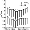
Results: The high-strength diffusion-weighting perpendicular to the cord suppressed signals from tissues outside of the spinal cord, including muscle and CSF. The parallel ADC acquired from filtered DWI at the level of injury relative to the most cranial region showed a greater decrease (38.71%) compared with the decrease in axial diffusivity acquired by DTI (17.68%).

Conclusions: The results demonstrated that filtered DWI is feasible in the acute setting of spinal cord injury and reveals spinal cord diffusion characteristics not evident with conventional DTI.


Figures

FIG 1.
FIG 1.
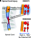
Schematic representation of the fDWI/DDE MR imaging technique for spinal cord injury. Traumatic injury to the spinal cord (A) results in microscopic damage to axons, illustrated here as beading and end-bulbs that reflect the underlying acute pathology (B). A prominent edema response (light blue) is typical surrounding the injury site. Traditional DWI/DTI derives measures reflecting the bulk sum of all features. With fDWI/DDE, a high-strength diffusion-weighting perpendicular to the spinal cord suppresses extracellular edema (and CSF) to estimate tissue-specific diffusivity metrics less confounded by edema.
FIG 2.
FIG 2.
fADC and AD maps for the healthy spinal cord. Single-subject fADC (A) and AD maps (B) at C4 for a healthy individual on a 3T system. Comparison of the mean white matter fADC or AD values (C) and SNR (D).
FIG 3.
FIG 3.
fADC and AD maps for an individual (subject 3) with an acute spinal cord injury. A T2-weighted image for an individual with an acute spinal cord injury on a 1.5T system. Single slices above, at, and below the injury site (as labeled in the T2 image) for fADC and AD maps. Ax GRE indicates axial gradient recalled-echo.
FIG 4.
FIG 4.
fADC and AD compared at each individual section for acute spinal cord injury (n = 8). There is a large, unidirectional decrease in fADC at the injury site compared with a lesser, multidirectional decrease in AD values. The asterisk indicates significance compared with the first section (P < .05).
FIG 5.
FIG 5.
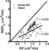
Correlations of fADC and AD at each individual section for the intact spinal cord and acute spinal cord injury. Correlations are significant for the intact spinal cord; however, a lower correlation and nonrandom residuals for the acute SCI setting indicate that fADC and AD do not have a simple linear relationship, suggesting that they provide differing information.

References

    1. Zhao C, Rao JS, Pei XJ, et al. . Longitudinal study on diffusion tensor imaging and diffusion tensor tractography following spinal cord contusion injury in rats. Neuroradiology 2016;58:607–14 10.1007/s00234-016-1660-7 - DOI - PubMed
    1. Kim JH, Loy DN, Wang Q, et al. . Diffusion tensor imaging at 3 hours after traumatic spinal cord injury predicts long-term locomotor recovery. J Neurotrauma 2010;27:587–98 10.1089/neu.2009.1063 - DOI - PMC - PubMed
    1. Skinner NP, Kurpad SN, Schmit BD, et al. . Rapid in vivo detection of rat spinal cord injury with double-diffusion-encoded magnetic resonance spectroscopy. Magn Reson Med 2017;77:1639–49 10.1002/mrm.26243 - DOI - PMC - PubMed
    1. Patel SP, Smith TD, VanRooyen JL, et al. . Serial diffusion tensor imaging in vivo predicts long-term functional recovery and histopathology in rats following different severities of spinal cord injury. J Neurotrauma 2016;33:917–28 10.1089/neu.2015.4185 - DOI - PMC - PubMed
    1. Chiang CW, Wang Y, Sun P, et al. . Quantifying white matter tract diffusion parameters in the presence of increased extra-fiber cellularity and vasogenic edema. Neuroimage 2014;101:310–19 10.1016/j.neuroimage.2014.06.064 - DOI - PMC - PubMed

Publication types

MeSH terms