. 2021 Oct 19;118(42):e2113015118.
doi: 10.1073/pnas.2113015118.

A mutation-mediated evolutionary adaptation of Zika virus in mosquito and mammalian host

Affiliations

A mutation-mediated evolutionary adaptation of Zika virus in mosquito and mammalian host

Xi Yu et al. Proc Natl Acad Sci U S A. .

Abstract

Zika virus (ZIKV) caused millions of infections during its rapid and expansive spread from Asia to the Americas from 2015 to 2017. Here, we compared the infectivity of ZIKV mutants with individual stable substitutions which emerged throughout the Asian ZIKV lineage and were responsible for the explosive outbreaks in the Americas. A threonine (T) to alanine (A) mutation at the 106th residue of the ZIKV capsid (C) protein facilitated the transmission by its mosquito vector, as well as infection in both human cells and immunodeficient mice. A mechanistic study showed that the T106A substitution rendered the C a preferred substrate for the NS2B-NS3 protease, thereby facilitating the maturation of structural proteins and the formation of infectious viral particles. Over a complete "mosquito-mouse-mosquito" cycle, the ZIKV C-T106A mutant showed a higher prevalence of mosquito infection than did the preepidemic strain, thus promoting ZIKV dissemination. Our results support the contribution of this evolutionary adaptation to the occasional widespread reemergence of ZIKV in nature.

Keywords: Zika virus; evolutionary adaptation; host; mosquito; mutation.


Conflict of interest statement

The authors declare no competing interest.

Figures
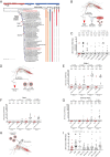
Fig. 1.
Fig. 1.
The effect of C-T106A substitution on the infectivity and prevalence of A. aegypti mosquitoes. (A) Phylogenetic analysis. Evolutionary mutations among ZIKV strains are indicated at the branch points of the phylogenetic tree. Asian lineage strains FSS13025 and PRVABC59, representing preepidemic and contemporary isolates, are indicated by red labels. The complete open reading frame nucleotide sequences of 47 representative ZIKV strains were aligned and analyzed using the maximum likelihood method with 1,000 bootstrap replicates in the MEGA X program. (B and C) Infectivity of ZIKV mutants in mosquitoes. Vero cells were infected with viruses at a multiplicity of infection (MOI) of 0.1, and the culture supernatant was collected 4 dpi. ZIKV-infected Vero cell supernatants (500 μL) were mixed with fresh human blood (500 μL) for the membrane feeding of A. aegypti. The number at the top of each column represents the infected number/total number. (DG) Vector competence of A. aegypti to the ZIKV FSS13025 strain or FSS-C T106A mutant. Purified ZIKV virions (10 μL) were mixed with fresh human blood (990 μL) for the membrane feeding of A. aegypti. A final concentration of 1 × 10⁶ p.f.u. ⋅ mL−1 ZIKV was used for mosquito oral infection. The mosquito midguts, heads, and salivary glands were dissected at 4, 7, 10, and 14 d postfeeding. The ZIKV infection (E), dissemination (F), and transmission (G) rates were determined by taking the number of infected midguts (E), heads (F), and salivary glands (G) divided by the total number of engorged mosquitoes. (H and I) The infectivity of ZIKV with single–amino acid mutations in mosquito hemocoel tissues. A. aegypti mosquitoes were infected intrathoracically with 50 p.f.u. of purified ZIKV particles. (C, EG, and I) Each dot represents a mosquito. P values were determined by the two-sided Fisher’s exact test (C and EG) or the two-tailed Mann–Whitney U test (I). (C, E–G, and I) P > 0.5 not significant (n.s.). P values were adjusted using the Benjamini–Hochberg procedure (C) or the Dunnett’s test (I) to account for multiple comparisons. (C and I) The P value represents a comparison between the FSS13025 and the other groups. (C, EG, and I) The limit of detection is illustrated by dotted lines. (C, EG, and I) Experiments consisted of at least three biological replicates with similar results. FSS and PRV represent the ZIKV FSS13025 and PRVABC59 strains, respectively.
Fig. 2.
Fig. 2.
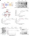
The effect of C-T106A substitution on infectivity in mammalian hosts. (AC) Replication kinetics of FSS13025 and FSS-C T106A viruses on THP-1 cells (A), hu-moDC (B), and hu-moMØ (C). The cells were infected with 0.1 MOI of the indicated viruses. The infected cells were detected at 24, 48, and 72 h postinfection by qRT-PCR, and the ZIKV quantities were normalized against human GAPDH. (DF) Comparison of A129 mouse infection by FSS13025 and FSS-C T106A viruses. The 6-wk-old A129 mice were infected with 50 p.f.u. of FSS13025 or FSS-C T106A virus by footpad inoculation. (D) Viraemia detection. The viremia from infected A129 mice (n = 6 per group pooled from three independent biological replicates) was measured over a time course using a plaque assay. (E) Viral loads in mice tissues. At day 7 postinfection, the animals were euthanized, and the viral load in tissues was measured by qRT-PCR. (F) Animal mortality was recorded daily in the animals infected with either the FSS13025 or FSS-C T106A viruses (n = 10 mice per group pooled from three independent biological replicates). (AE) The data are presented as the means ± SEM. (AE) The two-tailed Mann–Whitney U test was performed for statistical analysis. (F) The survival rates of the infected mice were plotted using a Kaplan–Meier curve and were statistically analyzed using the log-rank (Mantel–Cox) test. (A–F) P > 0.5 not significant (n.s.). (AE) Experiments consisted of at least three biological replicates with similar results. FSS represents the ZIKV FSS13025 strain.
Fig. 3.
Fig. 3.
The C-T106A substitution promotes ZIKV infection through more efficient C maturation. (A) Schematic representation of ZIKV structural proteins and the viral Ser protease NS2B-NS3 cleavage site on the C protein. (B) Expression of viral proteins in FSS13025- or FSS-C T106A–infected Vero cells. The cells were infected with 0.1 MOI of each virus. At 24, 48, and 72 h postinfection, the infected cells were collected for SDS-PAGE and Western blot analysis. (C) Schematic representation of the ZIKV virion assembly resulting from NS2B-NS3 Ser protease and host signalase cleavage. (DF) The C-T106A mutation promotes the efficient production of infectious virions. Vero cells were infected with 0.1 MOI of FSS or FSS-C T106A virus. The culture supernatant was collected at the indicated time points postinfection. (D) The concentration of the ZIKV E protein in the collected supernatant was determined by ELISA. (E) The quantity of the ZIKV genome in the collected supernatant was detected by qRT-PCR. (F) The quantity of ZIKV infectious virions in the collected supernatant was detected by the plaque assay. (G and H) The C-T106A substitution promoted NS2B-NS3 Ser protease-mediated cleavage activity. (G) Purification of a recombinant NS2B-NS3 protein with an E. coli expression system. The protein was detected by staining with Coomassie blue in an SDS-PAGE gel and by Western blotting using an anti-T7 tag antibody. (H) The protease activity of recombinant NS2B-NS3 was measured using Abz/Dnp-labeled FSS13025 or FSS-C T106A peptide substrates. The assays were performed in triplicate at 37 °C using a 10-nM enzyme concentration with varying substrate concentrations starting from 500 μM. The Michaelis–Menten kinetics were plotted using a nonlinear regression function. The catalytic rates, the binding affinity of the substrate, and catalytic efficiency are mentioned in the table inset in the graph. (I) Cleavage of C-prM by the NS2B-NS3 protease. The ZIKV NS2B-NS3 protease and ZIKV FSS C-prM proteins with either Thr-106 or Ala-106 substitution were ectopically coexpressed in 293T cells, and the cell lysates were analyzed using Western blotting at 48-h posttransfection. GAPDH expression was used as a loading control. (DF) The data are presented as the means ± SEM. (DF) The two-tailed Mann–Whitney U test was performed for statistical analysis. (B, DG, and I) Experiments consisted of at least three biological replicates with similar results. (H) The data were pooled from four independent biological replicates, and the data are presented as the means ± SD. (H) The P values were determined by the Student’s unpaired t test. (D–F and H) P > 0.5 not significant (n.s.). FSS represents the ZIKV FSS13025 strain.
Fig. 4.
Fig. 4.
The C-T106A substitution promotes both ZIKVs during the mosquito-host-mosquito lifecycle. (A) Schematic representation of the study design. (B) Detection of ZIKV viremia by the plaque assay (n = 3 mice per group). (C and D) Comparison of the infection rate (C) and mosquito infectivity (D) of the ZIKV strains (n = 3 mice per group). (D) The number at the top of each column represents the infected number/total number. (D) Each dot represents a mosquito. (B and C) The data are the means ± SEM. The P values were determined by the two-tailed Mann–Whitney U test (B and D) or the two-sided Fisher’s exact test (C). P values were adjusted using the Dunnett’s test (B) or the Benjamini–Hochberg procedure (C and D) to account for multiple comparisons. The black P value represents a comparison between FSS13025 and PRVABC59. The gray P value represents a comparison between FSS13025 and FSS-C T106A. *P < 0.05; **P < 0.01; ***P < 0.001; ****P < 0.0001; and n.s., not significant. (D) The limit of detection is illustrated by dotted lines. (BD) Experiments consisted of at least three biological replicates with similar results. FSS and PRV represent the ZIKV FSS13025 and PRVABC59 strains, respectively.

References

    1. Musso D., Nilles E. J., Cao-Lormeau V.-M., Rapid spread of emerging Zika virus in the Pacific area. Clin. Microbiol. Infect. 20, O595–O596 (2014). - PubMed
    1. Marchette N. J., Garcia R., Rudnick A., Isolation of Zika virus from Aedes aegypti mosquitoes in Malaysia. Am. J. Trop. Med. Hyg. 18, 411–415 (1969). - PubMed
    1. Musso D., Gubler D. J., Zika virus. Clin. Microbiol. Rev. 29, 487–524 (2016). - PMC - PubMed
    1. Faye O., et al. ., Molecular evolution of Zika virus during its emergence in the 20th century. Int. J. Infect. Dis. 21, 2–3 (2014). - PMC - PubMed
    1. Cao-Lormeau V.-M., et al. ., Zika virus, French polynesia, South Pacific, 2013. Emerg. Infect. Dis. 20, 1085–1086 (2014). - PMC - PubMed

Publication types