. 2021 Oct 7;12(1):5868.
doi: 10.1038/s41467-021-26178-y.

ChAdOx1 nCoV-19 (AZD1222) protects Syrian hamsters against SARS-CoV-2 B.1.351 and B.1.1.7

Affiliations

ChAdOx1 nCoV-19 (AZD1222) protects Syrian hamsters against SARS-CoV-2 B.1.351 and B.1.1.7

Robert J Fischer et al. Nat Commun. .

Abstract

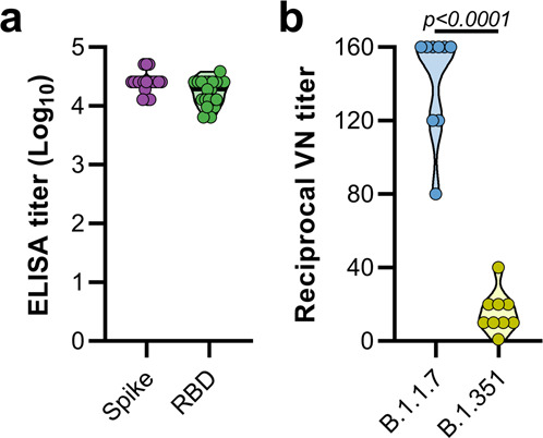
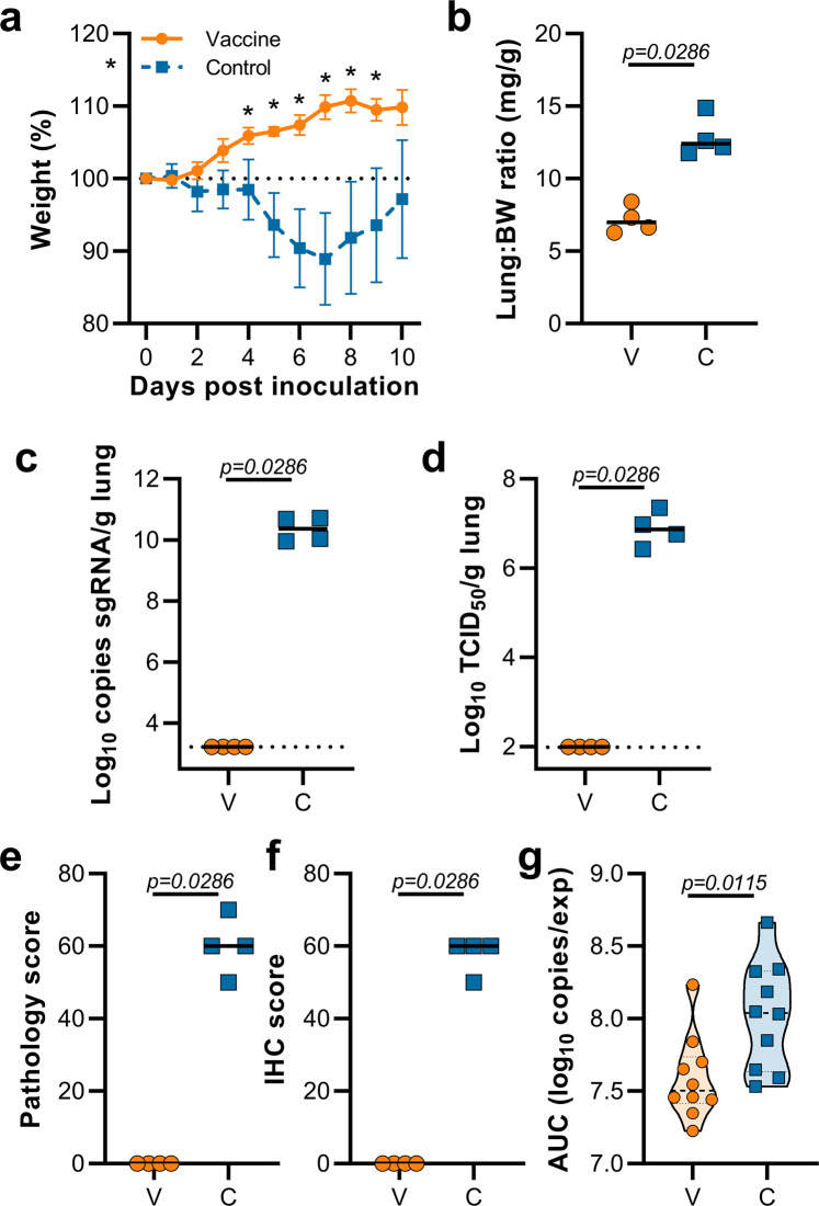
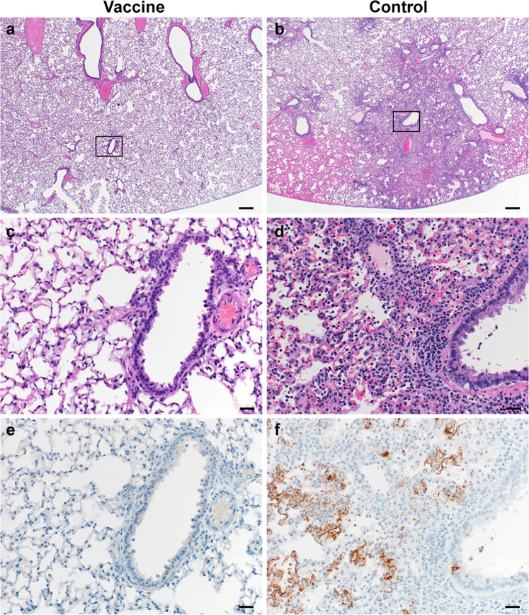
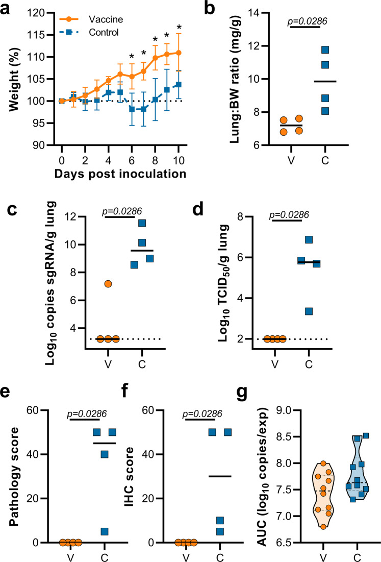
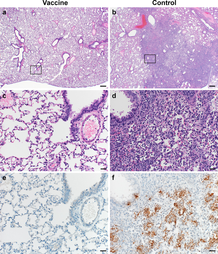
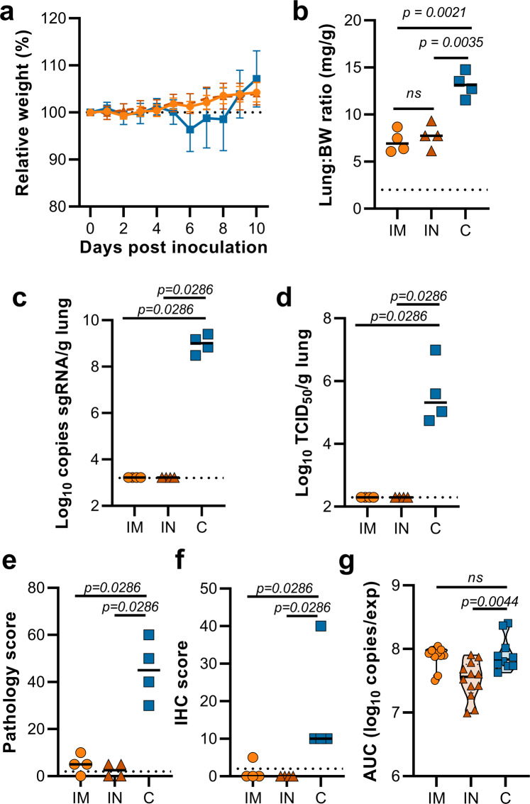
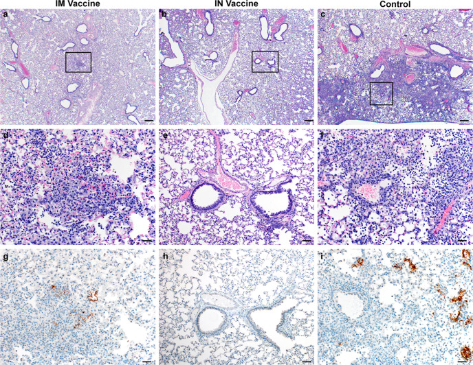
We investigated ChAdOx1 nCoV-19 (AZD1222) vaccine efficacy against SARS-CoV-2 variants of concern (VOCs) B.1.1.7 and B.1.351 in Syrian hamsters. We previously showed protection against SARS-CoV-2 disease and pneumonia in hamsters vaccinated with a single dose of ChAdOx1 nCoV-19. Here, we observe a 9.5-fold reduction of virus neutralizing antibody titer in vaccinated hamster sera against B.1.351 compared to B.1.1.7. Vaccinated hamsters challenged with B.1.1.7 or B.1.351 do not lose weight compared to control animals. In contrast to control animals, the lungs of vaccinated animals do not show any gross lesions. Minimal to no viral subgenomic RNA (sgRNA) and no infectious virus can be detected in lungs of vaccinated animals. Histopathological evaluation shows extensive pulmonary pathology caused by B.1.1.7 or B.1.351 replication in the control animals, but none in the vaccinated animals. These data demonstrate the effectiveness of the ChAdOx1 nCoV-19 vaccine against clinical disease caused by B.1.1.7 or B.1.351 VOCs.


Conflict of interest statement

S.C.G. is a board member of Vaccitech and named as an inventor on a patent covering the use of ChAdOx1-vector-based vaccines and a patent application covering a SARS-CoV-2 (nCoV-19) vaccine (UK patent application no. 2003670.3). T.L. is named as an inventor on a patent application covering a SARS-CoV-2 (nCoV-19) vaccine (UK patent application no. 2003670.3). The University of Oxford and Vaccitech, having joint rights in the vaccine, entered into a partnership with AstraZeneca in April 2020 for further development, large-scale manufacture, and global supply of the vaccine. Equitable access to the vaccine is a key component of the partnership. Neither Oxford University nor Vaccitech will receive any royalties during the pandemic period or from any sales of the vaccine in developing countries. All other authors declare no competing interests.

Figures

Fig. 1
Fig. 1. Vaccination of Syrian hamsters with ChAdOx1 nCoV-19 elicits binding and neutralizing antibodies against B.1.1.7 and B.1.351.
a Violin plot of binding antibodies against spike protein or RBD of SARS-CoV-2 (clade A) in serum obtained 25 days post vaccination with ChAdOx1 nCoV-19. b Violin plot of virus neutralizing antibody titers against B.1.1.7 or B.1.351 in serum obtained 25 days post vaccination with ChAdOx1 nCoV-19. Statistical significance determined via Mann–Whitney test, n = 9 biologically independent animals.
Fig. 2
Fig. 2. Vaccination of Syrian hamsters with ChAdOx1 nCoV-19 prevents lower respiratory tract infection with SARS-CoV-2 VOC B.1.1.7.
a Relative weight upon intranasal challenge with 104 TCID50 of B.1.1.7. Shown is the geometric mean with a 95% confidence interval (CI). *p-value < 0.05, corrected for multiple comparisons using the Holm–Šidák correction. b Lung:body weight (BW) ratio (mg:g) of hamsters euthanized at 5 DPI. Line = median. c sgRNA viral load in lung tissue obtained at 5 DPI. Line = median. Dotted line = limit of detection. d Infectious SARS-CoV-2 titer in lung tissue obtained at 5 DPI. Line = median. Dotted line = limit of detection. e Percentage affected lung tissue per animal as determined via histology. Line = median. f Percentage of lung tissue positive for SARS-CoV-2 antigen per animal. Line = median. g Truncated violin plot of the area under the curve (AUC) analysis of shedding as measured by sgRNA analysis in swabs collected on 1–5 DPI. Dashed line = median. Dotted line = quartiles. Statistical significance determined via multiple student t-tests with an n of 10 biologically independent animals (a), or two-tailed Mann–Whitney test (be) with an n of 4 biologically independent animals and (g) with an n of 10 biologically independent animals. p-values for days 4, 5, 6, 7, 8, 9, and 10 are 0.001251, < 0.000001, < 0.000001, < 0.000001, < 0.000001, < 0.000001, and < 0.000001, respectively. V = ChAdOx1 nCoV-19 vaccinated; C = ChAdOx1 GFP vaccinated; Orange circle = Hamsters vaccinated with ChAdOx1 nCoV-19, Blue square = Hamsters vaccinated with ChAdOx1 GFP.
Fig. 3
Fig. 3. Pulmonary effects of direct intranasal challenge with SARS-CoV-2 variant B.1.1.7 in Syrian hamsters at 5 DPI.
a, b H&E staining, 20x, scale bar = 200 µm; a no pathology. b Focally extensive areas of bronchointerstitial pneumonia. c, d H&E staining, 200x, scale bar = 20 µm; c no pathology. d Bronchointerstitial pneumonia with alveolar histiocytosis, fibrin and edema. e, f IHC staining against N protein SARS-CoV-2 (brown) 200x, scale bar = 20 µm. e No staining. f Staining of bronchiolar epithelial cells, type I&II pneumocytes, and rare macrophages. Micrographs are representative of what was observed viewing 100% of a single 5 µm section of the complete lungs containing all lobes for each of four animals.
Fig. 4
Fig. 4. Vaccination of Syrian hamsters with ChAdOx1 nCoV-19 prevents lower respiratory tract infection with SARS-CoV-2 VOC B.1.351.
a Relative weight upon intranasal challenge with 104 TCID50 of B.1.351. Shown is the geometric mean with a 95% confidence interval (CI). *p-value < 0.005, corrected for multiple comparisons using the Holm–Šidák correction. b Lung:body weight (BW) ratio (mg:g) of hamsters euthanized at 5 DPI. Line = median. c sgRNA viral load in lung tissue obtained at 5 DPI. Line = median. Dotted line = limit of detection. d Infectious SARS-CoV-2 titer in lung tissue obtained at 5 DPI. Line = median. Dotted line = limit of detection. e Percentage affected lung tissue per animal as determined via histology. Line = median. f Percentage of lung tissue positive for SARS-CoV-2 antigen per animal. Line = median. g Truncated violin plot of area under the curve (AUC) analysis of shedding as measured by sgRNA analysis in swabs collected on 1–5 DPI. Dashed line = median. Dotted line = quartiles. Statistical significance determined via multiple student t-tests with an n of 10 biologically independent animals (a), or two-tailed Mann–Whitney test (be) with an n of 4 biologically independent animals and (g) with an n of 10 biologically independent animals. p-values for days 6, 7, 8, 9, and 10 are 0.000004, <0.000001, <0.000001, <0.000001 and 0.000007, respectively. V = ChAdOx1 nCoV-19 vaccinated; C = ChAdOx1 GFP vaccinated; Orange circle = Hamsters vaccinated with ChAdOx1 nCoV-19, Blue square = Hamsters vaccinated with ChAdOx1 GFP.
Fig. 5
Fig. 5. Pulmonary effects of direct intranasal challenge with SARS-CoV-2 variant B.1.351 in Syrian hamsters at 5 DPI.
ab H&E staining, 20x, scale bar = 200 µm; a no pathology. b Focally extensive areas of bronchointerstitial pneumonia. c, d H&E staining, 200x, scale bar = 20 µm; c no pathology. d Bronchointerstitial pneumonia with alveolar histiocytosis, fibrin, and edema. e, f IHC staining against SARS-CoV-2 (brown) 200x, scale bar = 20 µm; e no staining. f Staining of bronchiolar epithelial cells, type I&II pneumocytes, and rare macrophages. Micrographs are representative of what was observed viewing 100% of a single 5 µm section of the complete lungs containing all lobes for each of four animals.
Fig. 6
Fig. 6. Vaccination of Syrian hamsters with ChAdOx1 nCoV-19 via the IM or IN route prevents lower respiratory tract infection with SARS-CoV-2 VOC B.1.351-2.
Hamsters were vaccinated by the IM or the IN routes 60 days prior to challenge with B.1.351-2. a Relative weight upon intranasal challenge with 104 TCID50 of B.1.351. Shown is the geometric mean with a 95% confidence interval (CI). There was no statistical significance between the groups on any day. b Lung:body weight (BW) ratio (mg:g) of hamsters euthanized at 5 DPI. Line = median. c sgRNA viral load in lung tissue obtained at 5 DPI. Line = median. Dotted line = limit of detection. d Infectious SARS-CoV-2 titer in lung tissue obtained at 5 DPI. Line = median. Dotted line = limit of detection. e Percentage affected lung tissue per animal as determined via histology. Line = median. f Percentage of lung tissue positive for SARS-CoV-2 antigen per animal. Line = median. g Truncated violin plot of area under the curve (AUC) analysis of shedding as measured by sgRNA analysis in swabs collected on 1–5 DPI. Dashed line = median. Dotted line = quartiles. Statistical significance determined via mixed-effect analyses n = 12 biologically independent animals (vaccinated groups) and n = 10 (control group) (a), or two-tailed Mann–Whitney test (be) with an n of 8 biologically independent animals and (g) with an n of 12 biologically independent animals (vaccinated groups) and n = 10 (control group). IM = ChAdOx1 nCoV-19 vaccinated via intramuscular route; IN = ChAdOx1 nCoV-19 vaccinated via intranasal route; C = naïve animals; Orange circle = Hamsters vaccinated with ChAdOx1 nCoV-19 via intramuscular route, Orange triangle = Hamsters vaccinated with ChAdOx1 nCoV-19 via intranasal route, Blue square = Naïve hamsters.
Fig. 7
Fig. 7. Pulmonary effects of direct IN challenge with SARS-CoV-2 variant B.1.351 in Syrian hamsters which received an IN or IM vaccine at 5 DPI.
ac H&E 20x, scale bar = 200 µm; a no pathology. b, c general prevalence of interstitial pneumonia. df H&E 200x, scale bar = 20 µm; d No pathology e rare focus of interstitial pneumonia. f moderate interstitial pneumonia. gi IHC staining against N protein SARS-CoV-2, 200x, scale bar = 20 µm. g No viral antigen. h Rare foci of viral antigen in type I pneumocytes. i Viral antigen within the larger area of interstitial pneumonia in type I pneumocytes. Micrographs are representative of what was observed viewing 100% of a single 5 µm section of the complete lungs containing all lobes for each of four animals.

Update of

References

    1. Chand, M. et al. Investigation of Novel SARS-COV-2 variant Variant of Concern 202012/01https://assets.publishing.service.gov.uk/government/uploads/system/uploa... (2020).
    1. Davies, N. G. et al. Estimated transmissibility and impact of SARS-CoV-2 lineage B.1.1.7 in England. Science10.1126/science.abg3055 (2021). - PMC - PubMed
    1. Tegally, H. et al. Emergence And Rapid Spread of a New Severe Acute Respiratory Syndrome-Related Coronavirus 2 (SARS-CoV-2) Lineage with Multiple Spike Mutations in South Africa. 10.1101/2020.12.21.20248640 (2020).
    1. van Doremalen, N. et al. Intranasal ChAdOx1 nCoV-19/AZD1222 Vaccination Reduces Shedding of SARS-CoV-2 D614G in Rhesus Macaques. 10.1101/2021.01.09.426058 (2021). - PubMed
    1. Port, J. R. et al. SARS-CoV-2 Disease Severity and Transmission Efficiency is Increased for Airborne But Not Fomite Exposure in Syrian Hamsters.10.1101/2020.12.28.424565 (2020). - PMC - PubMed

Publication types