Review
. 2021 Oct 7;7(1):73.
doi: 10.1038/s41572-021-00307-9.

Histiocytic disorders

Affiliations
Review

Histiocytic disorders

Kenneth L McClain et al. Nat Rev Dis Primers. .

Abstract

The historic term 'histiocytosis' meaning 'tissue cell' is used as a unifying concept for diseases characterized by pathogenic myeloid cells that share histological features with macrophages or dendritic cells. These cells may arise from the embryonic yolk sac, fetal liver or postnatal bone marrow. Prior classification schemes align disease designation with terminal phenotype: for example, Langerhans cell histiocytosis (LCH) shares CD207+ antigen with physiological epidermal Langerhans cells. LCH, Erdheim-Chester disease (ECD), juvenile xanthogranuloma (JXG) and Rosai-Dorfman disease (RDD) are all characterized by pathological ERK activation driven by activating somatic mutations in MAPK pathway genes. The title of this Primer (Histiocytic disorders) was chosen to differentiate the above diseases from Langerhans cell sarcoma and malignant histiocytosis, which are hyperproliferative lesions typical of cancer. By comparison LCH, ECD, RDD and JXG share some features of malignant cells including activating MAPK pathway mutations, but are not hyperproliferative. 'Inflammatory myeloproliferative neoplasm' may be a more precise nomenclature. By contrast, haemophagocytic lymphohistiocytosis is associated with macrophage activation and extreme inflammation, and represents a syndrome of immune dysregulation. These diseases affect children and adults in varying proportions depending on which of the entities is involved.


Figures

Figure 1.
Figure 1.. Mutations found in histiocytic disorders
Bar graphs showing the genes and mutations that have been associated with LCH, ECD, JXG, and RDD (panel A) and HLH (panel B) and the percentage of patients carrying these variants. DIAP: Dysregulated Immune Activation and Proliferation-Associated Genes , PIDD: primary immune deficiency disease; WT: wild type (no mutations found) Data from Durham and Chinn
Figure 2.
Figure 2.. Physiopathology of Langerhans cell histiocytosis
(A): The BRAFV600E mutation is detected in about 50% of LCH lesions. Interestingly, this mutation is also detectable in pluripotent CD34+ hematopoietic progenitor cells (HPC) in the bone marrow and most particularly in CMP and GMP that are the bone marrow precursors of dendritic cells and monocytes. In the blood from LCH patients, the mutation can be mostly detectable in monocytes and dendritic cells but not in lymphocytes and neutrophils. This myeloid skewing has been further explored using mouse models. The BRAFV600E mutation was recently expressed in murine haematopoietic stem cells. Strikingly, the mice developed a LCH disease, suggesting that the expression of the BRAFV600E mutation in multipotent HPC is sufficient to drive to LCH lesion formation. Moreover, the mutated HPC acquired a senescence programme, so called senescence associated secretory phenotype (SASP),that is characterized by cell cycle arrest and secretion of cytokines, in particular IL-1 and IL-6. These cytokines partially participated in an autocrine and paracrine fashion in the myeloid skewing of the HPC observed in LCH. Thus, the myeloid skewing observed in LCH is driven by extrinsic and intrinsic cues. (B): The senescence programme that is started in the bone marrow is conserved in tissues. Mutated mononuclear phagocytes do also secrete a lot of cytokines as part of the SASP. For example, IL-6, IL-8 and IL-1 participate in recruiting inflammatory cells to the tissue, leading to granuloma formation. Matrix metalloproteinases have a role in the tissue destruction observed in LCH lesions. Moreover, mutated mononuclear phagocytes are trapped in the tissue because of CCR7 down-regulation that impairs their migration and because of BCL-2 and BCL-xL up-regulation that make them resistant to apoptosis. The MAPK pathway is an evolutionarily conserved signaling cascade that transmits signals from cell surface receptors that normally execute programs related to cell cycle progression, differentiation, protein translation and resistance to cell death. Mutations such as the BRAFV600E lead to constitutive activation of the pathway and uncontrolled cell growth as well as other effect mentioned above. HSC: hematopoietic stem cell; MPP, multipotent progenitors; Mep, megakaryocyte erythroid progenitors; Cmp, common myeloid progenitors; Gmp, granulocyte macrophage progenitors; Clp, common lymphoid progenitors.References:, ,
Figure 3.
Figure 3.. Clinical presentations of LCH, ECD, JXG and RDD
The organ systems known to be involved in histiocytic disorders. Percentages of patients reporting a specific manifestation are derived from large series studies for LCH:; ECD: , , ; JXG: , ; RDD ,
Figure 4.
Figure 4.. Manifestations of LCH
A. Erythematous, scaly LCH rash; B. Multiple lytic skull lesions (arrows) in a patient with LCH; C. PET scan showing involvement of skull base, occiput, left fifth rib, left iliac bone, and left hemisacrum. Although the intense uptake in long bones is physiological, the scan shows hypermetabolic splenomegaly and bilateral cervical/upper abdominal lymphadenopathy.); D Pulmonary cysts and nodules (arrows) from LCH; E. Brain MRI with intense T2 signal in the dentate nuclei characteristic of neurodegenerative LCH (arrow).
Figure 5.
Figure 5.. ECD clinical presentations.
A.The classical ECD finding of “hairy kidney“ (a ring of ECD tissue around the kidneys seen on CT scan of the abdomen with contrast; arrow). B. Typical sheathing of the aorta (arrow) illustrated on this contrast-enhanced CT scan of the chest.
Figure 6.
Figure 6.. JXG Rash
The rashes of JXG patients are typically yellowish, orange, or purplish papules, which can appear anywhere on the body. Most patients have fewer than a dozen scattered lesions, but some can have many more, like this patient. JXG involves the visceral organs (lung, liver, kidney, spleen, brain, pancreas, adrenal glands, or intestines) in <5% of patients and causes bone lesions in <3%.
Figure 7.
Figure 7.. RDD clinical Presentation
Massive cervical adenopathy
Figure 8.
Figure 8.. Diagnostic algorithm for HLH.
Flow diagram of clinical decision making when confronted with a patient who has signs and symptoms of HLH. aSome patients with genetic predisposition to HLH may develop atypical disease presentations; bIf safe to obtain; cIf not previously performed; dFor example, consider HSV, VZV, HHV6, HIV, Parvovirus, leishmaniasis, tick-borne illnesses, mosquito-borne illnesses, histoplasmosis and TB, amongst others.
Figure 9.
Figure 9.. HLH clinical presentations
The organ systems known to be involved in HLH, with percentages of patients reporting a specific manifestation. The image shows brain MRI of an HLH patient with CNS involvement, with T2 signal hyperintensity and micro-cystic encephalomalacia of the cerebral white matter and cerebral cortex.
Figure 10.
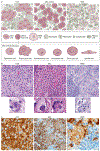
Figure 10.. Histology characteristics of LCH, JXG and RDD
A. Although Langerhans cell histiocytosis (LCH), histiocytoses of the xanthogranuloma family (including Erdheim Chester Disease (ECD) and juvenile xanthogranuloma (JXG),), and Rosai Dorfman Disease (RDD) have certain classic morphologies and defining phenotypes, our understanding of histiocytic lesions continues to evolve and may be best conceptualized along a spectrum, sometimes with morphology and phenotypes overlapping in certain instances and often with similar mutation profiles. In fact, ECD and JXG share similar morphological cell types under the microscope, including xanthomatous cells with variable admixture of Touton giant cells, vacuolated cells, spindled cells, oncocytic cells, and epithelioid cells. B. H&E images for LCH, xanthogranuloma family, and RDD (original magnification at 400x). C Immunostaining specific for BRAF-V600E (known as BRAF-VE1, brown) Although the mutant specific immunohistochemistry for BRAF-VE1 is more commonly expressed in LCH and ECD, with strong granular cytoplasmic staining of the histiocytes, rare examples in pediatric JXG family (limited to the central nervous system of children) and isolated case reports of RDD have been identified to also harbour the mutation and express the mutant protein, which reacts to the immunostaining. Original magnification at 1000x for LCH, 400x for CNS-JXG, and 1000x for RDD. Photos made with assistance of Mr Chris Woods Cincinnati Children’s Department of Pathology.
Figure 11.
Figure 11.. Histology of HLH
Bone marrow aspirate shows a large activated histiocyte with cytoplasmic phagocytosis. (Wright-Giemsa stain, original magnification 1000x).

References

    1. Favara BE et al. Contemporary classification of histiocytic disorders. The WHO Committee On Histiocytic/Reticulum Cell Proliferations. Reclassification Working Group of the Histiocyte Society. Med. Pediatr. Oncol 29, 157–166 (1997). - PubMed
    1. Emile JF et al. Revised classification of histiocytoses and neoplasms of the macrophage-dendritic cell lineages. Blood 127, 2672–2681 (2016). - PMC - PubMed
    1. Aschoff L. & Kiyono K. Frage der grossen Mononulearn. Folia Haematol 15, 383–390 (1913).
    1. Steinman RM & Cohn ZA Identification of a novel cell type in peripheral lymphoid organs of mice. I. Morphology, quantitation, tissue distribution. J Exp. Med 137, 1142–1162 (1973). - PMC - PubMed
    1. Steinman RM & Cohn ZA Identification of a novel cell type in peripheral lymphoid organs of mice. II. Functional properties in vitro. J Exp. Med 139, 380–397 (1974). - PMC - PubMed

Publication types

MeSH terms