. 2021 Oct 7;11(1):19979.
doi: 10.1038/s41598-021-99470-y.

sFlt-1 and CA 15.3 are indicators of endothelial damage and pulmonary fibrosis in SARS-CoV-2 infection

Affiliations

sFlt-1 and CA 15.3 are indicators of endothelial damage and pulmonary fibrosis in SARS-CoV-2 infection

Marilena Greco et al. Sci Rep. .

Abstract

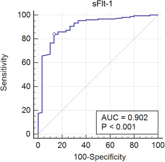
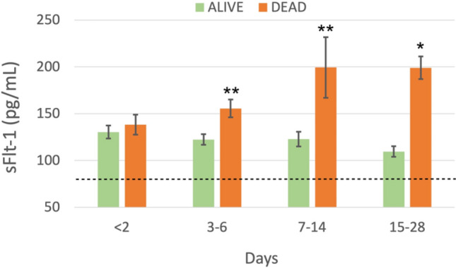
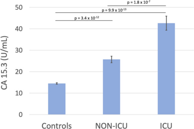
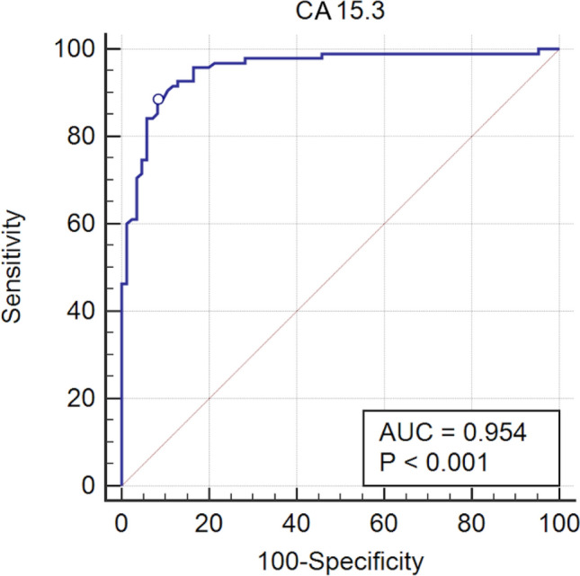
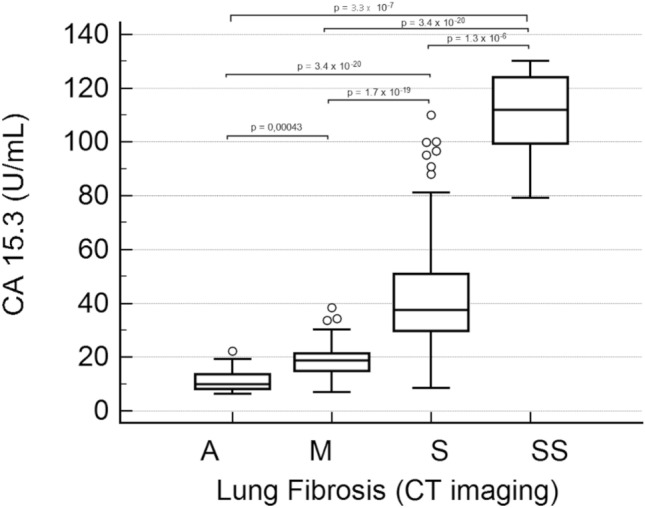
COVID-19 pandemic led to a worldwide increase of hospitalizations for interstitial pneumonia with thrombosis complications, endothelial injury and multiorgan disease. Common CT findings include lung bilateral infiltrates, bilateral ground-glass opacities and/or consolidation whilst no current laboratory parameter consents rapidly evaluation of COVID-19 risk and disease severity. In the present work we investigated the association of sFLT-1 and CA 15.3 with endothelial damage and pulmonary fibrosis. Serum sFlt-1 has been associated with endothelial injury and sepsis severity, CA 15.3 seems an alternative marker for KL-6 for fibrotic lung diseases and pulmonary interstitial damage. We analysed 262 SARS-CoV-2 patients with differing levels of clinical severity; we found an association of serum sFlt-1 (ROC AUC 0.902, decision threshold > 90.3 pg/mL, p < 0.001 Sens. 83.9% and Spec. 86.7%) with presence, extent and severity of the disease. Moreover, CA 15.3 appeared significantly increased in COVID-19 severe lung fibrosis (ICU vs NON-ICU patients 42.6 ± 3.3 vs 25.7 ± 1.5 U/mL, p < 0.0001) and was associated with lung damage severity grade (ROC AUC 0.958, decision threshold > 24.8 U/mL, p < 0.0001, Sens. 88.4% and Spec. 91.8%). In conclusion, serum levels of sFlt-1 and CA 15.3 appeared useful tools for categorizing COVID-19 clinical stage and may represent a valid aid for clinicians to better personalise treatment.


Conflict of interest statement

The authors declare no competing interests.

Figures

Figure 1
Figure 1
Receiver operating characteristic (ROC) analysis of sFlt-1 serum levels between SARS-CoV-2 infected and non-infected patients; the circle indicates the cut-off point (90.3 pg/mL) with 83.89% Sensitivity and 86.67% Specificity for positive patients. AUC denotes area under the curve.
Figure 2
Figure 2
Longitudinal analysis of serum levels of sFlt-1 during hospitalisation of SARS-CoV-2 patients, divided in Alive and Dead according to the outcome of the disease. Patients were monitored for four weeks (less than 2 days, 3–6 days, 7–14 days, 15–28 days from first positive detection of SARS-CoV-2 RNA). The dotted line indicates sFlt-1 level observed in Controls (78.9 ± 2.5 pg/mL); asterisks indicate statistically significant difference: *p < 0.05; **p < 0.01.
Figure 3
Figure 3
CA 15.3 serum levels in SARS-CoV-2 infected patients, divided according to their hospitalisation in Intensive Care Unit (ICU) or other in different wards (NON-ICU), and control patients.
Figure 4
Figure 4
Receiver operating characteristic (ROC) analysis of sFlt-1 serum levels between severe and non-severe SARS-CoV-2 infected patients; the circle indicates the cut-off point (24.8 U/mL) with 88.4% Sensitivity and 91.8% Specificity for positive patients. AUC denotes area under the curve.
Figure 5
Figure 5
CA 15.3 serum levels in SARS-CoV-2 infected patients according to lung fibrosis and damage severity grade obtained by CT imaging. A, absence of lung fibrosis; M, moderate grade; S, severe grade; SS; critic grade. Boxes represent first and third quartiles (25 to 75 percentile), bands within boxes represent the median value, whiskers display ranges of 1.5 interquartile ranges from the end of the box, outside values are displayed as separate points.

References

    1. Hoffmann M, et al. SARS-CoV-2 cell entry depends on ACE2 and TMPRSS2 and is blocked by a clinically proven protease inhibitor. Cell. 2020;181:271–280. doi: 10.1016/j.cell.2020.02.052. - DOI - PMC - PubMed
    1. Grillo F, Barisione E, Ball L, Mastracci L, Fiocca R. Lung fibrosis: An undervalued finding in COVID-19 pathological series. Lancet Infect. Dis. 2021;4:e72. doi: 10.1016/S1473-3099(20)30582-X. - DOI - PMC - PubMed
    1. Varga Z, et al. Endothelial cell infection and endotheliitis in COVID-19. Lancet. 2020;395:1417–1418. doi: 10.1016/S0140-6736(20)30937-5. - DOI - PMC - PubMed
    1. Ferrario CM, et al. Effect of angiotensin-converting enzyme inhibition and angiotensin II receptor blockers on cardiac angiotensin-converting enzyme 2. Circulation. 2005;111:2605–2610. doi: 10.1161/CIRCULATIONAHA.104.510461. - DOI - PubMed
    1. Bonetti PO, Lerman LO, Lerman A. Endothelial dysfunction—a marker of atherosclerotic risk. Arterioscl. Throm. Vas. 2003;23:168–175. doi: 10.1161/01.ATV.0000051384.43104.FC. - DOI - PubMed