. 2021 Oct 7;4(1):1157.
doi: 10.1038/s42003-021-02665-2.

Functional characterization of a bioengineered liver after heterotopic implantation in pigs

Affiliations

Functional characterization of a bioengineered liver after heterotopic implantation in pigs

Brett D Anderson et al. Commun Biol. .

Erratum in

Abstract

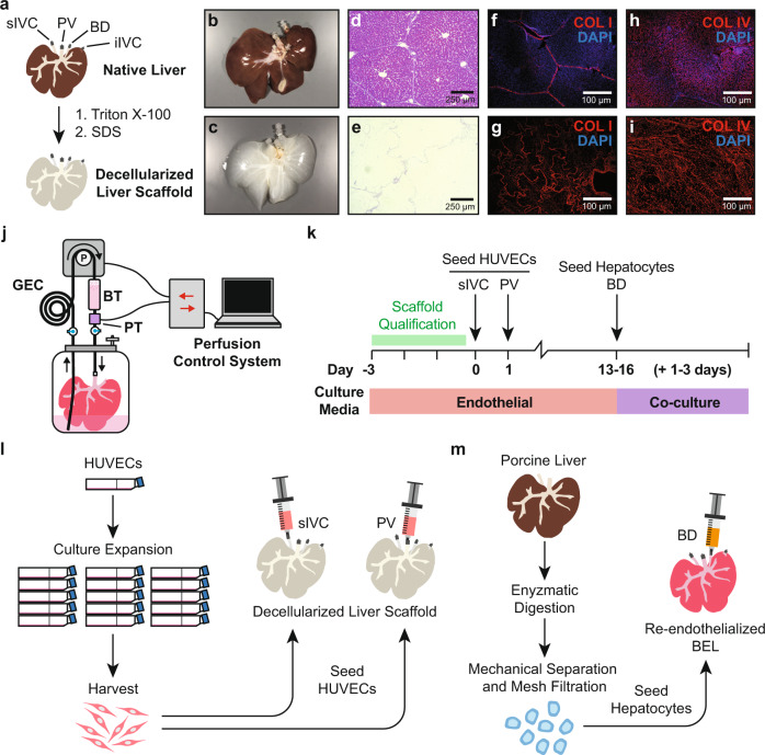
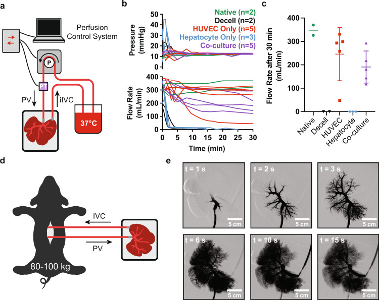
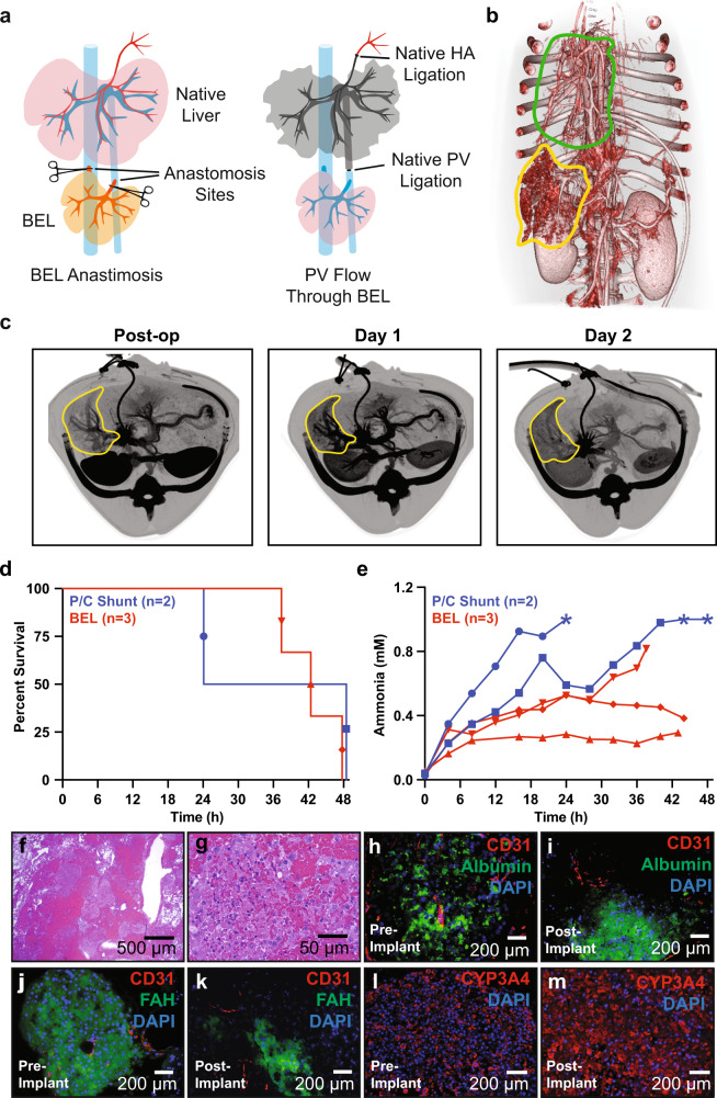
Organ bioengineering offers a promising solution to the persistent shortage of donor organs. However, the progression of this technology toward clinical use has been hindered by the challenges of reconstituting a functional vascular network, directing the engraftment of specific functional cell types, and defining appropriate culture conditions to concurrently support the health and phenotypic stability of diverse cell lineages. We previously demonstrated the ability to functionally reendothelialize the vasculature of a clinically scaled decellularized liver scaffold with human umbilical vein endothelial cells (HUVECs) and to sustain continuous perfusion in a large animal recovery model. We now report a method for seeding and engrafting primary porcine hepatocytes into a bioengineered liver (BEL) scaffold previously reendothelialized with HUVECs. The resulting BELs were competent for albumin production, ammonia detoxification and urea synthesis, indicating the presence of a functional hepatocyte compartment. BELs additionally slowed ammonia accumulation during in vivo perfusion in a porcine model of surgically induced acute liver failure. Following explant of the graft, BEL parenchyma showed maintenance of canonical endothelial and hepatocyte markers. Taken together, these results support the feasibility of engineering a clinically scaled functional BEL and establish a platform for optimizing the seeding and engraftment of additional liver specific cells.


Conflict of interest statement

The authors declare the following competing interests: B.D.A., A.A.K., A.M., B.G.S., A.R.S., V.L.N., R.N.P., T.W.G., D.S.D., and J.J.R. are employees of Miromatrix Inc. Miromatrix Inc. is a publicly funded company and owns the exclusive patent rights for the perfusion decellularization and recellularization technologies utilized in this study. The remaining authors declare no competing interests.

Figures

Fig. 1
Fig. 1. Overview of approach to bioengineering a porcine-derived whole liver construct.
a Overview of vessel cannulation and detergent perfusion methods for whole liver decellularization. b, c Photographs of representative porcine (b) native and (c) decellularized whole livers. d, e Hematoxylin and eosin staining of histological sections from (d) native and (e) decellularized porcine liver tissue demonstrating the efficient removal of cellular material while preserving the native tissue architecture. fi Representative immunofluorescence micrographs from native (f, h) and decellularized (g, i) liver tissue demonstrating retention of Collagen I and Collagen IV, respectively, in cell-free scaffolds. The absence of DAPI staining in the decellularized scaffolds demonstrates removal of cellular DNA. j Schematic of perfusion bioreactor design. BELs are suspended in a vessel with culture media. An extended coil of silicone tubing (GEC) is included in the perfusion circuit to facilitate gas exchange. A bubble trap is positioned directly upstream of the BEL perfusion inlet. During media perfusion, a pressure transducer provides real-time feedback to a controller which in turn adjusts the flow rate on a peristaltic pump to maintain a constant perfusion pressure. k Schematic depicting bioreactor culture and cell seeding events. Decellularized scaffolds were pre-qualified in antibiotic-free media for 3 days prior to HUVEC seedings. HUVEC-seeded liver constructs were cultured in endothelial cell growth media until a 24 h average glucose consumption rate of 50–90 mg/h was reached (typically 13–16 days), at which point hepatocytes were seeded into the scaffold. BELs were maintained in co-culture media formulated for simultaneous culture of endothelial cells and hepatocytes for the remainder of the experiment (typically 1–3 additional days). l Overview of HUVEC culture and seeding approach to generate a reendothelialized liver construct. Cells were infused first through the cannulated iIVC followed by a second cell infusion through the PV 24 h later. m Schematic of porcine donor liver hepatocyte isolation and seeding of bioengineered liver constructs. Whole porcine livers are enzymatically digested, and dissociated cells were filtered through a series of mesh sieves to remove large debris and cell aggregates. Hepatocytes were enriched through multiple low speed (70 × g) centrifugation and washing steps. DAPI—4′,6′-diamidino-2-phenylindole; BEL—bioengineered liver; GEC—gas exchange coil; BT—bubble trap; iIVC—infrahepatic inferior vena cava; PV—portal vein.
Fig. 2
Fig. 2. Histological and functional characterization of BEL constructs.
a Representative photograph of a BEL seeded with HUVECs and porcine hepatocytes. b Hematoxylin and eosin staining of representative co-culture BEL tissue sections fixed 48 h after seeding hepatocytes. ce Immunofluorescent staining of cell lineage markers in non-serial tissue sections 48 h after seeding hepatocytes: (c) CD31 & albumin; (d) CD31 & FAH; (e) CD31 and LYVE1. f Schematic depicting seeding and culture timeline for HUVEC only, hepatocyte only, and co-culture BEL constructs used in (g, h, j, k). g vWF production in grafts before and after hepatocyte seeding. Data from independent HUVEC only (n = 5), hepatocyte only (n = 7), and co-culture (n = 7) BEL constructs are shown. Error bars denote the mean and standard deviation at each time point. h 24-h average albumin production in co-culture grafts 48 h following hepatocyte seeding. Data from independent HUVEC only (n = 5), hepatocyte only (n = 7), and co-culture (n = 7) BELs are shown. Error bars denote the mean and standard deviation. i Schematic of in vitro ammonia clearance and urea production assay. Ammonium chloride is added to the bioreactor media at a concentration of 0.8 mM. Ammonia and urea levels are measured in media samples taken at t = 0, 1, 2 7, and 23 h following the addition of ammonium chloride. Error bars denote the mean and standard deviation each time point. j, k Ammonia clearance (j) and urea production kinetics (k) following the addition of ammonium chloride to the bioreactor perfusion media. Data from independent HUVEC only (n = 5), hepatocyte only (n = 7), and co-culture (n = 7) BELs, media only controls (n = 4) are shown. Error bars denote the mean and standard deviation each time point. BEL—bioengineered liver; FAH—fumarylacetoacetate hydrolase; CD31—cluster of differentiation 31; LYVE1—lymphatic vessel endothelial hyaluronan receptor 1; vWF—von Willebrand factor.
Fig. 3
Fig. 3. Acute blood perfusion studies to assess vascular patency in BELs.
a Schematic of in vitro blood perfusion circuit. 37 °C porcine blood is perfused at 12 mmHg through the PV with a peristaltic pump and returned to a reservoir through the IVC. b Summary plots of pressures and flow rates measured over 60 minutes during in vitro blood perfusion studies using HUVEC only, hepatocyte only, or co-culture BELs. Freshly explanted (Native) porcine livers and decellularized scaffolds (Decell) were included as benchmarks for idealized perfusion and rapid thrombosis, respectively. c Violin plots summarizing BEL flow rates from (b) after 30 min of perfusion. d Schematic of ex vivo blood perfusion model. A synthetic perfusion circuit is established by cannulating the PV and sIVC within an anesthetized pig. Blood flow is diverted from the animal’s cannulated PV to the BEL PV and returned from the BEL sIVC into the animal’s cannulated iIVC. e Real-time angiography time lapse imaging following contrast infusion. Imaging was performed after 30 min of continuous blood perfusion. PV—portal vein; BEL—bioengineered liver; sIVC—suprahepatic inferior vena cava, iIVC—infrahepatic inferior vena cava.
Fig. 4
Fig. 4. Heterotopic implantation of co-culture BELs in a large animal model.
a Schematic of heterotopic BEL implant surgical model. See text for details. b Post-operative 3D-reconstruction from CT imaging demonstrating BEL perfusion and devascularization of native liver. BEL is outlined in yellow and native liver is outlined in green. c Representative axial CT imaging of recipient animal at post-op, 24 h, and 48 h time points. BEL is outlined in yellow. d Kaplan–Meier curves showing animal survival times within portocaval shunt and BEL implant groups. Symbols are matched to ammonia values in (d). e Post-operative blood ammonia levels measured in BEL implant recipient animals (n = 3) and portocaval shunt animals (n = 2) over the duration of the experiment. Asterisks (*) denote data points that were above the upper limit of quantification of the assay (1 mM). f, g Representative histological section of BEL tissue explanted 48 h post-implant showing viable hepatocytes and endothelialized vasculature. h, i Representative immunostaining BEL tissue (h) pre-implant and (i) explanted 48 h post-implant showing maintenance of CD31 and albumin expression. j, k Representative immunostaining BEL tissue (j) pre-implant and (k) explanted 48 h post-implant showing maintenance of CD31 and FAH expression. l, m Representative immunostaining BEL tissue (l) pre-implant and (m) explanted 48 h post-implant showing maintenance of CYP3A4 expression. HA—hepatic artery; BEL—bioengineered liver; P/C—portocaval; CT—computed tomography; FAH—fumarylacetoacetate hydrolase; CYP3A4—cytochrome p450 3A4.

Comment in

References

    1. Ott HC, et al. Perfusion-decellularized matrix: using nature’s platform to engineer a bioartificial heart. Nat. Med. 2008;14:213–221. doi: 10.1038/nm1684. - DOI - PubMed
    1. Uygun BE, et al. Organ reengineering through development of a transplantable recellularized liver graft using decellularized liver matrix. Nat. Med. 2010;16:814–820. doi: 10.1038/nm.2170. - DOI - PMC - PubMed
    1. Baptista PM, et al. The use of whole organ decellularization for the generation of a vascularized liver organoid. Hepatology. 2011;53:604–617. doi: 10.1002/hep.24067. - DOI - PubMed
    1. Nichols JE, et al. Production and transplantation of bioengineered lung into a large-animal model. Sci Transl Med. 2018;10:eaao3926. doi: 10.1126/scitranslmed.aao3926. - DOI - PubMed
    1. Bonandrini B, et al. Recellularization of well-preserved acellular kidney scaffold using embryonic stem cells. Tissue Eng. Part A. 2014;20:1486–1498. doi: 10.1089/ten.tea.2013.0269. - DOI - PMC - PubMed