. 2021 Oct 7;4(1):1168.
doi: 10.1038/s42003-021-02693-y.

Widespread bacterial diversity within the bacteriome of fungi

Affiliations

Widespread bacterial diversity within the bacteriome of fungi

Aaron J Robinson et al. Commun Biol. .

Abstract

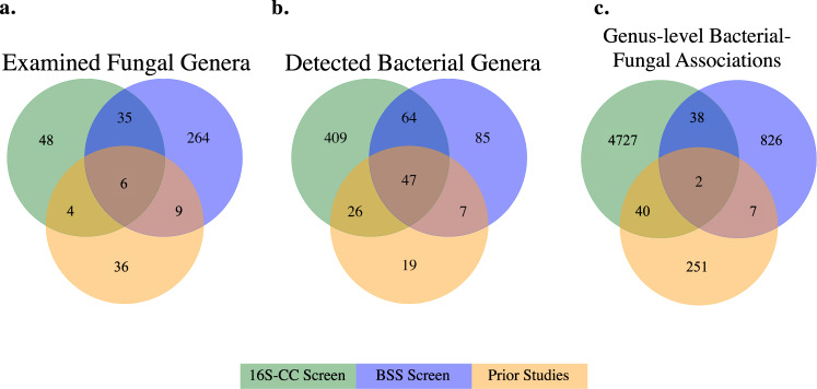
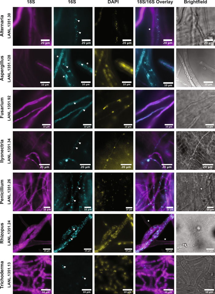
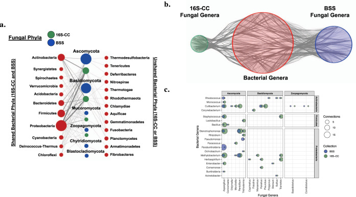
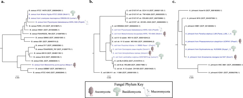
Knowledge of associations between fungal hosts and their bacterial associates has steadily grown in recent years as the number and diversity of examinations have increased, but current knowledge is predominantly limited to a small number of fungal taxa and bacterial partners. Here, we screened for potential bacterial associates in over 700 phylogenetically diverse fungal isolates, representing 366 genera, or a tenfold increase compared with previously examined fungal genera, including isolates from several previously unexplored phyla. Both a 16 S rDNA-based exploration of fungal isolates from four distinct culture collections spanning North America, South America and Europe, and a bioinformatic screen for bacterial-specific sequences within fungal genome sequencing projects, revealed that a surprisingly diverse array of bacterial associates are frequently found in otherwise axenic fungal cultures. We demonstrate that bacterial associations with diverse fungal hosts appear to be the rule, rather than the exception, and deserve increased consideration in microbiome studies and in examinations of microbial interactions.


Conflict of interest statement

The authors declare no competing interests.

Figures

Fig. 1
Fig. 1. Fungal taxa, bacterial taxa, and genus-level associations found among the culture collection screen (16S-CC), the bioinformatic screen of fungal genome sequencing projects (BSS) and prior studies.
Each diagram displays overlaps in either a examined fungal genera, b bacterial genera detected, or c genus-level bacterial–fungal associations found in the two screens conducted as part of this work and in prior studies.
Fig. 2
Fig. 2. Expanded taxonomic diversity of bacterial–fungal associations.
Prior findings of bacterial–fungal associations were compared with both the 16S-CC and BSS screens completed in this study. The upper panel presents the taxonomic diversity of fungal hosts examined in a prior studies, b the culture collections included in the 16S-CC screen, and c the fungal genome projects used in the BSS screen. The lower panel displays the corresponding taxonomic diversity of bacteria associated with these fungi, observed in d prior studies, e the 16S-CC screen, and f the BSS screen. Terminal nodes represent distinct genera and edges are colored by phyla. Abbreviations for the fungal hosts: M Mucoromycota, Z Zoopagomycota, Bl Blastocladiomycota, C Chytridiomycota. Abbreviations for the bacterial associates: Acido/A Acidobacteria, Cyano Cyanobacteria, T Tenericutes). Several bacterial phyla not previously described as fungal associates, but identified in our 16S-CC and the BSS screens are represented in gray (Aquificae, Armatimonadetes, Calditrichaeota, Chlamydiae, Chloroflexi, Deinococcus-Thermus, Fibrobacteres, Fusobacteria, Gemmatimonadetes, Nitrospirae, Planctomycetes, Rhodothermaeota, Spirochaetes, Synergistetes, Thermodesulfobacteria, Thermotogae, and Verrucomicrobia).
Fig. 3
Fig. 3. Diversity of bacteria found among fungi from culture collections and public fungal genome sequencing projects.
The internal cladogram is colored by fungal phyla and terminal nodes represent fungal genera. The innermost ring indicates the screen used to detect these bacterial–fungal associations, and the outer five rings represent the top five bacterial lineages (from inner to outer ring: Alphaproteobacteria, Betaproteobacteria, Gammaproteobacteria, Actinobacteria, and Bacilli) most frequently detected in our screens. These rings are shaded from dark (many interactions detected) to white (no interactions observed).
Fig. 4
Fig. 4. Visualization of bacterial associates in diverse fungal hosts using 16S ribosomal RNA staining by fluorescence in situ hybridization.
Fungal ribosomes were stained using a universal eukaryotic 18S rRNA probe (magenta); bacteria were co-stained with a universal 16S rRNA probe (cyan); DAPI was used as a global nuclear stain (yellow). Overlays of the 18S and 16S fluorescence show positive correlation of bacteria along or within hyphae. Observed bacterial signal was variable among fungal samples, and displayed coccoid and rod-shaped phenotypes. Individual structures resembling bacterial cells were observed (arrowheads) as were biofilm-like growth patterns along hyphae (asterisks). Bacterial structures absent in neighboring hyphae indicate variability in spatial distributions (stars).
Fig. 5
Fig. 5. Fluorescence in situ hybridization staining with Lacunisphaera specific probes in an Aspergillus isolate.
FISH staining was conducted on an Aspergillus isolate using a universal 18S rRNA probe (magenta) and a Lacunisphaera genus-specific 16S rRNA probe set (cyan). Coccoid structures attributed to the bacterial associate are shown with arrowheads. Non-specific DAPI DNA staining displays co-localization with 16S signals from the bacteria that appear distinct from the nuclear morphology of the fungus (asterisks). A non-hybridizing probe (Non-Eub338) was used as a negative control.
Fig. 6
Fig. 6. Phylum and genus level bacterial–fungal associations identified in fungal culture collections and genome sequencing projects.
a Tripartite network showing associations (gray edges) between bacterial phyla (red) and fungal phyla from either the 16S-CC (green) or BSS (blue) screen. Bacterial phyla that were identified in both screens are shown on the left, while phyla only identified in one screen are shown on the right. Nodes are sized by the number of genera represented in each phylum and edges are weighted by the number of distinct associations among bacterial and fungal genera. b Network of associations (gray edges) between bacterial genera (red nodes) and fungal genera from either the 16S-CC screen (green nodes) or the BSS screen (blue nodes) generated using Cytoscape, which demonstrates the complexity of the overall network of potential associations detected across both screens at the genus level. c Genus-level bacterial–fungal associations identified in both the 16S-CC and BSS screen. The size of each circle corresponds to the total number of genera detected in each screen.
Fig. 7
Fig. 7. Genome-wide single nucleotide polymorphism (SNP)-based phylogenetic analysis of fungal-derived bacterial sequences.
Phylogenetic trees generated by analysis with PhaME for fungal-derived sequences of a Bacillus cereus, b Escherichia coli, and c Acinetobacter johnsonii. Fungal-derived sequences are annotated in blue lettering and include the corresponding JGI project ID (shown in parentheses) and icons next to their annotation indicate the phylum of the fungal host. Non-fungal-derived reference genomes are indicated in black. NCBI accession IDs are included for these reference genomes (shown in parenthesis). Branches with bootstrap values (1000 bootstrap replicates) <60% are marked with an asterisk (*). All trees are midpoint rooted.

References

    1. Simon J, Marchesi JR, Mougel C, Selosse MA. Host-microbiota interactions: from holobiont theory to analysis. Microbiome. 2019;7:1–5. doi: 10.1186/s40168-019-0619-4. - DOI - PMC - PubMed
    1. Peay KG, Kennedy PG, Talbot JM. Dimensions of biodiversity in the Earth mycobiome. Nat. Rev. Microbiol. 2016;14:434–447. doi: 10.1038/nrmicro.2016.59. - DOI - PubMed
    1. Cregger MA, et al. The Populus holobiont: dissecting the effects of plant niches and genotype on the microbiome. Microbiome. 2018;6:31. doi: 10.1186/s40168-018-0413-8. - DOI - PMC - PubMed
    1. Ross AA, Hoffmann AR, Neufeld JD. The skin microbiome of vertebrates. Microbiome. 2019;7:1–14. doi: 10.1186/s40168-019-0694-6. - DOI - PMC - PubMed
    1. Huseyin CE, O’Toole PW, Cotter PD, Scanlan PD. Forgotten fungi—the gut mycobiome in human health and disease. FEMS Microbiol. Rev. 2017;41:479–511. doi: 10.1093/femsre/fuw047. - DOI - PubMed

Publication types