. 2021 Sep 21:12:732344.
doi: 10.3389/fpls.2021.732344. eCollection 2021.

Variables Affecting Shoot Growth and Plantlet Recovery in Tissue Cultures of Drug-Type Cannabis sativa L

Affiliations

Variables Affecting Shoot Growth and Plantlet Recovery in Tissue Cultures of Drug-Type Cannabis sativa L

Janesse E Holmes et al. Front Plant Sci. .

Abstract

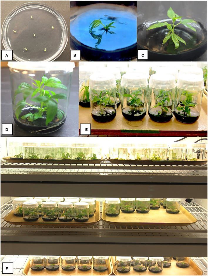
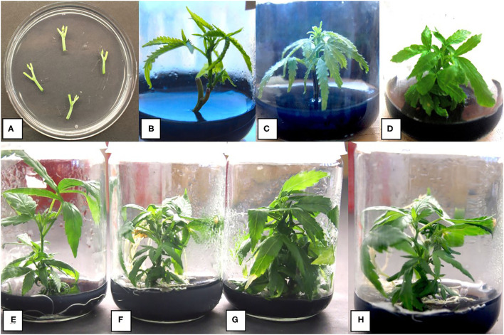
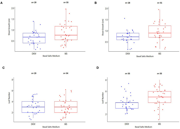
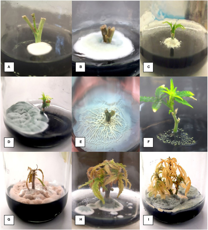
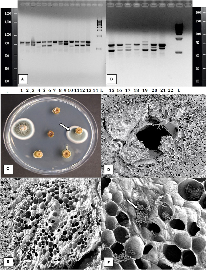
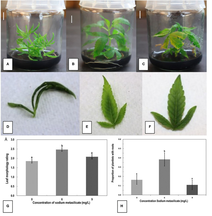
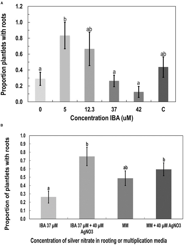
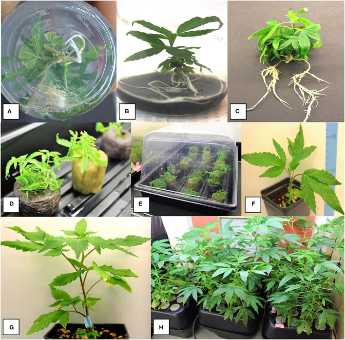
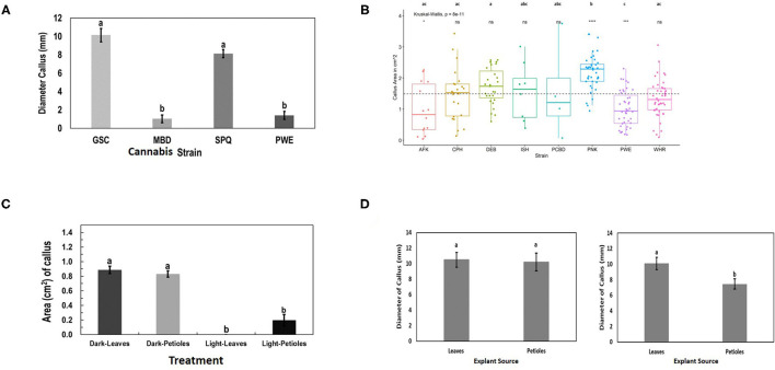
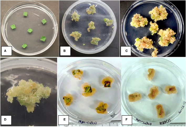
Tissue culture approaches are widely used in crop plants for the purposes of micropropagation, regeneration of plants through organogenesis, obtaining pathogen-free plantlets from meristem culture, and developing genetically modified plants. In this research, we evaluated variables that can influence the success of shoot growth and plantlet production in tissue cultures of drug-type Cannabis sativa L. (marijuana). Various sterilization methods were tested to ensure shoot development from nodal explants by limiting the frequency of contaminating endophytes, which otherwise caused the death of explants. Seven commercially grown tetrahydrocannabinol (THC)-containing cannabis genotypes (strains) showed significant differences in response to shoot growth from meristems and nodal explants on Murashige and Skoog (MS) medium containing thidiazuron (1 μM) and naphthaleneacetic acid (0.5 μM) plus 1% activated charcoal. The effect of Driver and Kuniyuki Walnut (DKW) or MS basal salts in media on shoot length and leaf numbers from nodal explants was compared and showed genotype dependency with regard to the growth response. To obtain rooted plantlets, shoots from meristems and nodal explants of genotype Moby Dick were evaluated for rooting, following the addition of sodium metasilicate, silver nitrate, indole-3-butyric acid (IBA), kinetin, or 2,4-D. Sodium metasilicate improved the visual appearance of the foliage and improved the rate of rooting. Silver nitrate also promoted rooting. Following acclimatization, plantlet survival in hydroponic culture, peat plugs, and rockwool substrate was 57, 76, and 83%, respectively. The development of plantlets from meristems is described for the first time in C. sativa and has potential for obtaining pathogen-free plants. The callogenesis response of leaf explants of 11 genotypes on MS medium without activated charcoal was 35% to 100%, depending on the genotype; organogenesis was not observed. The success in recovery of plantlets from meristems and nodal explants is influenced by cannabis genotype, degree of endophytic contamination of the explants, and frequency of rooting. The procedures described here have potential applications for research and commercial utility to obtain plantlets in stage 1 tissue cultures of C. sativa.

Keywords: Cannabis sativa L.; callogenesis; meristems; micropropagation; nodal explants; plantlet recovery; rooting; shoot growth.


Conflict of interest statement

The authors declare that the research was conducted in the absence of any commercial or financial relationships that could be construed as a potential conflict of interest.

Figures

Figure 1
Figure 1
Various stages of growth of shoots derived from meristems of drug-type cannabis genotypes grown on a multiplication medium (MM) containing activated charcoal. (A) Meristem explants placed on an agar medium in a 90-mm Petri dish to show their small size. (B) Early shoot growth after 4 weeks in culture from a meristem. (C) Shoot growth after 8 weeks. (D,E) Shoot growth after 10 weeks from meristems. (F) Baby food jars containing meristem explants at different stages of development in a controlled environment growth chamber. A number of different strains are shown. Conditions of incubation are described in the Methods section.
Figure 2
Figure 2
Stages of growth of shoots derived from nodal segments of drug-type cannabis genotypes grown on MM containing activated charcoal. (A) Nodal stem explants placed on an agar medium in a 90-mm Petri dish to show their size. (B) Shoot growth after 4 weeks from a nodal stem explant. The rate of growth is greater than that from a meristem. (C) Shoot growth after 6 weeks. (D) Shoot growth after 8 weeks. (E) Shoot growth after 8 weeks of genotype Death Bubba (DEB) from nodal explants. (F) Shoot growth after 8 weeks of genotype Pink Kush (PNK) from nodal explants. (G) Shoot growth after 8 weeks of genotype White Rhino (WHR) from nodal explants. (H) Shoot growth after 8 weeks of genotype Moby Dick (MBD) from nodal explants.
Figure 3
Figure 3
Comparison of shoot growth and leaf number from nodal stem explants of two cannabis genotypes placed on a basal salt medium containing either Driver and Kuniyuki Walnut (DKW) or Murashige and Skoog (MS) salts. (A) Shoot length of genotype Copenhagen Kush (CPH). (B) Shoot length of genotype Pennywise (PWE). (C) Leaf number of genotype Copenhagen Kush (CPH). (D) Leaf numbers of genotype Pennywise (PWE). The box of each dataset represents the interquartile range (IQR), which contains the third quartile (Q3–top side of the box), the median value of all of the data (the middle line), and the first quartile (Q1–bottom side of the box). The bars represent the maximum (Q3 + 1.5*IQR) and minimum (Q1–1.5*IQR) of the data. The numbers (n) above the bars depict explant numbers used.
Figure 4
Figure 4
Range of microbial contaminants emerging from surface-sterilized nodal stem explants at various times after placement on MM containing activated charcoal. (A) Early emergence of Penicillium species. (B) Large colony of Penicillium growing from a nodal explant. (C) Colony of Chaetomium emerging from a nodal explant. (D) Large colony of Penicillium growing out of a nodal stem explant. (E) Extensive Bacillus growth emerging from a nodal stem explant. (F) Growth of Pseudomonas from a stem explant. (G–I) Death of established shoots from nodal stem segments due to contamination by microbes appearing later during plantlet growth.
Figure 5
Figure 5
Molecular detection and identification of tissue culture contaminants. (A,B) PCR detection of fungal DNA present in naturally contaminated nodal explants. Upper bands at 750 bp size are plant DNA. Lower bands at ca. 650 bp are fungal DNA. These bands were cut and sequenced to determine the corresponding fungus present. Lanes 1–13 (lower bands) are as follows: clean, Simplicillium lasoniveum, Trichoderma harzianum. Clean, Beauveria bassiana, Fusarium oxysporum. Clean, Trametes versicolor, Lecanicillium fungicola, Chaetomium globosum, F. oxysporum, F. oxysporum, L. fungicola. Lane 14 = water control. L = molecular weight standards. Lanes 15–21 (lower bands) = Penicillium chrysogenum, Penicillium copticola, C. globosum, Penicillium olsonii, P. olsonii, T. versicolor. Lane 22 = water control, L= molecular weight standards. (C) Growth of Penicillium colonies emerging from the center of pith tissues in cut nodal segments. (D) Cross-section of a cannabis nodal stem explant showing the central pith surrounded by pith cells. (E) Close-up of pith cells as viewed in the scanning electron microscope. (F) Magnified view of pith cells in the scanning electron microscope showing fungal sporulation inside pith cells, likely of Penicillium sp. (arrow).
Figure 6
Figure 6
The effect of sodium metasilicate at 0 (A), 6 (B), and 9 (C) mg/L in the MM on shoot growth of genotype MBD from nodal segments. Optimal growth and rooting can be seen on 6 mg/L. (D–F) Leaf morphology rating scale applied to plantlets during growth in tissue culture as an indication of plantlet health. (D) A rating of 1 shows thin curled leaves, light green in color. (E) A rating of 2 shows leaves light green in color with some curling. (F) A rating of 3 shows dark green, flat leaves with serrated margins. (G) Average leaf morphology rating after 4 weeks of growth on MM with added sodium metasilicate. (H) Proportion of plantlets that developed roots on MM supplemented with sodium metasilicate at 6 mg/L was significantly different to the proportion of roots that developed from plantlets on MM, MM + sodium metasilicate 9 mg/L, and MMC. Bars followed by different letters indicate significant differences.
Figure 7
Figure 7
The effect of additives to the MS salts medium on root development in plantlets of genotype MBD derived from nodal stem segments after 4 weeks of growth. Additives were (A) indole-3-butyric acid IBA, (B) silver nitrate (AgNO3), compared with MM as a control. Statistical analysis was performed by Tukey's HSD test with significance at p < 0.05. Bars followed by different letters indicate significant differences.
Figure 8
Figure 8
Root development on shoots after 8–10 weeks of growth in tissue culture and acclimatization to produce plants. (A,B) Spontaneous development of roots on nodal explants. (C) Plantlets were removed from tissue culture and transferred to coco growth media and placed under high humidity conditions for 2 weeks. (D,E) Acclimatization of plantlets from meristems of genotype MBD in different growth substrates. Plantlets were removed from tissue culture jars after 12 weeks of growth on the MM and placed into rockwool or peat under humid conditions for 14 days. Rockwool or peat plugs were soaked in a fertilizer mix of 1 ml/L of pH Perfect® Sensi Grow A&B and CALiMAGIc (General Hydroponics, Santa Rosa, CA, United States) in ~pH 5.8 water. (F,G) Growth of plants on a coco potting medium under ambient conditions following successful acclimatization. (H) Hydroponic system filled with 8 L of the fertilizer mix with vigorous growth of plants derived from meristem tissue cultures.
Figure 9
Figure 9
Response of cannabis genotypes to callus development on MM without activated charcoal (MM-AC). (A) Callus diameter of four genotypes developing from 1 cm2 leaf explants. (B) Callus area from leaf explants of 8 cannabis genotypes compared with the mean callus area across all strains (represented by the dotted line). The “*”represents significance level and “ns” represents not significant relationships between each genotype and across all genotype means (dotted line). A Kruskal–Wallis test resulting in a p of 8 × 10⁻¹¹ and a Games–Howell post-hoc test were performed. Significant differences identified in the Games–Howell post-hoc test are shown using letters above the boxplots of each strain. (C) Growth of Cheesequake (CHQ) callus from leaf and petiole explants after 1 month under 24-h dark and 24-h light conditions on MM. Statistical analysis was performed by Tukey's HSD test with significance at p < 0.05. Bars followed by different letters indicate significant differences. (D) Growth of callus from leaves and petioles of genotype Girl Scout Cookie (GSC) (left) and Space Queen (SPQ) (right). Measurements of diameter were made after 4 weeks. Statistical analysis was performed by Tukey's HSD test with significance at p < 0.05. Bars followed by different letters indicate significant differences.
Figure 10
Figure 10
Callus development on leaf and petiole explants of different cannabis genotypes on MM-AC. (A) Leaf explants at the start of the experiment from a donor plant. (B) Callus from leaf explants of genotype SPQ after 8 weeks of growth. Variation in callus growth between explants can be seen. (C) Callus growth of genotype GSC from leaf explants after 8 weeks of growth. (D) Callus from leaf explants of genotype SPQ after 8 weeks of growth. (E) Callus from leaf explants of cannabis genotype Pink Kush after 8 weeks in the dark. (F) Callus from petiole explants of cannabis genotype Pure CBD after 8 weeks in the dark. No evidence of shoot development was observed in any of the callus cultures.

References

    1. Adhikary D., Kulkarni M., El-Mezawy A., Mobini S., Elhiti M., Gjuric R., et al. . (2021). Medical cannabis and industrial hemp tissue culture: present status and future potential. Front. Plant Sci. 12:275. 10.3389/fpls.2021.627240 - DOI - PMC - PubMed
    1. Barnett S. E., Cala A. R., Hansen J. L., Crawford J., Viands D. R., Smart L. B., et al. . (2020). Evaluating the microbiome of hemp. Phytobiomes J. 4, 351–363. 10.1094/PBIOMES-06-20-0046-R - DOI
    1. Braemer R., Paris M. (1987). Biotransformation of cannabinoids by a cell suspension culture of Cannabis sativa L. Plant Cell Rep. 6, 150–152. - PubMed
    1. Chandra S., Lata H., Khan I. A., ElSohly M. A. (2017). “Cannabis sativa L.: botany and horticulture,” in Cannabis sativa L. Botany and Biotechnology, eds S. Chandra, H. Lata, M. ElSohly M (Cham: Springer; ). 10.1007/978-3-319-54564-6 - DOI
    1. Cheong E. J., Mock R., Li R. (2012). Elimination of five viruses from sugarcane using in vitro culture of axillary buds and apical meristems. Plant Cell Tissue Organ. Cult. 109, 439–445. 10.1007/s11240-011-0108-3 - DOI

LinkOut - more resources