. 2021 Sep 28:2021:3995958.
doi: 10.1155/2021/3995958. eCollection 2021.

Neuroprotective Effect of Danggui Shaoyao San via the Mitophagy-Apoptosis Pathway in a Rat Model of Alzheimer's Disease

Affiliations

Neuroprotective Effect of Danggui Shaoyao San via the Mitophagy-Apoptosis Pathway in a Rat Model of Alzheimer's Disease

Zhenyan Song et al. Evid Based Complement Alternat Med. .

Abstract

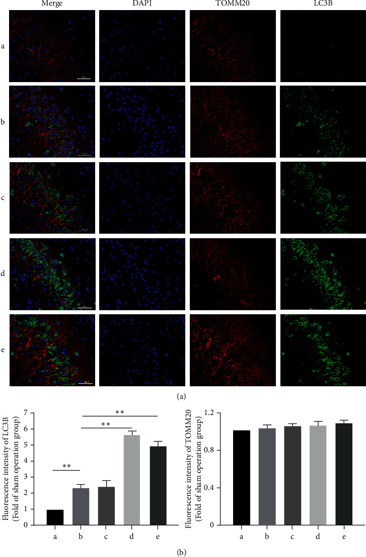
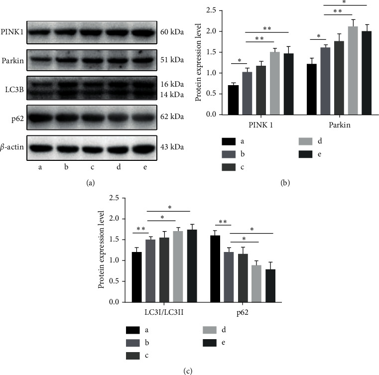
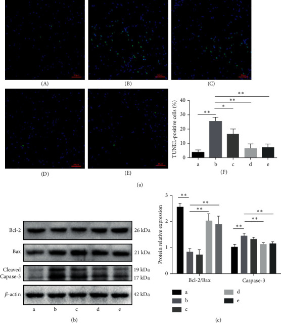
Alzheimer's disease (AD) is a serious neurodegenerative disease. While the main pathological characteristic of AD is widely believed to be the accumulation of amyloid-beta (Aβ) in neurons around neurofibrillary plaques, the molecular mechanism of pathological changes is not clear. Traditional Chinese medicine offers many treatments for AD. Among these, Danggui Shaoyao San (DSS) is a classic prescription. In this study, an AD model was established by injecting Aβ 1-42 into the brains of rats, which were then treated with different concentrations of Danggui Shaoyao San (sham operation; model; and Danggui Shaoyao San high-dose, medium-dose, and low-dose intervention groups). The Morris water maze test was used to assess the learning and memory abilities of the animals in each group. Nissl staining was used to detect neurons. Mitophagy was evaluated by transmission electron microscopy and immunofluorescence colocalization. Apoptosis was assessed by terminal deoxynucleotidyl transferase dUTP nick end labeling (TUNEL) assay. The expression levels of autophagy- and apoptosis-related proteins were measured by western blot. Compared to the model group, the groups of AD rats administered medium and high doses of Danggui Shaoyao San showed significantly increased learning and memory abilities (P < 0.05), as well as significantly increased autophagosomes in the hippocampus. Moreover, the expression of PTEN-induced kinase 1 (PINK1), Parkin, and microtubule-associated protein light chain 3 (LC3-I/LC3-II) was increased, while that of p62 was significantly decreased (P < 0.05). The neuronal apoptosis rate was also significantly decreased, the Bcl-2/Bax ratio was significantly increased, and the cleaved caspase-3 protein expression was significantly decreased (P < 0.05). Therefore, Danggui Shaoyao San inhibited neuronal apoptosis in AD rats via a mechanism that may be related to the activation of the PINK1-Parkin-mediated mitophagy signaling pathway.


Conflict of interest statement

The authors declare that there are no conflicts of interest regarding the publication of this paper.

Figures

Figure 1
Figure 1
Danggui Shaoyao San effectively improves the learning and memory function of AD rats. A: sham operation group; B: model group; C: DSS low-dose group; D: DSS medium-dose group; E: DSS high-dose group. (a) Escape time in the memory learning phase of the Morris water maze positioning navigation test. (b) Morris water maze space exploration test of the number of crossings on the hidden platform area. (c) Morris water maze space exploration test of the target quadrant stay time. (d) The trajectories of the rats looking for hidden platforms on day 5. N = 6 animals/group, P < 0.05, and ∗∗P < 0.01. AD: Alzheimer's disease; DSS: Danggui Shaoyao San.
Figure 2
Figure 2
Nissl staining of the hippocampus. (a) Sham operation group. (b) Model group. (c) DSS low-dose group. (d) DSS medium-dose group. (e) DSS high-dose group. DSS: Danggui Shaoyao San.
Figure 3
Figure 3
Danggui Shaoyao San promotes mitophagy in hippocampal neurons of AD rats by transmission electron microscope. Formation and distribution of autophagosomes in rat hippocampus. The red arrow indicates the autophagosome. (a) Sham operation group. (b) Model group. (c) DSS low-dose group. (d) DSS medium-dose group. (e) DSS high-dose group. (f) Statistics of the number of autophagosomes. N = 3 animals/group; five fields were selected from each group to calculate the number of autophagosomes, ∗∗P < 0.01. AD: Alzheimer's disease; DSS: Danggui Shaoyao San.
Figure 4
Figure 4
Danggui Shaoyao San promotes mitophagy in hippocampal neurons of AD Rats. A: sham operation group; B: model group; C: DSS low-dose group; D: DSS medium-dose group; E: DSS high-dose group. (a) Immunofluorescence colocalization of mitochondrial membrane TOMM20 (red) and autophagy LC3B (green) markers. (b) Representative images of the fluorescence intensities of LC3B and TOMM20 with or without DSS treatment. N = 3 animals/group, ∗∗P < 0.01. AD: Alzheimer's disease; DSS: Danggui Shaoyao San.
Figure 5
Figure 5
Danggui Shaoyao San activates the PINK1-Parkin pathway to promote mitophagy. A: sham operation group; B: model group; C: DSS low-dose group; D: DSS medium-dose group; E: DSS high-dose group. (a) Detection of PINK1, Parkin, LC3, and p62 protein expression by WB. (b) Statistical results of the relative expression levels of PINK1 and Parkin proteins. (c) Statistical results of the relative expression levels of LC3-I/LC3-II and p62. N = 3 animals/group, P < 0.05 and ∗∗P < 0.01. DSS: Danggui Shaoyao San; WB: western blot.
Figure 6
Figure 6
Danggui Shaoyao San inhibits neuronal apoptosis by regulating apoptosis-related proteins. (a) TUNEL-detected neuronal apoptosis. A: sham operation group; B: model group; C: DSS low-dose group; D: DSS medium-dose group; E: DSS high-dose group. F: statistical results of TUNEL-positive cells. (b) Detection of Bcl-1, Bax, and caspase-3 protein expression by WB. (c) Statistical results of the relative expression levels of Bcl-1, Bax, and caspase-3. N = 3 animals/group, P < 0.05 and ∗∗P < 0.01. TUNEL: terminal deoxynucleotidyl transferase dUTP nick end labeling; WB: western blot.

References

    1. Marasco R. A. Current and evolving treatment strategies for the Alzheimer disease continuum. American Journal of Managed Care . 2020;26:S167–167S176. doi: 10.37765/ajmc.2020.88481. - DOI - PubMed
    1. Jia J., Wei C., Chen S., et al. The cost of Alzheimer’s disease in China and re‐estimation of costs worldwide. Alzheimer’s and Dementia . 2018;14(4):483–491. doi: 10.1016/j.jalz.2017.12.006. - DOI - PubMed
    1. Alzheimer’s Association. Alzheimer’s disease facts and figures. Alzheimer’s & Dementia: The Journal of the Alzheimer’s Association . 2020;16 - PubMed
    1. Hampel H., Caraci F., Cuello A. C., et al. A path toward precision medicine for neuroinflammatory mechanisms in alzheimer’s disease. Frontiers in Immunology . 2020;11:p. 456. doi: 10.3389/fimmu.2020.00456. - DOI - PMC - PubMed
    1. Pradeepkiran J. A., Reddy P. H. Defective mitophagy in Alzheimer’s disease. Ageing Research Reviews . 2020;64 doi: 10.1016/j.arr.2020.101191.101191 - DOI - PMC - PubMed