. 2021 Sep 1;131(17):e145472.
doi: 10.1172/JCI145472.

Ubiquitination of ATF6 by disease-associated RNF186 promotes the innate receptor-induced unfolded protein response

Affiliations

Ubiquitination of ATF6 by disease-associated RNF186 promotes the innate receptor-induced unfolded protein response

Kishu Ranjan et al. J Clin Invest. .

Abstract

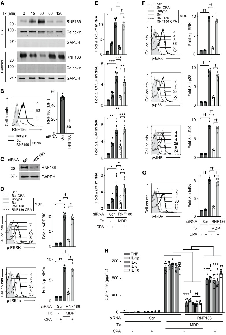
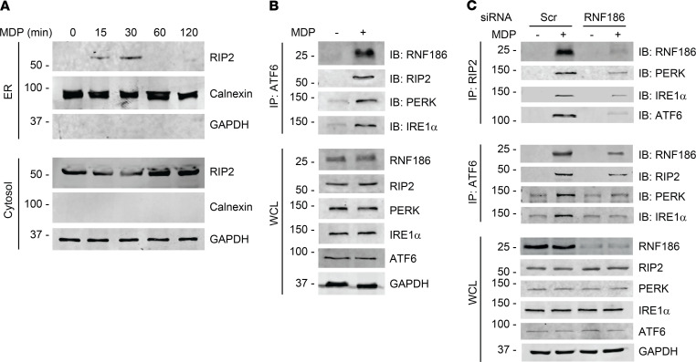
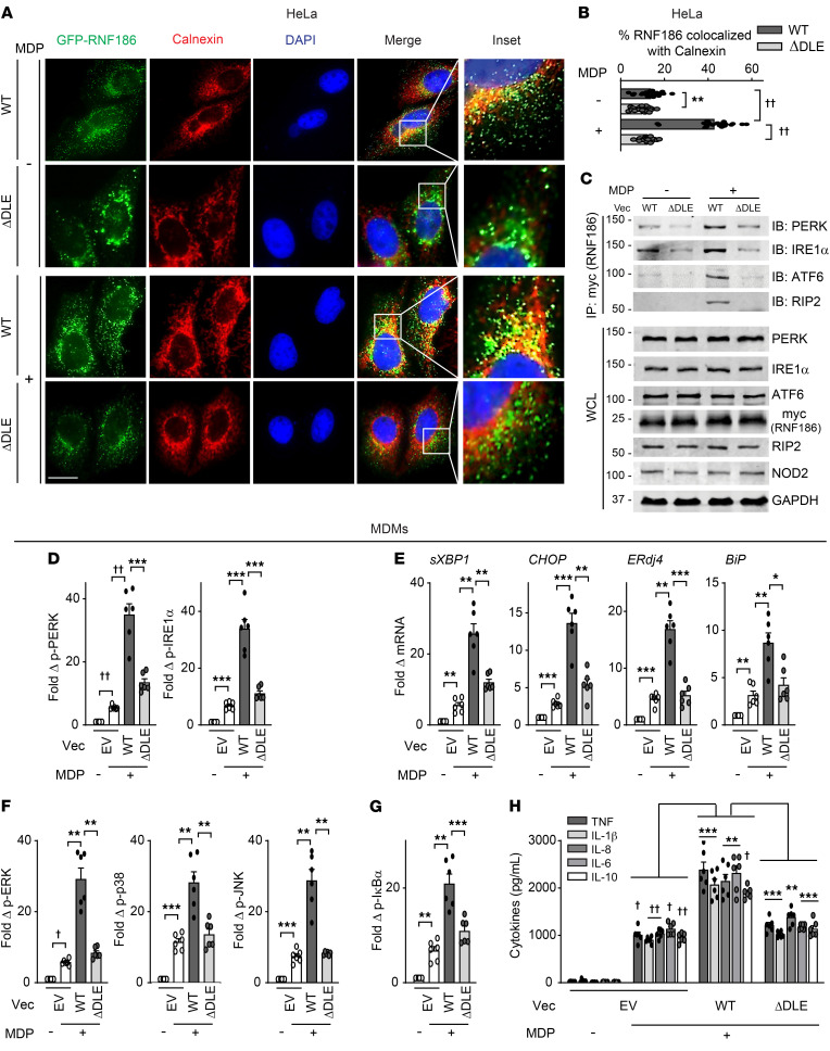
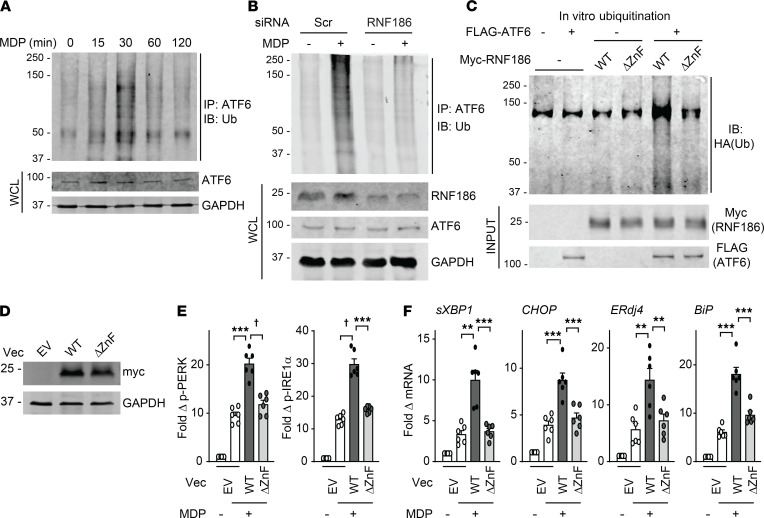
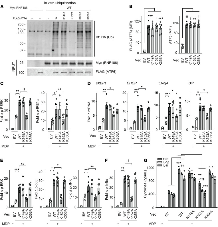
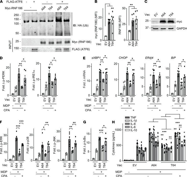
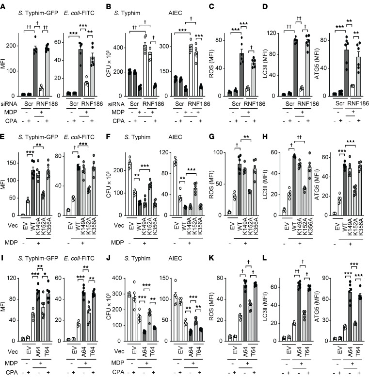
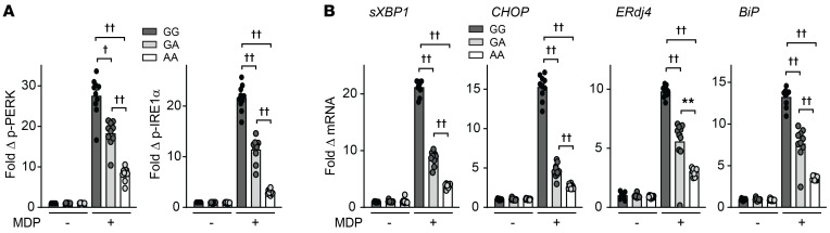
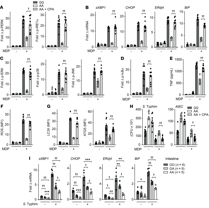
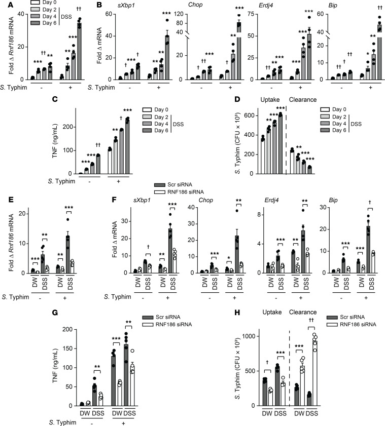
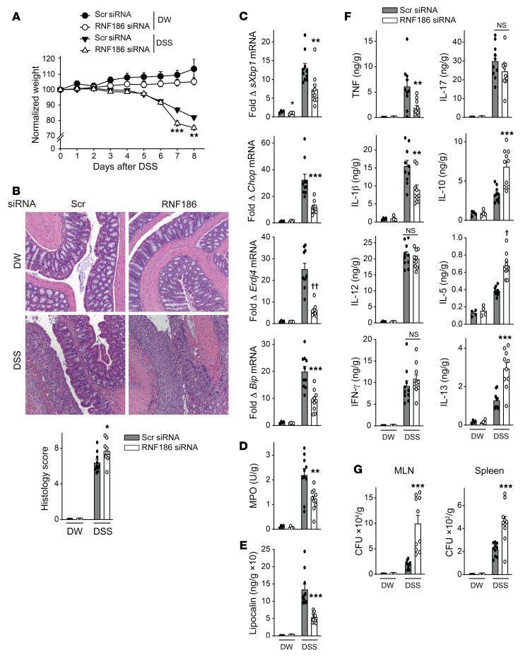
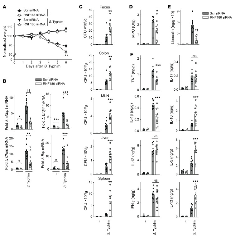
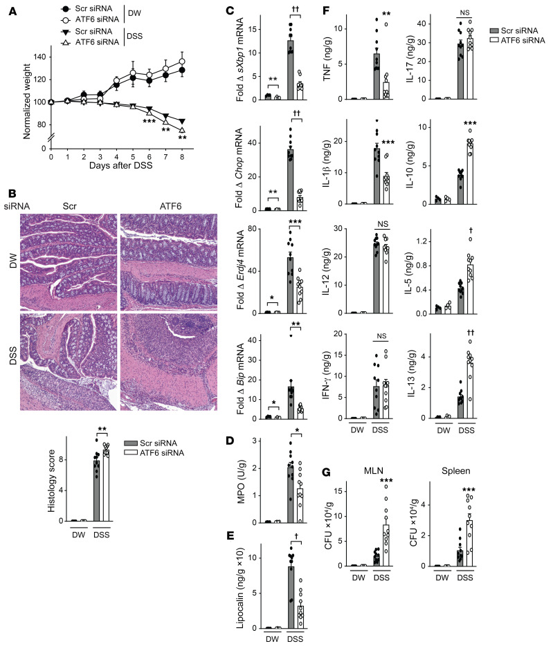
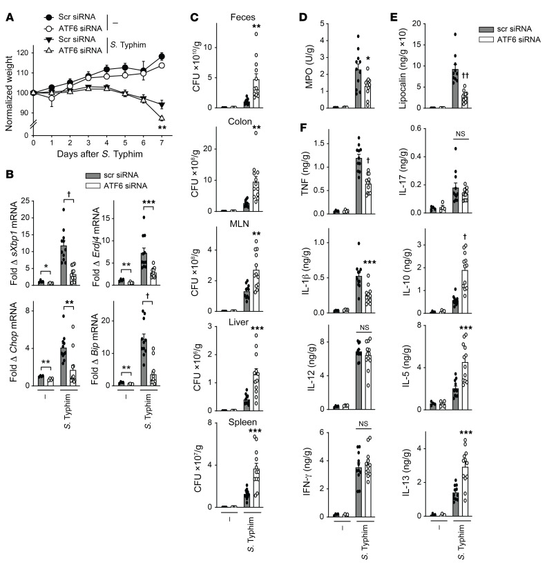
Properly balancing microbial responses by the innate immune system through pattern recognition receptors (PRRs) is critical for intestinal immune homeostasis. Ring finger protein 186 (RNF186) genetic variants are associated with inflammatory bowel disease (IBD). However, functions for the E3 ubiquitin ligase RNF186 are incompletely defined. We found that upon stimulation of the PRR nucleotide-binding oligomerization domain containing 2 (NOD2) in human macrophages, RNF186 localized to the ER, formed a complex with ER stress sensors, ubiquitinated the ER stress sensor activating transcription factor 6 (ATF6), and promoted the unfolded protein response (UPR). These events, in turn, led to downstream signaling, cytokine secretion, and antimicrobial pathway induction. Importantly, RNF186-mediated ubiquitination of K152 on ATF6 was required for these outcomes, highlighting a key role for ATF6 ubiquitination in PRR-initiated functions. Human macrophages transfected with the rare RNF186-A64T IBD risk variant and macrophages from common rs6426833 RNF186 IBD risk carriers demonstrated reduced NOD2-induced outcomes, which were restored by rescuing UPR signaling. Mice deficient in RNF186 or ATF6 demonstrated a reduced UPR in colonic tissues, increased weight loss, and less effective clearance of bacteria with dextran sodium sulfate-induced injury and upon oral challenge with Salmonella Typhimurium. Therefore, we identified that RNF186 was required for PRR-induced, UPR-associated signaling leading to key macrophage functions; defined that RNF186-mediated ubiquitination of ATF6 was essential for these functions; and elucidated how RNF186 IBD risk variants modulated these outcomes.

Keywords: Immunology; Innate immunity; Macrophages.


Conflict of interest statement

Conflict of interest: The authors have declared that no conflict of interest exists.

Figures

Figure 1
Figure 1. RNF186 localizes to the ER and is required for NOD2-induced UPR pathway activation.
(A) MDMs were treated with 100 μg/mL MDP for the indicated times. ER and cytosolic fractions were assessed for RNF186 expression by Western blot. Markers for ER (calnexin) and cytosolic (GAPDH) fractions are shown. (B and C) MDMs were transfected with scrambled or RNF186 siRNA. RNF186 protein expression by (B) flow cytometry with representative histogram with MFI values shown with horizontal lines and summary graph (n = 6 donors; similar results were observed in an additional n = 6) and (C) Western blot. (DH) MDMs were transfected with scrambled or RNF186 siRNA and then treated with 100 μg/mL MDP ± 10 μM cyclopiazonic acid (CPA). (D) Fold phospho-PERK and phospho-IRE1α induction at 30 minutes (n = 6; similar results in an additional n = 6). (E) Fold change mRNA expression at 6 hours (n = 6; similar results in an additional n = 6). (F and G) Fold phospho-protein induction at 15 minutes (n = 6; similar results in an additional n = 6). (H) Cytokines at 12 hours (n = 6; similar results in an additional n = 6). Mean + SEM. Marker positions are shown (kDa) for Western blots. *P < 0.05; **P < 0.01; ***P < 0.001; P < 1 × 10⁻⁴; ††P < 1 × 10⁻⁵ determined by 2-tailed Student’s t test with a Bonferroni-Holm correction for multiple comparisons. Scr, scrambled; Tx, treatment.
Figure 2
Figure 2. NOD2 stimulation induces an RNF186 complex with UPR sensors.
(A) MDMs were treated with 100 μg/mL MDP for the indicated times. ER and cytosolic fractions were assessed for RIP2 by Western blot. Markers for ER (calnexin) and cytosolic (GAPDH) fractions are shown. Representative of 2 independent experiments. (B) MDMs were treated with 100 μg/mL MDP for 30 minutes. ATF6 was immunoprecipitated followed by immunoblotting (IB) of the indicated proteins. Representative of 2 independent experiments. (C) MDMs were transfected with scrambled or RNF186 siRNA and then treated with 100 μg/mL MDP for 30 minutes. RIP2 or ATF6 was immunoprecipitated, and recruitment of the indicated proteins was assessed by IB. Representative of 2–3 independent experiments. Expression of the respective proteins and GAPDH in whole-cell lysates (WCLs) served as loading controls. Marker positions are shown (kDa). Scr, scrambled.
Figure 3
Figure 3. RNF186 localization to the ER is required for NOD2-induced UPR signaling and downstream outcomes.
(A and B) HeLa cells were transfected with GFP-tagged RNF186-WT or RNF186-ΔDLE and NOD2 and then treated with 100 μg/mL MDP for 30 minutes. Cells were immunostained for ER (calnexin, red) and nucleus (DAPI, blue). (A) Representative micrographs; scale bar: 10 μm. Inset represents 3× enlarged images from the merged panel. (B) Summary graph of percentage of RNF186 colocalized with calnexin (ER) (25 cells quantified). Representative of 2 independent experiments. (C) HeLa cells were transfected with myc-RNF186 WT or ΔDLE and NOD2 followed by treatment with 100 μg/mL MDP for 30 minutes. Myc (RNF186) was immunoprecipitated, and the recruitment of the indicated proteins was assessed by immunoblotting (IB). Whole-cell lysates (WCLs) of the indicated proteins served as loading controls. Marker positions are shown (kDa). Representative of 3 independent experiments. (DH) MDMs (rs6426833 AA low RNF186-expressing carriers) (n = 6) were transfected with empty vector (EV) or myc-tagged RNF186 WT or ΔDLE and then treated with 100 μg/mL MDP. (D) Fold phospho-PERK and phospho-IRE1α induction at 30 minutes. (E) Fold change mRNA expression at 4 hours. (F and G) Fold phospho-protein induction at 15 minutes. (H) Cytokines at 24 hours. Mean + SEM. *P < 0.05; **P < 0.01; ***P < 0.001; P < 1 × 10⁻⁴; ††P < 1 × 10⁻⁵ determined by 2-tailed Student’s t test with a Bonferroni-Holm correction for multiple comparisons. Vec, vector.
Figure 4
Figure 4. RNF186 promotes NOD2-induced ubiquitination of the ATF6 complex, and RNF186 E3 ubiquitin ligase activity is required for the NOD2-induced UPR.
(A) MDMs were treated with 100 μg/mL MDP for the indicated times. ATF6 was immunoprecipitated, and ubiquitinated proteins (Ubs) were assessed by Western blot (representative of 3 independent experiments). (B) MDMs were transfected with scrambled or RNF186 siRNA and then treated with 100 μg/mL MDP for 30 minutes. ATF6 was immunoprecipitated, and Ubs were assessed by Western blot (representative of 2 independent experiments). Whole-cell lysates (WCLs) of the indicated proteins served as loading controls. (C) In vitro ubiquitination was assessed as per Methods with purified HA-ubiquitin with (+) or without (-) purified FLAG-ATF6 with/without purified myc-RNF186 WT or ΔZnF. Ubiquitin protein (α-HA) was detected by Western blot (representative of 2 independent experiments). Marker positions are shown (kDa). (DF) MDMs were transfected with empty vector (EV) or myc-tagged RNF186 WT or ΔZnF. (D) RNF186 protein expression (myc) by Western blot. (E and F) Transfected MDMs (n = 6) were treated with 100 μg/mL MDP. (E) Fold phospho-PERK and phospho-IRE1α induction at 30 minutes. (F) Fold change mRNA expression at 4 hours. Mean + SEM. **P < 0.01; ***P < 0.001; P < 1 × 10⁻⁴ determined by 2-tailed Student’s t test with a Bonferroni-Holm correction for multiple comparisons. Scr, scrambled; Vec, vector; ZnF, zinc finger.
Figure 5
Figure 5. Lysine 152 in ATF6 is critical for RNF186-dependent ubiquitination of ATF6 and PRR-induced, RNF186-dependent downstream outcomes.
(A) In vitro ubiquitination was assessed as per Methods with purified HA-ubiquitin with/without purified myc-RNF186 WT with/without purified FLAG-tagged WT or lysine mutants (K149A, K152A, K350A, K356A) of ATF6. Ubiquitin protein (α-HA) was detected by Western blot (representative of 3 independent experiments). Marker positions are shown (kDa). (BG) MDMs were transfected with empty vector (EV) or FLAG-tagged WT or mutants of ATF6. (B) ATF6 protein expression as detected by flow cytometry (n = 6). (CG) Transfected MDMs (n = 6) were treated with 100 μg/mL MDP. (C) Fold phospho-PERK and phospho-IRE1α induction at 30 minutes. (D) Fold change mRNA expression at 4 hours. (E and F) Fold phospho-protein induction at 15 minutes. (G) Cytokines at 24 hours. Similar results in an additional n = 6 for BG. Mean + SEM. *P < 0.05; **P < 0.01; ***P < 0.001; P < 1 × 10⁻⁴; ††P < 1 × 10⁻⁵ determined by 2-tailed Student’s t test with a Bonferroni-Holm correction for multiple comparisons. Vec, vector.
Figure 6
Figure 6. The rare RNF186 A64T IBD risk variant demonstrates lower levels of NOD2-induced, UPR-dependent outcomes.
(A) In vitro ubiquitination with purified HA-ubiquitin with/without purified FLAG-ATF6 with/without purified myc-RNF186-A64 or myc-RNF186-T64. Ubiquitin protein (α-HA) was detected by Western blot. Representative of 2 independent experiments. Marker positions are shown (kDa). (BH) MDMs (rs6426833 AA low RNF186-expressing carriers) were transfected with empty vector (EV) or myc-tagged RNF186-A64 (WT) or RNF186-T64 (risk variant). RNF186 protein expression as detected by (B) flow cytometry (n = 6) or (C) Western blot. (DH) Transfected MDMs were treated with 100 μg/mL MDP (n = 6) ± 10 μM CPA. (D) Fold phospho-PERK and phospho-IRE1α induction at 30 minutes. (E) Fold change mRNA expression at 6 hours. (F and G) Fold phospho-protein induction at 15 minutes. (H) Cytokines at 12 hours. Mean + SEM. *P < 0.05; **P < 0.01; ***P < 0.001; P < 1 × 10⁻⁴ determined by 2-tailed Student’s t test with a Bonferroni-Holm correction for multiple comparisons. Vec, vector.
Figure 7
Figure 7. RNF186-dependent UPR signaling is required for NOD2-induced bacterial clearance and antimicrobial pathways.
(AD) MDMs (n = 6) were transfected with scrambled or RNF186 siRNA, then treated with 100 μg/mL MDP for 48 hours ± 10 μM CPA. (EH) MDMs (n = 6; similar results in an additional n = 6) were transfected with empty vector (EV) or FLAG-tagged WT or lysine mutants (K149A, K152A, or K356A) of ATF6 and then treated with 100 μg/mL MDP for 48 hours. (IL) MDMs (n = 6) (rs6426833 AA low RNF186-expressing carriers) were transfected with EV or myc-tagged RNF186-A64 (WT) or RNF186-T64 (risk variant) and then treated with 100 μg/mL MDP for 48 hours ± 10 μM CPA. (A, E, and I) Bacterial uptake. (B, F, and J) Intracellular bacterial clearance. (C, G, and K) ROS production. (D, H, and L) LC3II and ATG5 expression. Mean + SEM. *P < 0.05; **P < 0.01; ***P < 0.001; P < 1 × 10⁻⁴; ††P < 1 × 10⁻⁵ determined by 2-tailed Student’s t test with a Bonferroni-Holm correction for multiple comparisons. Scr, scrambled; Vec, vector.
Figure 8
Figure 8. IBD risk rs6426833 AA disease risk carriers demonstrate reduced PRR-induced UPR signaling.
MDMs from rs6426833 GG, GA, or AA carriers (n = 10 donors/genotype) were left untreated or treated with 100 μg/mL MDP. (A) Fold phospho-PERK and phospho-IRE1α induction at 30 minutes. (B) Fold change mRNA expression at 4 hours. Mean + SEM. **P < 0.01; P < 1 × 10⁻⁴; ††P < 1 × 10⁻⁵ determined by 2-tailed Student’s t test with a Bonferroni-Holm correction for multiple comparisons.
Figure 9
Figure 9. Complementation of the UPR in MDMs from IBD risk rs6426833 AA carriers increases NOD2-induced, RNF186-dependent outcomes to levels observed in GG carrier MDMs.
(AH) MDMs from rs6426833 GG and AA carriers (n = 10 donors/genotype) were left untreated or treated with 100 μg/mL MDP ± 10 μM CPA. (A) Fold phospho-PERK and phospho-IRE1α induction at 30 minutes. (B) Fold change mRNA expression at 4 hours. (C and D) Fold phospho-protein induction at 15 minutes. (E) Cytokines at 12 hours. (F) ROS at 48 hours. (G) LC3II and ATG5 expression at 48 hours. (H) After 48 hours, intracellular bacterial clearance was assessed. (I) Intestinal myeloid cells were cocultured with S. Typhimurium. Fold change mRNA at 2 hours stratified on rs6426833 genotype. Mean + SEM. *P < 0.05; **P < 0.01; ***P < 0.001; P < 1 × 10⁻⁴; ††P < 1 × 10⁻⁵. Significance is between AA carriers with/without CPA in AH determined by 2-tailed Student’s t test and with a Bonferroni-Holm correction for multiple comparisons in I.
Figure 10
Figure 10. RNF186 promotes bacterially induced UPR pathways and cytokines and bacterial clearance in mouse intestinal macrophages during intestinal injury.
(AD) WT mice were given 2.5% DSS in drinking water (DW) for the indicated times (n = 4/time point). (EH) Mice given scrambled or RNF186 siRNA (i.p.) were administered 2.5% DSS in DW for 4 days (n = 5). (AH) Colonic macrophages were isolated, cocultured with S. Typhimurium, and assessed for (A, B, E, and F) fold change mRNA expression at 4 hours; (C and G) TNF secretion at 24 hours; (D and H) bacterial uptake and intracellular bacterial clearance. Mean + SEM. Scr, scrambled. *P < 0.05; **P < 0.01; ***P < 0.001; P < 1 × 10⁻⁴; ††P < 1 × 10⁻⁵ determined by 2-tailed Student’s t test.
Figure 11
Figure 11. RNF186 promotes clearance of resident intestinal microbiota during DSS-induced injury.
Mice given scrambled or RNF186 siRNA (i.p.) were administered 2.5% DSS in drinking water (DW) (n = 4–10). (A) Body weights. (B) Representative H&E of colon sections and histology scores; original magnification, 100×. (C) Colon mRNA expression. (D) Colon myeloperoxidase. (E) Fecal lipocalin. (F) Colon cytokines. (G) Bacteria in MLN and spleen. (AG) DSS for 6 days and then water for an additional 2 days; independent experiment for AE, and G with DSS for 7 days. Mean + SEM. NS, not significant. *P < 0.05; **P < 0.01; ***P < 0.001; P < 1 × 10⁻⁴; ††P < 1 × 10⁻⁵ determined by 2-tailed Student’s t test.
Figure 12
Figure 12. RNF186 is required for optimal clearance of S. Typhimurium upon oral infection in mice.
Mice given scrambled or RNF186 siRNA (i.p.) were orally inoculated with S. Typhimurium (n = 4–12). Six days later mice were assessed. (A) Body weights. (B) Colon mRNA expression. (C) S. Typhimurium CFU in the indicated tissues. (D) Colon myeloperoxidase. (E) Fecal lipocalin. (F) Colon cytokines. Mean + SEM. NS, not significant. *P < 0.05; **P < 0.01; ***P < 0.001; ††P < 1 × 10⁻⁵ determined by 2-tailed Student’s t test.
Figure 13
Figure 13. ATF6 promotes clearance of resident intestinal microbiota during DSS-induced injury.
Mice given scrambled or ATF6 siRNA (i.p.) were administered 2.5% DSS in drinking water (DW) (n = 4–10). (A) Body weights. (B) Representative H&E of colon sections and histology scores; original magnification, 100×. (C) Colon mRNA expression. (D) Colon myeloperoxidase. (E) Fecal lipocalin. (F) Colon cytokines. (G) Bacteria in MLN and spleen. (AG) DSS for 6 days and then water for an additional 2 days; independent experiment for AE and G with DSS for 8 days. Mean + SEM. NS, not significant. *P < 0.05; **P < 0.01; ***P < 0.001; P < 1 × 10⁻⁴; ††P < 1 × 10⁻⁵ determined by 2-tailed Student’s t test.
Figure 14
Figure 14. ATF6 is required for optimal clearance of S. Typhimurium upon oral infection in mice.
Mice given scrambled or ATF6 siRNA (i.p.) were orally inoculated with S. Typhimurium (n = 4–12). Seven days later mice were assessed. (A) Body weights. (B) Colon mRNA expression. (C) S. Typhimurium CFU in the indicated tissues. (D) Colon myeloperoxidase. (E) Fecal lipocalin. (F) Colon cytokines. Mean + SEM. NS, not significant. *P < 0.05; **P < 0.01; ***P < 0.001; P < 1 × 10⁻⁴, ††P < 1 × 10⁻⁵ determined by 2-tailed Student’s t test.

References

    1. Abraham C, Medzhitov R. Interactions between the host innate immune system and microbes in inflammatory bowel disease. Gastroenterology. 2011;140(6):1729–1737. doi: 10.1053/j.gastro.2011.02.012. - DOI - PMC - PubMed
    1. Abraham C, Cho JH. Inflammatory bowel disease. N Engl J Med. 2009;361(21):2066–2078. doi: 10.1056/NEJMra0804647. - DOI - PMC - PubMed
    1. Lahiri A, et al. Human LACC1 increases innate receptor-induced responses and a LACC1 disease-risk variant modulates these outcomes. Nat Commun. 2017;8:15614. - PMC - PubMed
    1. Yan J, et al. An inflammatory bowel disease-risk variant in INAVA decreases pattern recognition receptor-induced outcomes. J Clin Invest. 2017;127(6):2192–2205. doi: 10.1172/JCI86282. - DOI - PMC - PubMed
    1. Hedl M, et al. Pattern recognition receptor signaling in human dendritic cells is enhanced by ICOS ligand and modulated by the Crohn’s disease ICOSLG risk allele. Immunity. 2014;40(5):734–746. doi: 10.1016/j.immuni.2014.04.011. - DOI - PMC - PubMed

Publication types

MeSH terms