. 2021 Oct 15;6(64):eabl4509.
doi: 10.1126/sciimmunol.abl4509. Epub 2021 Sep 2.

Adaptive immune determinants of viral clearance and protection in mouse models of SARS-CoV-2

Affiliations

Adaptive immune determinants of viral clearance and protection in mouse models of SARS-CoV-2

Benjamin Israelow et al. Sci Immunol. .

Abstract

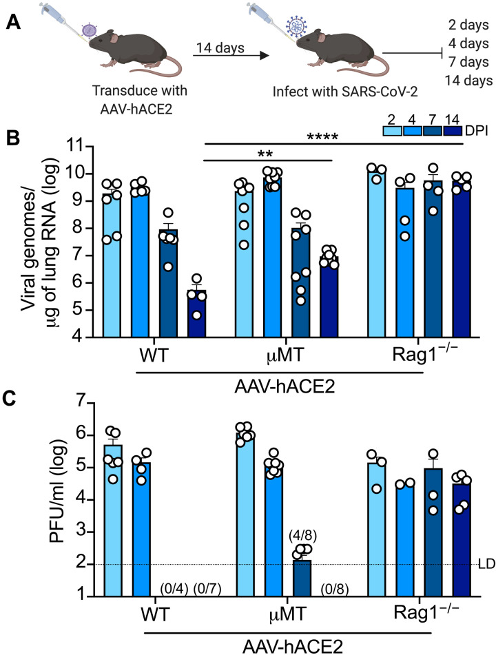
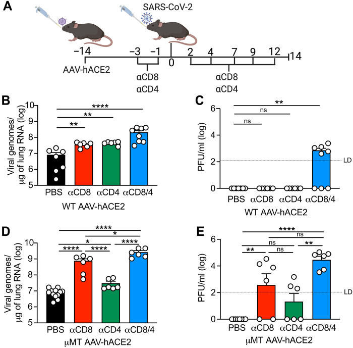
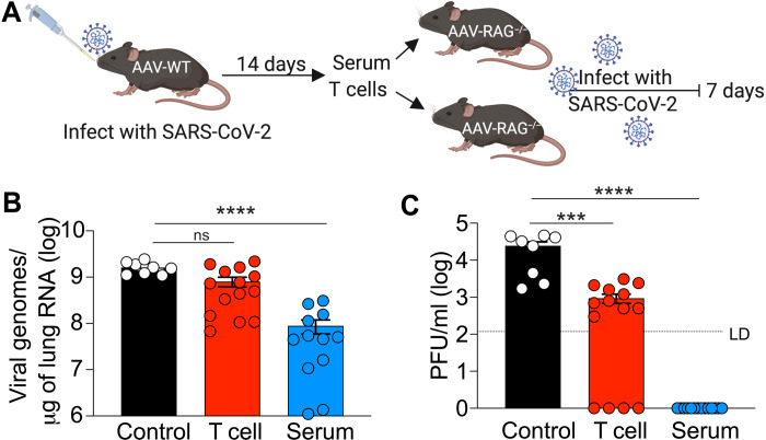
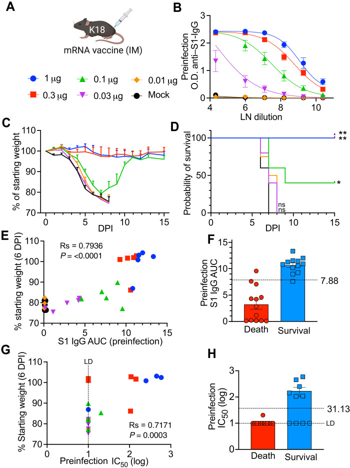
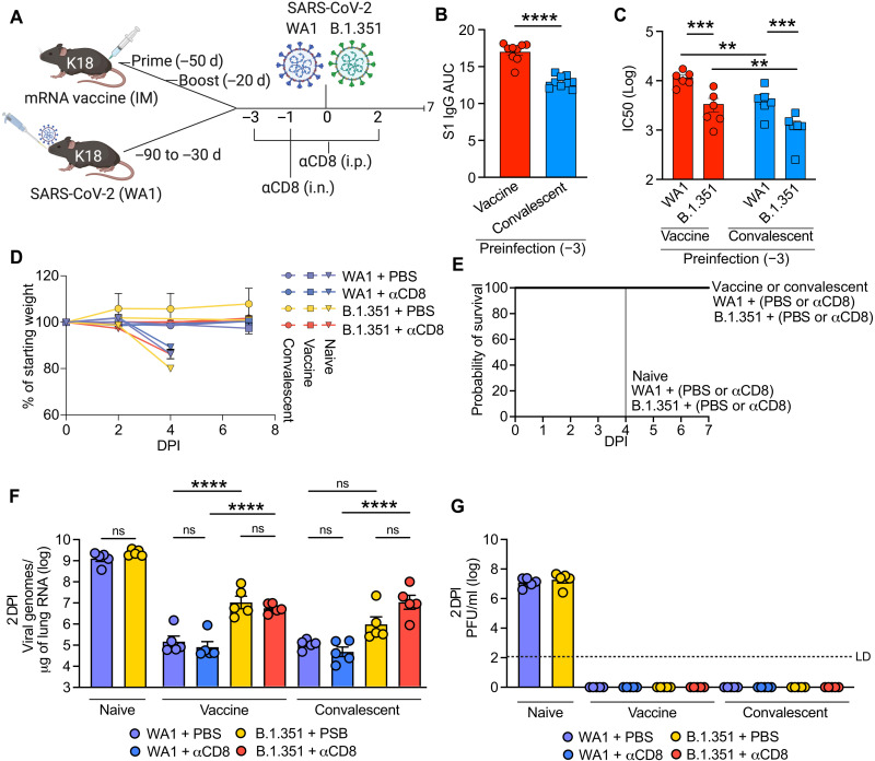
Severe acute respiratory syndrome coronavirus 2 (SARS-CoV-2) has caused more than 160 million infections and more than 3 million deaths worldwide. Although effective vaccines are currently being deployed, the adaptive immune determinants that promote viral clearance and confer protection remain poorly defined. Using mouse models of SARS-CoV-2, we demonstrate that both humoral and cellular adaptive immunity contribute to viral clearance in the setting of primary infection. Furthermore, we find that either convalescent mice or mice that receive mRNA vaccination are protected from both homologous infection and infection with a variant of concern, B.1.351. In addition, we find that this protection is largely mediated by antibody response and not cellular immunity. These results highlight the in vivo protective capacity of antibodies generated to both vaccine and natural infection.


Figures

Fig. 1.
Fig. 1.. SARS-CoV-2 clearance in RAG−/− and μMT mice.
(A) Experimental schematic. C57Bl/6J (WT), B6.129S7-Rag1tm1Mom/J (Rag1−/−), and B6.129S2-Ighmtm1Cgn/J (μMT) mice were intratracheally transduced with 1011 GC (genomic copies) of AAV encoding human ACE2 (AAV-hACE2) and allowed to recover for 14 days. Mice were then infected with WA1 strain SARS-CoV-2 at 106 PFU intranasally, and lungs samples are collected at 2, 4, 7, and 14 DPI and assessed by (B) quantitative PCR and (C) plaque assay. Values noted on x axis (C) indicate numbers of samples tested positive/number of samples. LD, limit of detection. Individual values noted as dots and bars indicate means ± SEM from three to eight samples of two to three independent experiments. P values were calculated by two-way ANOVA with Tukey’s multiple comparison. *P < 0.05, **P < 0.01, ***P < 0.001, and ****P < 0.001.
Fig. 2.
Fig. 2.. SARS-CoV-2 clearance in CD4+ and CD8+ T cell–depleted WT and μMT mice.
(A) Experimental design. AAV-hACE2 transduced WT (B and C) and μMT (D and E) were injected (intraperitoneal) with 200 mg of anti-mouse CD8a (clone 2.43) or 200 mg of anti-mouse CD4 (clone GK1.5) starting at 3 days before infection and given every 2 to 3 days as indicated until 12 DPI. Mice were infected with WA1 strain SARS-CoV-2 at 106 PFU intranasally, and lungs were collected at 14 DPI and assessed by quantitative PCR (B and D) and plaque assay (C and E). Individual values noted as dots and bars indicate means ± SEM from 6 to 12 samples of two to three independent experiments. P values were calculated by one-way ANOVA with Tukey’s multiple comparison. *P < 0.05, **P < 0.01, ***P < 0.001, and ****P < 0.001. ns, not significant.
Fig. 3.
Fig. 3.. Infection of RAG−/− mice after adoptive transfer of serum or T cells.
(A) Experimental design. AAV-hACE2–transduced WT mice were infected with 106 PFU WA1 strain SARS-CoV-2 via intranasal route. Fourteen DPI serum and mediastinal lymph node were collected. Total T cells were isolated by magnetic separation. AAV-hACE2–transduced RAG−/− mice received adoptive transfer of PBS (control), serum (200 ml), or 2 × 10⁶ total T cells at 1 day before infection with 106 PFU WA1 strain SARS-CoV-2. Lungs were collected at 7 DPI and assessed for (B) viral RNA by quantitative PCR and for (C) viral titer by plaque assay. Individual values noted as dots and bars indicate means ± SEM from 8 to 12 samples of two independent experiments. P values were calculated by one-way ANOVA with Tukey’s multiple comparison. *P < 0.05, **P < 0.01, ***P < 0.001, and ****P < 0.001.
Fig. 4.
Fig. 4.. Dose-dependent antibody response to mRNA vaccine reveals correlate of protection in K18 mice.
(A) B6.Cg-Tg(K18-ACE2)2Prlmn/J (K18-hACE2) mice received a single IM, intramuscular injection of Pfizer-BioNTech BNT162b2 mRNA vaccine at the indicated doses. (B) Twenty-eight days after injection, serum samples were harvested, and anti–SARS-CoV-2 anti-S1 antibody concentration was measured by ELISA at indicated dilutions (LN, natural log transformed). At 30 days after vaccination, mice were challenged with 4 × 10⁵ PFU WA1 SARS-CoV-2. O.D., optical density. (C) Weight loss (please see raw data file for calculated P values) and (D) Kaplan-Meier survival curve of animals monitored for 15 DPI. P values were calculated by log-rank (Mantel-Cox) test. *P < 0.05 and **P < 0.01. Individual preinfection serum from (B) was used to calculate (E) S1 IgG AUC or (G) IC50 with VSV pseudovirus neutralization assay and plotted against weight change at 6 DPI (Rs, Spearman correlation coefficient), and P values are indicated. Differences in (F) preinfection S1 IgG AUC and (H) VSV pseudovirus neutralization IC50 between mice that survived and mice that died from infection. Logistic regression (Prism) was used to calculate 50% survival titers indicated by dotted line. N = 5 mice per condition.
Fig. 5.
Fig. 5.. Response to homologous and B.1.351 challenge of convalescent and mRNA-vaccinated mice.
(A) Experimental design. K18 mice were either vaccinated with 1 μg of Pfizer/BioNTech mRNA via intramuscular injection via prime/boost with a 30-day interval or infected with 500 PFU WA1 strain SARS-CoV-2 via intranasal (i.n.) administration once. Preinfection serum was collected 3 days before infection, and (B) anti–SARS-CoV-2 antibody titer was assessed by ELISA, plotted as AUC, and (C) VSV pseudovirus neutralization assay performed with either pseudovirus expressing WA1 or B.1.351 spike and plotted as log IC50. Mice were injected (intraperitoneally, i.p.) with 200 mg of anti-mouse CD8a (clone 2.43) or PBS (control) at 3 and 1 days before infection and 2 days after infection, and mice received 100 mg of anti-mouse CD8a or PBS via intranasal administration at 1 day before infection. Mice were infected with 4 × 10⁵ PFU of with homologous strain (WA1) or VOC (B.1.351) via intranasal route and euthanized at 2, 4, or 7 DPI. (D) Weight loss and (E) Kaplan-Meier survival curve were monitored for 7 DPI. (F) Viral RNA was assessed by quantitative PCR and (G) viral titer assessed by plaque assay at 2 DPI. Individual values noted as dots and bars indicate means ± SEM from n = 5 samples per condition. P values were calculated by one-way ANOVA with Tukey’s multiple comparison, except in (B) where a Student’s t test was used and (C) where two-way ANOVA with Tukey’s multiple comparison was used. *P < 0.05, **P < 0.01, ***P < 0.001, and ****P < 0.001.

Update of

References

    1. Dan J. M., Mateus J., Kato Y., Hastie K. M., Yu E. D., Faliti C. E., Grifoni A., Ramirez S. I., Haupt S., Frazier A., Nakao C., Rayaprolu V., Rawlings S. A., Peters B., Krammer F., Simon V., Saphire E. O., Smith D. M., Weiskopf D., Sette A., Crotty S., Immunological memory to SARS-CoV-2 assessed for up to 8 months after infection. Science 371, eabf4063 (2021). - PMC - PubMed
    1. Grifoni A., Weiskopf D., Ramirez S. I., Mateus J., Dan J. M., Moderbacher C. R., Rawlings S. A., Sutherland A., Premkumar L., Jadi R. S., Marrama D., de Silva A. M., Frazier A., Carlin A. F., Greenbaum J. A., Peters B., Krammer F., Smith D. M., Crotty S., Sette A., Targets of T cell responses to SARS-CoV-2 coronavirus in humans with COVID-19 disease and unexposed individuals. Cell 181, 1489–1501.e15 (2020). - PMC - PubMed
    1. Baden L. R., El Sahly H. M., Essink B., Kotloff K., Frey S., Novak R., Diemert D., Spector S. A., Rouphael N., Creech C. B., McGettigan J., Khetan S., Segall N., Solis J., Brosz A., Fierro C., Schwartz H., Neuzil K., Corey L., Gilbert P., Janes H., Follmann D., Marovich M., Mascola J., Polakowski L., Ledgerwood J., Graham B. S., Bennett H., Pajon R., Knightly C., Leav B., Deng W., Zhou H., Han S., Ivarsson M., Miller J., Zaks T.; COVE study Group , Efficacy and safety of the mRNA-1273 SARS-CoV-2 vaccine. N. Engl. J. Med. 384, 403–416 (2021). - PMC - PubMed
    1. Rodda L. B., Netland J., Shehata L., Pruner K. B., Morawski P. A., Thouvenel C. D., Takehara K. K., Eggenberger J., Hemann E. A., Waterman H. R., Fahning M. L., Chen Y., Hale M., Rathe J., Stokes C., Wrenn S., Fiala B., Carter L., Hamerman J. A., King N. P., Gale M. Jr., Campbell D. J., Rawlings D. J., Pepper M., Functional SARS-CoV-2-specific immune memory persists after mild COVID-19. Cell 184, 169–183.e17 (2021). - PMC - PubMed
    1. Corbett K. S., Flynn B., Foulds K. E., Francica J. R., Boyoglu-Barnum S., Werner A. P., Flach B., O’Connell S., Bock K. W., Minai M., Nagata B. M., Andersen H., Martinez D. R., Noe A. T., Douek N., Donaldson M. M., Nji N. N., Alvarado G. S., Edwards D. K., Flebbe D. R., Lamb E., Doria-Rose N. A., Lin B. C., Louder M. K., O’Dell S., Schmidt S. D., Phung E., Chang L. A., Yap C., Todd J. M., Pessaint L., Van Ry A., Browne S., Greenhouse J., Putman-Taylor T., Strasbaugh A., Campbell T. A., Cook A., Dodson A., Steingrebe K., Shi W., Zhang Y., Abiona O. M., Wang L., Pegu A., Yang E. S., Leung K., Zhou T., Teng I. T., Widge A., Gordon I., Novik L., Gillespie R. A., Loomis R. J., Moliva J. I., Stewart-Jones G., Himansu S., Kong W. P., Nason M. C., Morabito K. M., Ruckwardt T. J., Ledgerwood J. E., Gaudinski M. R., Kwong P. D., Mascola J. R., Carfi A., Lewis M. G., Baric R. S., McDermott A., Moore I. N., Sullivan N. J., Roederer M., Seder R. A., Graham B. S., Evaluation of the mRNA-1273 vaccine against SARS-CoV-2 in nonhuman primates. N. Engl. J. Med. 383, 1544–1555 (2020). - PMC - PubMed

Publication types

Substances