. 2021 Jan-Dec:13:17590914211048260.
doi: 10.1177/17590914211048260.

The Mesencephalic Periaqueductal Gray, a Further Structure Involved in Breathing Failure Underlying Sudden Infant Death Syndrome

Affiliations

The Mesencephalic Periaqueductal Gray, a Further Structure Involved in Breathing Failure Underlying Sudden Infant Death Syndrome

Anna M Lavezzi et al. ASN Neuro. 2021 Jan-Dec.

Abstract

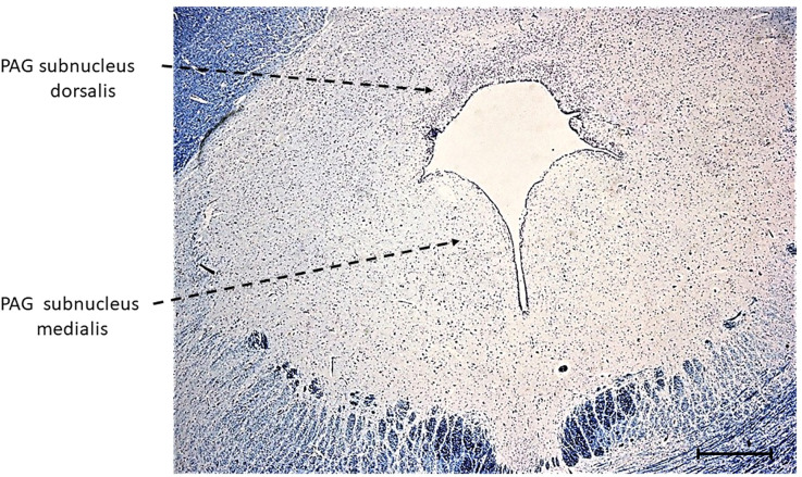
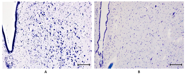
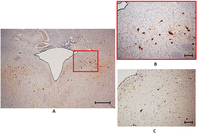
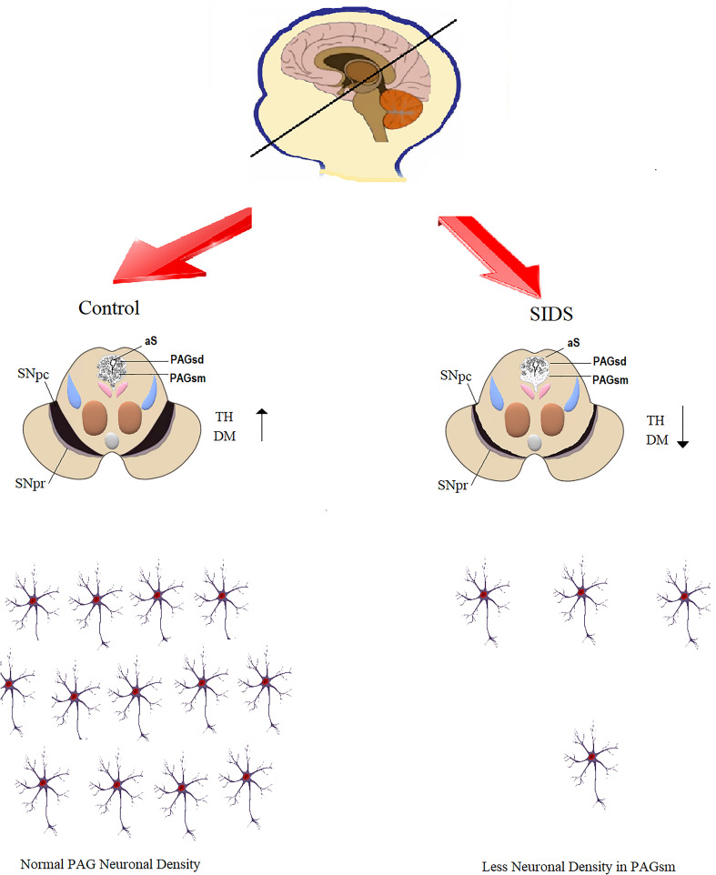
The aim of this study was to investigate the involvement of the periaqueductal gray (PAG), an area of gray matter surrounding the cerebral aqueduct of Sylvius, in the pathogenetic mechanism of SIDS, a syndrome frequently ascribed to arousal failure from sleep. We reconsidered the same samples of brainstem, more precisely midbrain specimens, taken from a large series of sudden infant deaths, namely 46 cases aged from 1 to about 7 months, among which 26 SIDS and 20 controls, in which we already highlighted significant developmental alterations of the substantia nigra, another mesencephalic structure with a critical role in breath and awakening regulation. Specific histological and immunohistochemical methods were applied to examine the PAG cytoarchitecture and the expression of the tyrosine hydroxylase, a marker of catecholaminergic neurons. Hypoplasia of the PAG subnucleus medialis was observed in 65% of SIDS but never in controls; tyrosine hydroxylase expression was significantly higher in controls than in SIDS. A significant correlation was found between these findings and those related to the substantia nigra, demonstrating a link between these neuronal centers and the brainstem respiratory network and a common involvement in the sleep-arousal phase failure leading to SIDS.

Keywords: brainstem; dopamine; neuropathology; periaqueductal gray; sudden infant death syndrome; tyrosine hydroxylase.


Conflict of interest statement

Declaration of conflicting interests: The authors declared no potential conflicts of interest with respect to the research, authorship, and/or publication of this article.

Figures

Figure 1.
Figure 1.
Photomicrographs of a transverse section of midbrain, at the level of inferior colliculus with the indication of the two subnuclei of the human periaqueductal gray (PAG): the subnucleus dorsalis and the subnucleus medialis. Klüver-Barrera stain; scale Bar: 200 µm.
Figure 2.
Figure 2.
Periaqueductal gray subnucleus dorsalis (PAGsd), characterized by a wide and dense cluster of small sized, round or fusiform cells, located back to the Sylvius aqueduct. Klüver-Barrera stain; scale Bar: 50 µm.
Figure 3.
Figure 3.
Periaqueductal gray subnucleus medialis (PAGsm). A) large elongated, darkly stained neurons with long processes in the ventrolateral area surrounding the Sylvius aqueduct (control case). B) rare neurons in the same area (SIDS case). Klüver-Barrera stain; scale Bar: 50 µm.
Figure 4.
Figure 4.
Tyrosine hydroxylase (TH) immunohistochemistry. (A) TH-immunopositivity in the PAGsm in a control case. The boxed area is shown at higher magnification in (B); here intensely stained neuronal bodies and processes are clearly visible. (C) Rare presence of weakly immunostained neurons in the PAGsm of a SIDS case. TH immunostaining; scale Bar: (A) 200 µm, (B) and (C) 50 µm.
Figure 5.
Figure 5.
Schematic representation of all the results obtained in this study showing that i the neuron number and the TH expression in the PAG are decreased in SIDS cases compared to controls. aS = aqueduct of Sylvius; DM = dopamine; PAGsd = periaqueductal grey subnucleus dorsalis; PAGsm = periaqueductal grey subnucleus medialis; SNpc = substantia nigra pars compacta; SNpr = substantia negra pars reticulata; TH = tyrosine hydroxylase.

Similar articles

Cited by

References

    1. Bandler R., Shipley M. T. (1994). Columnar organization in the midbrain periaqueductal gray: Modules for emotional expression? Trends in Neurosciences, 7(9), 379–389. https://doi.org/10.1016/0166-2236(94)90047-7 - DOI - PubMed
    1. Basbaum A. I., Fields H. L. (1978). Endogenous pain control mechanisms: Review and hypothesis. Annals of Neurology, 4(5), 451–462. 10.1002/ana.410040511 - DOI - PubMed
    1. Baskin D. S., Mehler W. R., Hosobuchi Y., Richardson D. E., Adams J. E., Flitter M. A. (1986). Autopsy analysis of the safety, efficacy and cartography of electrical stimulation of the central gray in humans. Brain Research, 371(2), 231–236. 10.1016/0006-8993(86)90358-6 - DOI - PubMed
    1. Beitz A. J. (1982). The organization of afferent projections to the midbrain periaqueductal gray of the rat. Neuroscience, 7, 133–159. 10.1016/0306-4522(82)90157-9. - DOI - PubMed
    1. Beitz A. J. (1985). The midbrain periaqueductal gray in the rat. I. Nuclear volume, cell number, density, orientation, and regional subdivisions . Journal of Comparative Neurology, 237(4), 445–459. 10.1002/cne.902370403 - DOI - PubMed

Substances