Review
. 2021 Oct 8:22:e933341.
doi: 10.12659/AJCR.933341.

Ileocolic Intussusception in a Woman: A Case Report and Literature Review

Affiliations
Review

Ileocolic Intussusception in a Woman: A Case Report and Literature Review

Alexandros A Moniakis et al. Am J Case Rep. .

Abstract

BACKGROUND Intussusception is a rare pathological entity in adults and remains a diagnostic challenge for clinicians, as it shares many clinical signs and symptoms with other morbid conditions (including appendicitis, abdominal hernias, colic, volvulus, and Meckel diverticulum). High clinical suspicion and use of appropriate imaging techniques are essential for early diagnosis and treatment of intussusception. Surgical intervention is the treatment of choice in cases of sustained and persistent invagination. CASE REPORT We present the case of a 65-year-old woman with a medical history of Crohn's disease, diabetes mellitus type II, hypertension, and rheumatoid arthritis. She was hospitalized for diarrhea, fatigue, and anemia. Computerized tomography of the abdomen and a colonoscopy revealed telescoping of the ileum, ileocecal valve, and part of the ascending colon inside the terminal segment of the ascending colon. The antegrade ileocolic intussusception was treated by performing a right hemicolectomy. The pathologic examination of the excised intestine showed mucosal lesions compatible with Crohn's disease, an inflammatory fibroid polyp at the terminal section of the ileum, and a low-grade appendiceal mucinous neoplasm. CONCLUSIONS Regardless of the etiology, when the normal motility of the intestine is altered, it can lead to invagination. Although intussusception is rare, it must always be part of the differential diagnosis for a patient presenting with constant abdominal pain.


Conflict of interest statement

Conflict of interest: None declared

Figures

Figure 1.
Figure 1.
Abdominal computed tomography scan: The red arrow shows invagination of the ileum, ileocecal valve, and part of the ascending colon inside the terminal section of the ascending colon. (Image processed with MS Paint, edition 6.1, 2009, Microsoft Corporation, Redmond, Washington, USA).
Figure 2.
Figure 2.
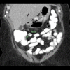
Coronal image of the abdominal computed tomography scan: The green arrow shows the parts involved in the intussusception. (Image processed with MS Paint, edition 6.1, 2009, Microsoft Corporation, Redmond, Washington, USA).
Figure 3.
Figure 3.
Image of the surgical specimen: The red arrow shows the point of invagination. (Image processed with MS Paint, edition 6.1, 2009, Microsoft Corporation, Redmond, Washington, USA).
Figure 4.
Figure 4.
Gross images of the polyps (All the images were processed with Adobe Photoshop CS3, Adobe Inc., San Jose, California, USA): (A) Adjacent to the ileocecal valve. (B) Close to the proximal margin. (C) White fleshy cut surface.
Figure 5.
Figure 5.
(A) Inflamed terminal ileum mucosa proximal to the larger polyp (Image processed with Adobe Photoshop CS3, Adobe Inc, San Jose, California, USA). (B) Microscopic image of the terminal ileum mucosa proximal to the larger polyp revealed chronic active inflammation, submucosal involvement, and serositis, as seen in Crohn’s disease (hematoxylin and eosin, 20×) (Image processed with Adobe Photoshop CS3, Adobe Inc, San Jose, California, USA).
Figure 6.
Figure 6.
Microscopic images of the polyps (All the images were processed with Adobe Photoshop CS3, Adobe Inc, San Jose, California, USA): (A) Submucosal mesenchymal lesion (hematoxylin and eosin, 20×). (B) Hypocellular neoplasm composed of spindle and stellate cells embedded in edematous stroma with admixed eosinophils and lymphocytes (hematoxylin and eosin, 40×). (C) Hypocellular neoplasm composed of spindle and stellate cells embedded in edematous stroma with admixed eosinophils and lymphocytes (hematoxylin and eosin, 100×).
Figure 7.
Figure 7.
(A) Neoplastic cells stained positive in CD34 (100×) (Image processed with Adobe Photoshop CS3, Adobe Inc, San Jose, California, USA). (B) Neoplastic cells stained negative in CD117 (400×) (Image processed with Adobe Photoshop CS3, Adobe Inc, San Jose, California, USA). (C) Neoplastic cells stained negative in S-100 (100×) (Image processed with Adobe Photoshop CS3, Adobe Inc, San Jose, California, USA). (D) Neoplastic cells stained negative in α-smooth muscle actin (400×) (Image processed with Adobe Photoshop CS3, Adobe Inc, San Jose, California, USA).
Figure 8.
Figure 8.
Low-grade appendiceal mucinous neoplasm: Neoplasm composed of mucinous filiform/villous epithelium with low-grade cytology (hematoxylin and eosin, 100×) (Image processed with Adobe Photoshop CS3, Adobe Inc., San Jose, California, USA).

References

    1. Zubaidi A, Al-Saif F, Silverman R. Adult intussusception: A retrospective review. Dis Colon Rectum. 2006;49(10):1546–51. - PubMed
    1. Wang N, Cui XY, Liu Y, et al. Adult intussusception: A retrospective review of 41 cases. World J Gastroenterol. 2009;15(26):3303–8. - PMC - PubMed
    1. Honjo H, Mike M, Kusanagi H, Kano N. Adult intussusception: A retrospective review. World J Surg. 2015;39(1):134–38. - PMC - PubMed
    1. McKay R. Ileocecal intussusception in an adult: The laparoscopic approach. JSLS. 2006;10(2):250–53. - PMC - PubMed
    1. Paramythiotis D, Goulas P, Moysidis M, et al. Bowel intussusception in adults: A report of three interesting cases and current trends for diagnosis and surgical management. Hippokratia. 2019;23(1):37–41. - PMC - PubMed