Review
. 2021 Oct 14;184(21):5309-5337.
doi: 10.1016/j.cell.2021.09.020. Epub 2021 Oct 7.

Hallmarks of response, resistance, and toxicity to immune checkpoint blockade

Affiliations
Review

Hallmarks of response, resistance, and toxicity to immune checkpoint blockade

Golnaz Morad et al. Cell. .

Erratum in

Abstract

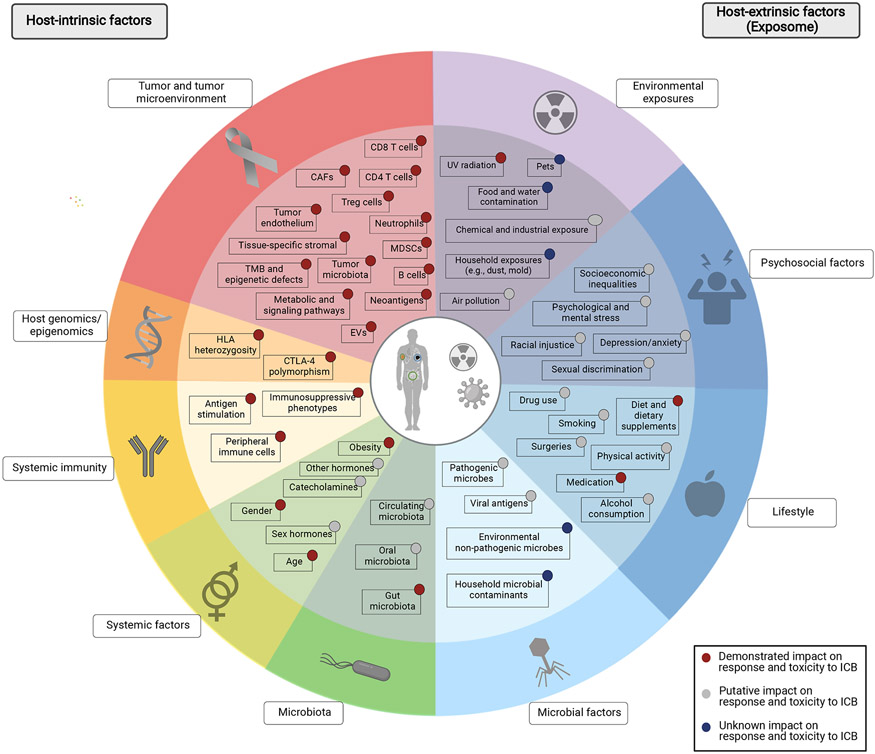
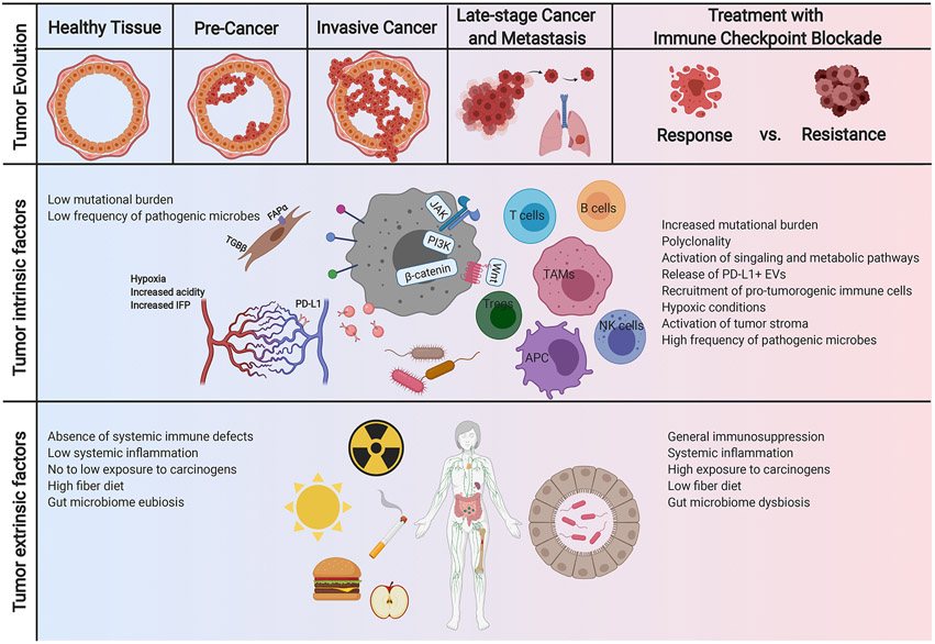
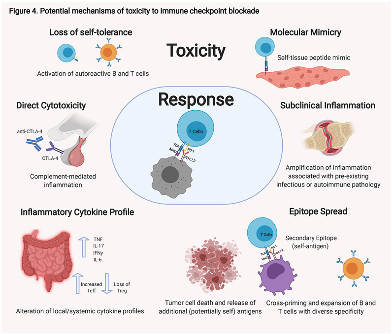
Unprecedented advances have been made in cancer treatment with the use of immune checkpoint blockade (ICB). However, responses are limited to a subset of patients, and immune-related adverse events (irAEs) can be problematic, requiring treatment discontinuation. Iterative insights into factors intrinsic and extrinsic to the host that impact ICB response and toxicity are critically needed. Our understanding of the impact of host-intrinsic factors (such as the host genome, epigenome, and immunity) has evolved substantially over the past decade, with greater insights on these factors and on tumor and immune co-evolution. Additionally, we are beginning to understand the impact of acute and cumulative exposures-both internal and external to the host (i.e., the exposome)-on host physiology and response to treatment. Together these represent the current day hallmarks of response, resistance, and toxicity to ICB. Opportunities built on these hallmarks are duly warranted.


Conflict of interest statement

Declaration of interests B.A.H. reports no conflicts of interest. G.M. is a co-inventor on US patents (PCT/US2019/022194, PCT/US2020/029556, PCT/US2020/046050) relating to extracellular vesicles. P.S. reports consulting, advisory roles, and/or stocks/ownership for Achelois, Adaptive Biotechnologies, Apricity Health, BioAlta, BioNTech, Codiak Biosciences, Constellation, Dragonfly Therapeutics, Forty-Seven Inc., Hummingbird, ImaginAb, Infinity Pharma, Jounce Therapeutics, Lave Therapeutics, Lytix Biopharma, Marker Therapeutics, Oncolytics, Phenomics, and Polaris Pharma and owns a patent licensed to Jounce Therapeutics. P.S. reports consulting, stock ownership, or advisory board membership for Achelois, Adaptive Biotechnologies, Apricity, BioAtla, BioNTech, Candel Therapeutics, Codiak, Dragonfly, Earli, Enable Medicine, Hummingbird, ImaginAb, Jounce, Lava Therapeutics, Lytix, Marker, PBM Capital, Phenomic AI, Polaris Pharma, Time Bioventures, and Trained Therapeutix and Venn Biosciences for an immediate family member. J.A.W. is a co-inventor on a US patent (PCT/US17/53,717) relating to the microbiome. J.A.W. reports speaker fees from Imedex, Dava Oncology, Omniprex, Illumina, Gilead, MedImmune, and BMS; consultant/advisor roles or advisory board membership for Roche-Genentech, Novartis, AstraZeneca, GSK, BMS, Merck/MSD, Biothera Pharma, and Microbiome DX; and receives clinical trial support from GSK, Roche-Genentech, BMS, and Novartis, all outside the current work.

Figures

Figure 1.
Figure 1.. Evolution of our understanding of cellular interactions contributing to tumor immunity.
The basic description of anti-tumor immunity encompasses tumor antigen presentation to T cells via antigen presenting cells (APCs) or tumor cells, followed by T cell activation against tumor cells, which involves a number of costimulatory and inhibitory molecules including CD28, CTLA-4, and PD-1. Over the years, our understanding of anti-tumor immunity has evolved tremendously, owing to the identification of several other regulatory molecules on these and other immune cell types. APC, antigen presenting cells; MDSCs, myeloid-derived suppressor cells; Treg, regulatory T cells; NK cells, natural killer cells; MHC, major histocompatibility complex; TCR, T cell receptor; CTLA4, cytotoxic T-lymphocyte-associated protein 4; PD-1, programmed cell death protein 1; PD-L1,2, programmed death-ligand 1,2; ICOS, Inducible T-cell COStimulator; ICOSL, ICOS ligand; GITR, Glucocorticoid-Induced TNFR-Related; GITRL, GITR ligand; LAG3, lymphocyte activation gene 3; BTLA, B- and T-lymphocyte attenuator; HVEM, Herpes Virus Entry Mediator ; VISTA, V-domain Ig suppressor of T cell activation; VISTAL, VISTA ligand; TIM3, T-cell immunoglobulin domain and mucin domain 3; CEACAM-1, carcinoembryonic antigen-related cell adhesion molecule 1; TGIT, T cell Ig and ITIM domain.
Figure 2.
Figure 2.. Factors impacting anti-tumor immunity and immunotherapy response.
Numerous factors regulate the dynamic process of tumor immunity and response to immune checkpoint blockade. Host-intrinsic factors including those inherent to the tumor cells and the tumor microenvironment (red), host genomics and epigenomics (orange), host immunity (yellow), as well as other immune-regulating factors (systemic factors, light green; microbiota, dark green) have been evaluated through a rapidly growing body of evidence. More recently, the importance of host-extrinsic factors, i.e., the exposome (shown in blue and purple) in modulating the tumor immunity and their potential impact on response to checkpoint blockade is being recognized increasingly and calls for comprehensive, albeit complicated, studies on this matter. TMB, tumor mutational burden; Treg, regulatory T cells; MDSCs, myeloid-derived suppressor cells; CAFs, cancer associated fibroblasts; EVs, extracellular vesicles; HLA, human leukocyte antigen; CTLA-4, cytotoxic T-lymphocyte-associated protein 4; UV, ultraviolet.
Figure 3.
Figure 3.. Coevolution of cancer and anti-tumor immunity.
This reciprocal evolution of tumor and the immune microenvironment has important clinical implications within the context of immunotherapy. As the tumor evolves, mechanisms of immune evasion can positively select the tumor subclones with low immunogenicity and disruption in antigen presentation. Furthermore, treatment with immune checkpoint blockade can also change the evolutionary landscape of the tumor, characterized by several factors such as reduction in mutational load in responders and can determine the mechanisms of resistance. TAM, tissue associated macrophages; NK cells, natural killer cells; APC, antigen presenting cells; Tregs, regulatory T cells; PD-L1, programmed death-ligand 1; TGF, tissue growth factor; FAP, fibroblast activation protein; IFP, interstitial fluid pressure; JAK, Janus kinase; PI3K, phosphatidylinositol-3-kinase; EVs, extracellular vesicles.
Figure 4.
Figure 4.. Potential mechanisms of toxicity to immune checkpoint blockade.
There are a number of possible mechanisms that have been proposed that contribute to the toxicities observed in some patients in response to immune checkpoint blockade. These possibilities are not mutually exclusive, and different mechanisms likely exist for different immune-related toxicities. Autoreactive T and B cells are thought to be key moieties in these processes. Autoreactive T cells could be generated de novo. These T cells are activated by professional APCs at the tumor site and reactive to tumor-specific antigens; however, they may coincidentally be reactive to peptides found on normal tissue that mimic the tumor-specific antigens. Alternatively, pre-existing autoreactive T and B cells that have escaped self-tolerance which were quiescent could be activated when self-peptides are presented through epitope spread by antigen presenting cells (APCs) at the tumor site. Immune-checkpoint blockade can result in alterations in the systemic immunity including changes in cytokine profiles. Changes in the cytokine profile within a given tissue can tip the existing balance towards inflammation. Alternative mechanisms also likely exist. For hypopituitarism, direct antibody-mediated cytotoxicity to CTLA-4 normally expressed on the pituitary gland is thought to play a role. Finally, amplification for pre-existing inflammatory or autoimmune pathologies are also possible. TNF, tumor necrosis factor; IFN, interferon; Teff, effector T cells; Treg, regulatory T cells.

References

    1. Abu-Sbeih H, and Wang Y (2020). Management Considerations for Immune Checkpoint Inhibitor-Induced Enterocolitis Based on Management of Inflammatory Bowel Disease. Inflamm Bowel Dis 26, 662–668. - PubMed
    1. Andreae S, Piras F, Burdin N, and Triebel F (2002). Maturation and activation of dendritic cells induced by lymphocyte activation gene-3 (CD223). J Immunol 168, 3874–3880. - PubMed
    1. Arbour KC, Mezquita L, Long N, Rizvi H, Auclin E, Ni A, Martinez-Bernal G, Ferrara R, Lai WV, Hendriks LEL, et al. (2018). Impact of Baseline Steroids on Efficacy of Programmed Cell Death-1 and Programmed Death-Ligand 1 Blockade in Patients With Non-Small-Cell Lung Cancer. J Clin Oncol 36, 2872–2878. - PubMed
    1. Ayers M, Lunceford J, Nebozhyn M, Murphy E, Loboda A, Kaufman DR, Albright A, Cheng JD, Kang SP, Shankaran V, et al. (2017). IFN-gamma-related mRNA profile predicts clinical response to PD-1 blockade. J Clin Invest 127, 2930–2940. - PMC - PubMed
    1. Baixeras E, Huard B, Miossec C, Jitsukawa S, Martin M, Hercend T, Auffray C, Triebel F, and Piatier-Tonneau D (1992). Characterization of the lymphocyte activation gene 3-encoded protein. A new ligand for human leukocyte antigen class II antigens. J Exp Med 176, 327–337. - PMC - PubMed

Publication types

Substances