. 2022 May 5;59(5):2003721.
doi: 10.1183/13993003.03721-2020. Print 2022 May.

Nestin promotes pulmonary fibrosis via facilitating recycling of TGF-β receptor I

Affiliations

Nestin promotes pulmonary fibrosis via facilitating recycling of TGF-β receptor I

Jiancheng Wang et al. Eur Respir J. .

Abstract

Background: Idiopathic pulmonary fibrosis (IPF) is a progressive fibrotic lung disease that is characterised by aberrant proliferation of activated myofibroblasts and pathological remodelling of the extracellular matrix. Previous studies have revealed that the intermediate filament protein nestin plays key roles in tissue regeneration and wound healing in different organs. Whether nestin plays a critical role in the pathogenesis of IPF needs to be clarified.

Methods: Nestin expression in lung tissues from bleomycin-treated mice and IPF patients was determined. Transfection with nestin short hairpin RNA vectors in vitro that regulated transcription growth factor (TGF)-β/Smad signalling was conducted. Biotinylation assays to observe plasma membrane TβRI, TβRI endocytosis and TβRI recycling after nestin knockdown were performed. Adeno-associated virus serotype (AAV)6-mediated nestin knockdown was assessed in vivo.

Results: We found that nestin expression was increased in a murine pulmonary fibrosis model and IPF patients, and that the upregulated protein primarily localised in lung α-smooth muscle actin-positive myofibroblasts. Mechanistically, we determined that nestin knockdown inhibited TGF-β signalling by suppressing recycling of TβRI to the cell surface and that Rab11 was required for the ability of nestin to promote TβRI recycling. In vivo, we found that intratracheal administration of AAV6-mediated nestin knockdown significantly alleviated pulmonary fibrosis in multiple experimental mice models.

Conclusion: Our findings reveal a pro-fibrotic function of nestin partially through facilitating Rab11-dependent recycling of TβRI and shed new light on pulmonary fibrosis treatment.


Conflict of interest statement

Conflict of interest: J. Wang has nothing to disclose. Conflict of interest: X. Lai has nothing to disclose. Conflict of interest: S. Yao has nothing to disclose. Conflict of interest: H. Chen has nothing to disclose. Conflict of interest: J. Cai has nothing to disclose. Conflict of interest: Y. Luo has nothing to disclose. Conflict of interest: Y. Wang has nothing to disclose. Conflict of interest: Y. Qiu has nothing to disclose. Conflict of interest: Y. Huang has nothing to disclose. Conflict of interest: X. Wei has nothing to disclose. Conflict of interest: B. Wang has nothing to disclose. Conflict of interest: Q. Lu has nothing to disclose. Conflict of interest: Y. Guan has nothing to disclose. Conflict of interest: T. Wang has nothing to disclose. Conflict of interest: S. Li has nothing to disclose. Conflict of interest: A.P. Xiang has nothing to disclose.

Figures
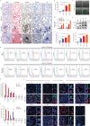
FIGURE 1
FIGURE 1
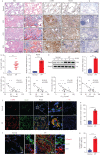
Nestin is upregulated in experimental pulmonary fibrosis, and localised mainly in lung myofibroblasts. a) Haematoxylin and eosin (H&E) staining, Masson's trichrome staining and immunohistochemistry images obtained using anti-α-smooth muscle actin (SMA), anti-nestin antibody of lung sections from C57/BL6 mice on days (d)7, 14 and 21 after bleomycin exposure (n=6 per group). b) Quantification of the area occupied by fibrotic stroma in the Masson's trichrome staining results presented in a) (n=6 per group). c) Two-photon fluorescent images of lung sections obtained from nestin–green fluorescent protein (GFP) mice (n=6 per group). Scale bars=100 µm. d) Quantitative (q)PCR analysis of α-SMA and nestin mRNA expression levels in lungs on days 7, 14 and 21 after bleomycin exposure (n=5 mice per group). e) Western blot analysis and f) quantification of nestin and α-SMA expression in lungs 7, 14 and 21 days after bleomycin exposure (n=5 mice per group). g) Flow cytometry was carried out to determine the co-expression of nestin and α-SMA and collagen I (myofibroblasts), NG2 (pericytes), calponin 1 (smooth muscle cells), CD31 (vascular endothelial cells), surfactant protein C (SPC) (type II alveolar epithelial cells), or aquaporin (AQP)5 (type I alveolar epithelial cells) in mice control and fibrotic lung samples. h) Statistical analysis of the number of nestin-positive cells in each positive cell type, as obtained from g) (n=3 mice per group). i) Immunofluorescence was carried out to determine the co-localisation of nestin–GFP (green) and α-SMA (myofibroblasts), collagen I (myofibroblasts), NG2 (pericytes), calponin 1 (smooth muscle cells), CD31 (vascular endothelial cells), SPC (type II alveolar epithelial cells) or AQP5 (type I alveolar epithelial cells) (red) in lungs. Scale bars=50 µm. NC: negative control. j) Semiquantitative scoring of double-positive cells as a percentage of nestin-positive cells, as obtained from immunofluorescent images (n=5 mice per group; five fields assessed per sample). ns: nonsignificant. Data are presented as mean±sd; **: p<0.01, ***: p<0.001; one-way ANOVA and Tukey's multiple comparisons test.
FIGURE 2
FIGURE 2
Nestin is upregulated in the lungs of patients with idiopathic pulmonary fibrosis (IPF). a) Haematoxylin and eosin (H&E) staining, Masson's trichrome staining and immunohistochemistry images of α-smooth muscle actin (SMA) and nestin in lung sections from IPF patients and healthy donors. Scale bars=200 µm. b) Quantification of nestin-positive cells of immunohistochemistry images from IPF patients and healthy donors (n=27 IPF patients, n=9 healthy donors; five fields assessed per sample). c) Quantitative (q)PCR analysis of nestin mRNA expression in the lungs from IPF patients and healthy donors (n=6 IPF patients, n=5 healthy donors). d) Western blot analysis and e) quantification of nestin expression in lung sections from IPF patients and healthy donors (n=3 IPF patients, n=3 healthy donors). Linear regression between nestin expression in patients with IPF and clinical parameters such as f) forced vital capacity (FVC) % pred, g) forced expiratory volume in 1 s (FEV1) % pred, h) diffusing capacity of the lung for carbon monoxide (DLCO) % pred and i) total lung capacity (TLC) % pred (n=35 IPF patients). j) Immunofluorescence staining of nestin and α-SMA in lung sections from IPF patients and healthy donors. Scale bars=50 µm. NC: negative control. k) Semiquantitative scoring of double-positive cells as a percentage of nestin-positive cells, as obtained from immunofluorescent images (n=6 IPF patients, n=6 healthy donors; five fields assessed per sample). l) Immunofluorescence staining of lung tissues from normal and IPF patients visualised using anti-nestin (green) and anti-collagen I (red). Scale bars=50 µm. m) Semiquantitative scoring of double-positive cells as a percentage of nestin-positive cells, as obtained from immunofluorescent images (n=6 IPF patients, n=6 healthy donors; five fields assessed per sample). IHC: immunohistochemistry. Data are presented mean±sd of three independent experiments; ***: p<0.001; one-way ANOVA and Tukey's multiple comparisons test.
FIGURE 3
FIGURE 3
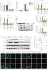
Nestin knockdown inhibits transforming growth factor (TGF)-β/Smad signalling. a) Nestin expression in primary mouse lung fibroblasts was analysed by quantitative (q)PCR (n=3 per group). b) Nestin-knockdown primary mouse lung fibroblasts were transfected with pGL3-SBE9-luciferase constructs and treated with or without TGF-β (5 ng·mL−1) for 24 h, and luciferase activity was measured (n=3 per group). c) qPCR analysis of α-smooth muscle actin (SMA) mRNA expression in primary mouse lung fibroblasts with nestin knockdown (n=3 per group). d) qPCR analysis of collagen I mRNA expression in primary mouse lung fibroblasts with nestin knockdown (n=3 per group). e) Western blot analysis and f) quantification of α-SMA and collagen I expression in nestin-knockdown primary mouse lung fibroblasts treated for 72 h with or without TGF-β (5 ng·mL−1) (n=3 per group). g) Western blot analysis and h) quantification of p-Smad2 and Smad2 expression in nestin-knockdown primary mouse lung fibroblasts treated with or without TGF-β (5 ng·mL−1) (n=3 per group). i) Immunofluorescence staining of nestin-knockdown primary mouse lung fibroblasts treated with or without TGF-β (5 ng·mL−1) and visualised using anti-nestin (green) and anti-p-Smad2 (red). Scale bars=10 µm. NC: negative control. Data are presented as the mean±sd of three independent experiments; **: p<0.01, ***: p<0.001; one-way ANOVA and Tukey's multiple comparisons test.
FIGURE 4
FIGURE 4
Nestin knockdown inhibits transforming growth factor (TGF)-β/Smad signalling via regulating the stability of TGF-β receptor I (TβRI) in mice. a) Quantitative (q)PCR analysis of TβRI and TβRII mRNA expression levels in nestin-knockdown primary mouse lung fibroblasts (n=3 per group). b) Western blot analysis and c) quantification of TβRI and TβRII expression in nestin-knockdown primary mouse lung fibroblasts (n=3 per group). d) Western blot analysis and e) quantification of TβRI expression in nestin-knockdown primary mouse lung fibroblasts at different time points after TGF-β (5 ng·mL−1) stimulation (n=3 per group). f) Half-life analysis and g) quantification of TβRI in nestin-knockdown primary mouse lung fibroblasts. All cell groups were treated with cycloheximide (CHX, 50 μg·mL−1) harvested at the indicated times (0, 4, 8, 12 h after CHX treatment) and subjected to immunoblotting (n=3 per group). h) Western blot analysis and i) quantification of Smad2 phosphorylation levels with nestin knockdown and TβRI overexpression treated with or without TGF-β (5 ng·mL−1) (n=3 per group). j) Immunofluorescence staining of p-Smad2 in primary mouse lung fibroblasts by nestin knockdown and overexpression of TβRI. Scale bars=20 µm. NC: negative control. k) Western blot analysis and l) quantification of α-SMA and collagen I expression levels in primary mouse lung fibroblasts by nestin knockdown and overexpression of TβRI (n=3 per group). m) Immunofluorescence staining of α-SMA in primary mouse lung fibroblasts by nestin knockdown and overexpression of TβRI. Scale bars=20 µm. ns: nonsignificant. Data are presented as the mean±sd of three independent experiments; ***: p<0.001; one-way ANOVA and Tukey's multiple comparisons test.
FIGURE 5
FIGURE 5
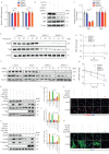
Nestin knockdown inhibits the recycling of transforming growth factor (TGF)-β receptor (TβR)I to the cell surface. a) Schematic overview of the biotinylation assay for quantifying plasma membrane TβRI level, TβRI endocytosis and recycling. b) Detection of the protein levels of TβRI on the plasma membrane in biotinylated serum-starved nestin-knockdown primary mouse lung fibroblasts after treatment with chloroquine (Chlq, 100 μM) for 4 h. c) Quantification of the surface TβRI protein levels in b) (n=3 per group). d) Live-cell fluorescence-activated cell sorting analysis of surface TβRI levels in nestin-knockdown primary mouse lung fibroblasts. e) Quantification of TβRI levels shown in panel d) (n=3 per group). f) Biotinylated serum-starved nestin-knockdown primary mouse lung fibroblasts were placed at 37°C for 30 min and then treated with glutathione after treated with Chlq (100 μM) for 4 h, and subjected to streptavidin agarose pulldown and Western blot analysis of TβRI. g) Quantification of the percentage of internalised TβRI in f) (n=3 per group); TβRI internalisation rate=total internalised TβRI at 30 min/plasma membrane TβRI×100%. h) Western blot analysis of recycled TβRI at 60 min. i) Quantification of the percentage of recycled TβRI in h) (n=3 per group); TβRI recycling rate=(internalised and recycled TβRI at 60 min−internalised TβRI at 60 min)/total internalised TβRI at 30 min×100%. j) Immunofluorescence staining of lysosome-associated membrane protein (LAMP)1 or LAMP2 and TβRI in nestin-knockdown primary mouse lung fibroblasts treated with TGF-β (5 ng·mL−1). Scale bars=5 µm. NC: negative control. k) Quantification of the percentage of TβRI colocalised with LAMP1 or LAMP2 (n=3; five fields assessed per sample). l) Western blot analysis and m) quantification of TβRI expression levels in nestin-knockdown primary mouse lung fibroblasts treated with or without Chlq (100 μM) (n=3 per group). n) Immunofluorescence staining of TβRI in nestin-knockdown primary mouse lung fibroblasts treated with or without Chlq (100 μM) for 4 h. Scale bars=20 µm. PM: plasma membrane; ns: nonsignificant. Data are presented as mean±sd of three independent experiments; ***: p<0.001; unpaired t-test and one-way ANOVA and Tukey's multiple comparisons test.
FIGURE 6
FIGURE 6
Rab11 is required for the ability of nestin to promote the recycling of transforming growth factor (TGF)-β receptor (TβR)I to the cell surface. a) Immunoprecipitation (IP) was performed using an anti-flag-nestin antibody, and immunoblotting of the protein levels of Rab11 and Rab4 in nestin-knockdown primary mouse lung fibroblasts. b) Immunofluorescence staining showed the colocalisation between nestin and Rab11, Rab4 in primary mouse lung fibroblasts. Scale bars=10 µm. NC: negative control. c) Quantification of the percentage of nestin co-localised with Rab11 and Rab4 (n=3; five fields assessed per sample). d) Rab11 activity assays were performed in nestin-knockdown primary mouse lung fibroblasts. e) Quantification of Rab11GTPase activity (n=3 per group). f) Immunofluorescence staining of Rab11 and TβRI in nestin-knockdown primary mouse lung fibroblasts. Scale bars=5 µm. g) Quantification of the percentage of TβRI colocalised with Rab11 (n=3; five fields assessed per sample). h) Immunoprecipitation was performed using an anti-TβRI antibody, and immunoblotting of the protein levels of Rab11 in nestin-knockdown primary mouse lung fibroblasts treated with chloroquine (Chlq) (100 μM) for 4 h. i) Immunoprecipitation was performed using an anti-Rab11 antibody, and immunoblotting of the protein levels of TβRI in nestin-knockdown primary mouse lung fibroblasts treated with Chlq (100 μM) for 4 h. j) Immunofluorescence staining of TβRI and Rab11 in nestin-knockdown primary mouse lung fibroblasts with overexpression of Rab11. Scale bars=5 µm. k) Quantification of the percentage of TβRI colocalised with Rab11 (n=3; five fields assessed per sample). Scale bars=5 µm. Data are presented as mean±sd of three independent experiments; ***: p<0.001; unpaired t-test and one-way ANOVA and Tukey's multiple comparisons test.
FIGURE 7
FIGURE 7
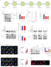
Downregulation of nestin attenuates bleomycin-induced pulmonary fibrosis and transforming growth factor (TGF)-β overexpression-induced pulmonary fibrosis in mice. a) Experimental design. 8-week-old C57BL/6 mice were injected intratracheally with bleomycin (3 U·kg−1) or PBS. 10 days later, the mice were injected intratracheally with Adeno-associated virus serotype (AAV)6-ShNES or AAV6-Scramble. Samples were collected for analysis 21 days after bleomycin administration. b) Quantitative (q)PCR analysis of nestin mRNA expression in the lungs of C57/BL6 mice from the different groups (n=6 mice per group). c) Western blot analysis and d) quantification of nestin expression levels in lungs from C57/BL6 mice of the different groups (n=3 per group). e) qPCR analysis of α-smooth muscle actin (SMA) and collagen I mRNA expression levels in lungs from C57/BL6 mice of the different groups (n=6 mice per group). f) Western blot analysis and g) quantification of α-SMA, collagen I, p-Smad2 and Smad2 expression levels in lungs from C57/BL6 mice of the different groups (n=3 per group). h) Hydroxyproline levels in lungs of C57/BL6 mice from the different groups (n=6 mice per group). i) Experimental design. 8-week-old C57BL/6 mice were injected intratracheally with AdTGF-β1 or Advector. 10 days later, the mice were injected intratracheally with AAV6-ShNES or AAV6-Scramble. Samples were collected for analysis 21 days after bleomycin administration. j) TGF-β1 levels in the lungs of C57/BL6 mice from the different groups (n=6 mice per group). k) Immunofluorescence staining with nestin, α-SMA, TβRI and p-Smad2 in lung slices from the different groups. Scale bars=70 µm. NC: negative control. Data are presented as the mean±sd of three independent experiments; *: p<0.05, **: p<0.01, ***: p<0.001; one-way ANOVA and Tukey's multiple comparisons test.
FIGURE 8
FIGURE 8
Nestin knockdown inhibits transforming growth factor (TGF)-β/Smad signalling in human fibroblasts and pulmospheres. a) Nestin expression in primary human lung fibroblasts was analysed by quantitative (q)PCR (n=3 per group). b) qPCR analysis of α-smooth muscle actin (SMA) mRNA expression in human lung fibroblasts with nestin knockdown (n=3 per group). c) qPCR analysis of collagen I mRNA expression in human lung fibroblasts with nestin knockdown (n=3 per group). d) Western blot analysis and quantification of e) α-SMA, f) collagen I, g) p-smad2, smad2 and h) TGF-β receptor (TβR)I expression in nestin-knockdown human lung fibroblasts treated for 72 h with or without TGF-β (5 ng·mL−1) (n=3 per group). i) Immunofluorescence staining of nestin-knockdown human lung fibroblasts treated with or without TGF-β (5 ng·mL−1) and visualised using anti-nestin (green) and anti-α-SMA (red). Scale bars=10 µm. NC: negative control. j) Immunofluorescence staining of nestin-knockdown human lung fibroblasts treated with or without TGF-β (5 ng·mL−1) and visualised using anti-nestin (green) and anti-p-Smad2 (red). Scale bars=10 µm. k) Overview of human pulmospheres preparation from normal and idiopathic pulmonary fibrosis (IPF) lung tissues and immunostaining. l) Immunofluorescence staining of nestin-knockdown human pulmospheres and visualised using anti-nestin (green) and anti-α-SMA (red). Scale bars=50 µm. m) Quantification of nestin-positive cells per pulmosphere in l). n) Quantification of α-SMA positive cells per pulmosphere in l). Data are presented as mean±sd of three independent experiments; *: p<0.05, **: p<0.01, ***: p<0.001; one-way ANOVA and Tukey's multiple comparisons test.

Comment in

Similar articles

Cited by

References

    1. Sheppard D. ROCKing pulmonary fibrosis. J Clin Invest 2013; 123: 1005–1006. doi:10.1172/JCI68417 - DOI - PMC - PubMed
    1. Travis WD, Costabel U, Hansell DM, et al. . An official American Thoracic Society/European Respiratory Society statement: update of the international multidisciplinary classification of the idiopathic interstitial pneumonias. Am J Respir Crit Care Med 2013; 188: 733–748. doi:10.1164/rccm.201308-1483ST - DOI - PMC - PubMed
    1. Raghu G, Weycker D, Edelsberg J, et al. . Incidence and prevalence of idiopathic pulmonary fibrosis. Am J Respir Crit Care Med 2006; 174: 810–816. doi:10.1164/rccm.200602-163OC - DOI - PubMed
    1. Ahluwalia N, Shea BS, Tager AM. New therapeutic targets in idiopathic pulmonary fibrosis. Aiming to rein in runaway wound-healing responses. Am J Respir Crit Care Med 2014; 190: 867–878. doi:10.1164/rccm.201403-0509PP - DOI - PMC - PubMed
    1. Selman M, King TE, Pardo A, et al. . Idiopathic pulmonary fibrosis: prevailing and evolving hypotheses about its pathogenesis and implications for therapy. Ann Intern Med 2001; 134: 136–151. doi:10.7326/0003-4819-134-2-200101160-00015 - DOI - PubMed

Publication types

LinkOut - more resources