Review
. 2021 Dec;17(12):726-744.
doi: 10.1038/s41574-021-00562-6. Epub 2021 Oct 8.

The evolving view of thermogenic adipocytes - ontogeny, niche and function

Affiliations
Review

The evolving view of thermogenic adipocytes - ontogeny, niche and function

Farnaz Shamsi et al. Nat Rev Endocrinol. 2021 Dec.

Abstract

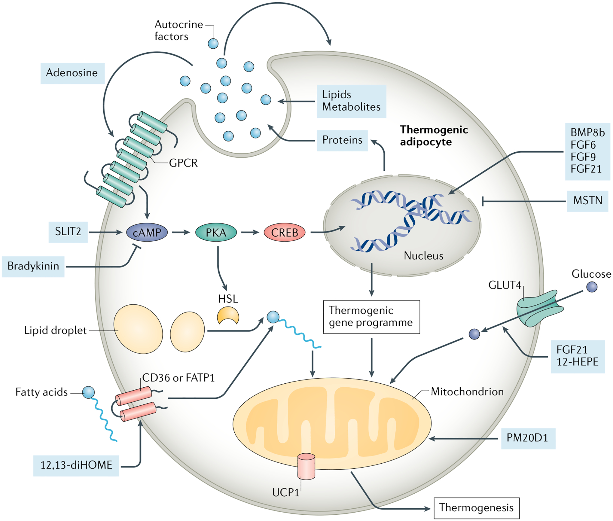
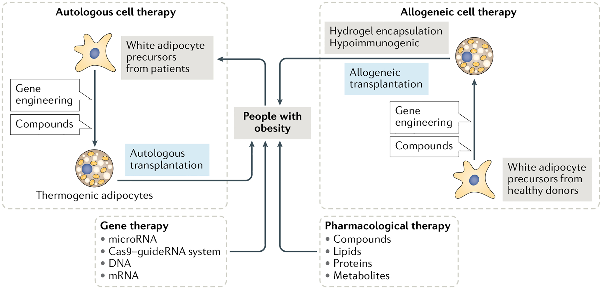
The worldwide incidence of obesity and its sequelae, such as type 2 diabetes mellitus, have reached pandemic levels. Central to the development of these metabolic disorders is adipose tissue. White adipose tissue stores excess energy, whereas brown adipose tissue (BAT) and beige (also known as brite) adipose tissue dissipate energy to generate heat in a process known as thermogenesis. Strategies that activate and expand BAT and beige adipose tissue increase energy expenditure in animal models and offer therapeutic promise to treat obesity. A better understanding of the molecular mechanisms underlying the development of BAT and beige adipose tissue and the activation of thermogenic function is the key to creating practical therapeutic interventions for obesity and metabolic disorders. In this Review, we discuss the regulation of the tissue microenvironment (the adipose niche) and inter-organ communication between BAT and other tissues. We also cover the activation of BAT and beige adipose tissue in response to physiological cues (such as cold exposure, exercise and diet). We highlight advances in harnessing the therapeutic potential of BAT and beige adipose tissue by genetic, pharmacological and cell-based approaches in obesity and metabolic disorders.


Conflict of interest statement

Competing interests

Y.-H.T. is an inventor on US Patent 7,576,052 related to BMP7 and US patent applications related to 12,13-diHOME and FGF6/9. The other authors declare no competing interests.

Figures

Fig. 1 |
Fig. 1 |. Cellular crosstalk between neurons and other adipose-resident cells.
Extensive communication between the neurons, thermogenic adipocytes and other cell types in adipose tissue is essential for brown adipose tissue (BAT) thermogenesis and cold-induced browning of white adipose tissue (WAT). The central nervous system (CNS) stimulates the release of neurotransmitters (pale blue), including noradrenaline (NE), neuropeptide Y (NPY) and ATP, from the sympathetic nerve terminals in BAT and WAT upon exposure to cold temperatures. Thermogenic adipocytes (here shown in beige and brown) release lipids and neurotrophic factors (pale yellow) that affect the growth, survival and function of sympathetic nerves in the adipose niche. These factors, whose production and release are stimulated by sympathetic nerve activity, promote the coordinated expansion of sympathetic innervation that is essential for maintaining tissue responsiveness to CNS signals. Both axon growth and angiogenic sprouting are regulated through a common array of attractive and repulsive cues. Nerve terminals release signalling molecules to guide and promote angiogenesis. Blood vessels also secrete factors (red) that attract and direct axons to innervate the vasculature. Sympathetic nerves regulate the development and function of immune cells by releasing neurotransmitters, such as NE and NPY. Several cytokines (grey) produced by adipose-resident immune cells also positively or negatively impact the survival and growth of sensory and sympathetic nerves. BMP, bone morphogenetic protein; NGF, nerve growth factor; NRG4, neuregulin 4; S100b, S100 calcium binding protein B; TGFβ1, transforming growth factor-β 1; VEGFA, vascular endothelial growth A factor.
Fig. 2 |
Fig. 2 |. Cellular crosstalk between vasculature and other adipose-resident cells.
Accumulation of pro-angiogenic factors combined with reduced levels of angiogenic inhibitors in the adipose niche promotes the expansion and remodelling of the vascular network in response to thermogenic stimuli. Thermogenic adipocytes (shown in beige and brown) and their progenitors secrete several angiogenic factors (pale yellow) to guide vascular cells to expand, regress or remodel depending on the requirements of the adipose tissue microenvironment. Reciprocally, endothelial cells release factors (red), such as endothelin1 (EDN1) and nitric oxide (NO), that promote the thermogenic function of brown and beige adipocytes. EDN1 and platelet-derived growth factor C (PDGF-C) also regulate adipogenesis in progenitors. Pro-angiogenic and anti-angiogenic cytokines (grey) and growth factors secreted by immune cells regulate vasculature function and remodelling. ANGPT1, angiopoietin 1; FGF, fibroblast growth factor; HGF, hepatocyte growth factor; NPY, neuropeptide Y; TGFβ1, transforming growth factor-β 1; TNF, tumour necrosis factor; VEGF, vascular endothelial growth factor.
Fig. 3 |
Fig. 3 |. Cellular crosstalk between immune cells and other adipose-resident cells.
Adipose tissue-resident immune cells are major contributors to the adipose niche by playing key roles in both the function of brown adipose tissue (BAT) and white adipose tissue (WAT) and homeostasis through extensive cellular crosstalk with other cell types. M1 macrophages (dark blue) secrete pro-inflammatory cytokines (pale red) that impair insulin signalling and suppress thermogenesis in adipocytes. Conversely, a type 2 immune response, which is mediated by M2 macrophages (yellow), eosinophils (red), innate lymphoid type 2 cells (ILC2, grey) and invariant natural killer T-cells (iNKTs, green), enhances BAT activation and browning of WAT (factors involved in the type 2 responses are shown in pale blue). Thermogenic adipocytes (shown in beige and brown) regulate the type 2 immune response by releasing ADIPOQ, CXCL14, GDF15, METRNL and CCL11 (adipocyte-released factors are shown in pale yellow). A multicellular communication axis between sympathetic nerves, adipocytes, eosinophils, adipocyte progenitors and macrophages also orchestrates the adaptive response of WAT to cold. Methionine-enkephalin (MET-ENK) are IL2C-derived secreted peptides that induce uncoupling protein 1 expression in white adipocytes. Sympathetic neuron-associated macrophages (SAMs) mediate the clearance of extracellular noradrenaline (NE, grey) and thereby negatively regulate NE availability and the thermogenic activity of BAT and beige adipose tissue. ADIPOQ, adiponectin; CCL11, chemokine (C-C motif) ligand 11; CXCL14, C-X-C motif chemokine ligand 14; FGF, fibroblast growth factor; GDF15, growth differentiation factor 15; METRNL, meteorin-like glial cell differentiation regulator; PDGF, platelet-derived growth factor; TNF, tumour necrosis factor.
Fig. 4 |
Fig. 4 |. Autocrine and paracrine factors regulating brown and beige adipocytes.
Several factors released by brown and beige adipocytes function in an autocrine and/or paracrine manner to regulate nutrient utilization and thermogenic gene expression in adipocytes. The secreted factors (pale blue circles and boxes) include members of bone morphogenetic protein (BMP) and transforming growth factor-β (TGFβ), such as BMP8b, myostatin (MSTN), fibroblast growth factor (FGF) families of growth factors (for example, FGF6, FGF9 and FGF21), bradykinin, SLIT2, PM20D1, adenosine and others. Additionally, thermogenic adipocyte-derived lipokines, such as 12,13-diHOME and 12-HEPE, promote fatty acid and glucose uptake to adipocytes, thereby controlling key aspects of fuel utilization and energy metabolism in brown adipose tissue and white adipose tissue. CREB, cAMP responsive element binding protein; FATP1, fatty acid transport protein 1; GPCR, G protein-coupled receptor; HSL, hormone-sensitive lipase; PKA, protein kinase A; PM20D1, the secreted enzyme peptidase M20 domain containing 1. UCP1, uncoupling protein 1.
Fig. 5 |
Fig. 5 |. Inter-organ communication of thermogenic adipose tissue and other tissues.
Thermogenic adipose tissue (shown here as beige and brown adipocytes) is recognized as an endocrine organ, which can secrete several factors (pale yellow) to regulate the gene expression or functions in distant organs, such as the heart, liver, muscle and white adipose tissue. These organs, as well as the brain and gut, can also communicate to thermogenic adipose tissue through the secretion of endocrine factors (pale blue) in response to different stimulations. This inter-organ communication highlights the importance of thermogenic adipose tissue in whole-body metabolism. ACTH, adrenocorticotropic hormone; BAIBA, β-aminoisobutyric acid; EPDR1, ependymin-related protein 1; EVs, extracellular vesicles; FGF, fibroblast growth factor; GCs, glucocorticoids; GLP1, glucagon-like peptide 1; METRNL, meteorin-like glial cell differentiation regulator; NE, noradrenaline; NPs, natriuretic peptides; NRG4, neuregulin 4; PM20D1, peptidase M20 domain containing 1; SLIT2, slit guidance ligand 2; TGFβ2, transforming growth factor-β2; TSH, thyroid-stimulating hormone.
Fig. 6 |
Fig. 6 |. Cell-based, gene-based and pharmacological therapies for activation and conversion of thermogenic adipose tissue.
In pharmacological therapies, several compounds, proteins, lipids or metabolites have been applied for treating obesity in human clinical trials through the regulation of thermogenic adipose tissue activity and energy expenditure. In gene therapy, the delivery of DNA (transgene), mRNA, microRNA or Cas9–guideRNA systems is used for increasing the expression of genes involved in the thermogenic pathway. Targeted delivery into thermogenic adipose tissue or white adipose tissue increases efficiency and specificity and minimizes the adverse effects on other cell types. Cell-based therapies include autologous and allogeneic cell therapy according to the source of the transplanted cells. In autologous cell therapy, precursor cells isolated from an individual with obesity can be engineered and differentiated to thermogenic adipose tissue, followed by transplanting back to the same individual. Allogeneic cell therapy uses precursor cells from healthy donors. Although this strategy overcomes the burden of cell source, preventing transplanted cells from immune rejection needs to be considered using hydrogel encapsulation or reducing immunogenicity before allogeneic cell transplantation into the recipients. Although gene-based and cell-based therapy have not yet been applied for treating obesity in humans, these might be potential strategies.

Comment in

References

    1. Peres Valgas da Silva C, Hernandez-Saavedra D, White JD & Stanford KI Cold and exercise: therapeutic tools to activate brown adipose tissue and combat obesity. Biology 8, 9 (2019). - PMC - PubMed
    1. Cannon B & Nedergaard J Brown adipose tissue: function and physiological significance. Physiol. Rev 84, 277–359 (2004). - PubMed
    1. Bartelt A et al. Brown adipose tissue activity controls triglyceride clearance. Nat. Med 17, 200–205 (2011). - PubMed
    1. Scheja L & Heeren J The endocrine function of adipose tissues in health and cardiometabolic disease. Nat. Rev. Endocrinol 15, 507–524 (2019). - PubMed
    1. Villarroya F, Cereijo R, Villarroya J & Giralt M Brown adipose tissue as a secretory organ. Nat. Rev. Endocrinol 13, 26–35 (2017). - PubMed

Publication types

MeSH terms