

Restoration of Immune Privilege in Human Dermal Papillae Controlling Epithelial-Mesenchymal Interactions in Hair Formation
- PMID: 34626334
- PMCID: PMC8782965
- DOI: 10.1007/s13770-021-00392-7
Restoration of Immune Privilege in Human Dermal Papillae Controlling Epithelial-Mesenchymal Interactions in Hair Formation
Abstract
Background: Hair follicles are among a handful of organs that exhibit immune privilege. Dysfunction of the hair follicle immune system underlies the development of inflammatory diseases, such as alopecia areata.
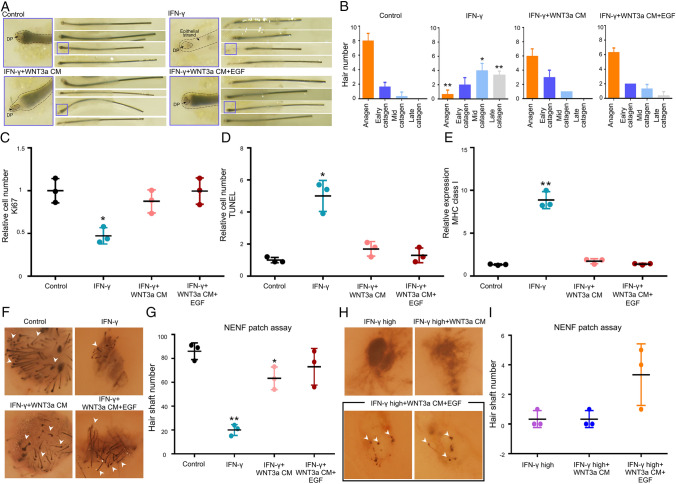
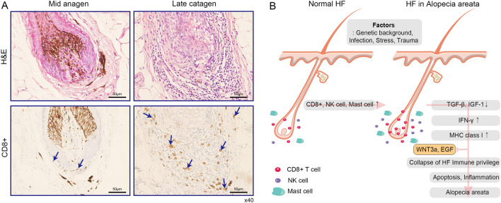
Methods: Quantitative reverse transcription PCR and immunostaining was used to confirm the expression of major histocompatibility complex class I in human dermal papilla cells. Through transcriptomic analyses of human keratinocyte stem cells, major histocompatibility complex class I was identified as differentially expressed genes. Organ culture and patch assay were performed to assess the ability of WNT3a conditioned media to rescue immune privilege. Lastly, CD8+ T cells were detected near the hair bulb in alopecia areata patients through immunohistochemistry.
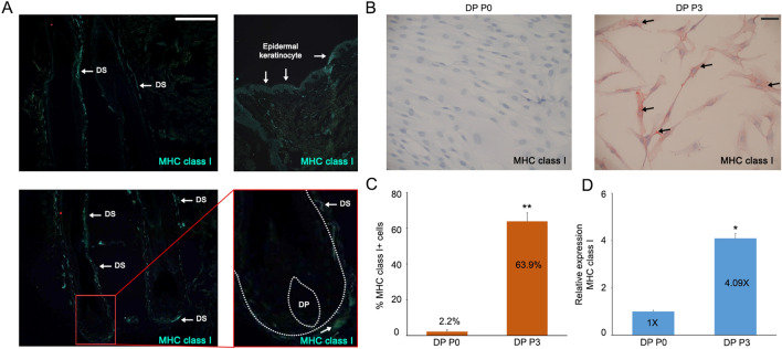
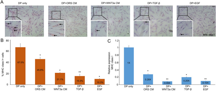
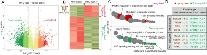
Results: Inflammatory factors such as tumor necrosis factor alpha and interferon gamma were verified to induce the expression of major histocompatibility complex class I proteins in dermal papilla cells. Additionally, loss of immune privilege of hair follicles was rescued following treatment with conditioned media from outer root sheath cells. Transcriptomic analyses found 58 up-regulated genes and 183 down-regulated genes related in MHC class I+ cells. Using newborn hair patch assay, we demonstrated that WNT3a conditioned media with epidermal growth factor can restore hair growth. In alopecia areata patients, CD8+ T cells were increased during the transition from mid-anagen to late catagen.
Conclusion: Identification of mechanisms governing epithelial and mesenchymal interactions of the hair follicle facilitates an improved understanding of the regulation of hair follicle immune privilege.
Keywords: Epithelial-mesenchymal interaction; Hair follicle; Immune privilege; MHC molecule.
© 2021. The Korean Tissue Engineering and Regenerative Medicine Society.
Conflict of interest statement
The authors declare that they have no conflict of interest.
Figures

Similar articles
-
Hair follicle immune privilege and its collapse in alopecia areata.Exp Dermatol. 2020 Aug;29(8):703-725. doi: 10.1111/exd.14155. Exp Dermatol. 2020. PMID: 32682334 Review.
-
Collapse and Restoration of Hair Follicle Immune Privilege Ex Vivo: A Model for Alopecia Areata.Methods Mol Biol. 2020;2154:133-141. doi: 10.1007/978-1-0716-0648-3_11. Methods Mol Biol. 2020. PMID: 32314213
-
The Effect of JAK Inhibitor on the Survival, Anagen Re-Entry, and Hair Follicle Immune Privilege Restoration in Human Dermal Papilla Cells.Int J Mol Sci. 2020 Jul 20;21(14):5137. doi: 10.3390/ijms21145137. Int J Mol Sci. 2020. PMID: 32698510 Free PMC article.
-
The hair follicle and immune privilege.J Investig Dermatol Symp Proc. 2003 Oct;8(2):188-94. doi: 10.1046/j.1087-0024.2003.00807.x. J Investig Dermatol Symp Proc. 2003. PMID: 14582671 Review.
-
The human hair follicle immune system: cellular composition and immune privilege.Br J Dermatol. 2000 May;142(5):862-73. doi: 10.1046/j.1365-2133.2000.03464.x. Br J Dermatol. 2000. PMID: 10809841
Cited by
-
Immune niches for hair follicle development and homeostasis.Front Physiol. 2024 Apr 22;15:1397067. doi: 10.3389/fphys.2024.1397067. eCollection 2024. Front Physiol. 2024. PMID: 38711955 Free PMC article. Review.
-
The Molecular Mechanism of Natural Products Activating Wnt/β-Catenin Signaling Pathway for Improving Hair Loss.Life (Basel). 2022 Nov 11;12(11):1856. doi: 10.3390/life12111856. Life (Basel). 2022. PMID: 36430990 Free PMC article. Review.
-
Proteins from Stressed Mesenchymal Stem Cells Can Repair Hair Follicles and Promote Hair Regeneration.ACS Pharmacol Transl Sci. 2025 May 24;8(6):1768-1777. doi: 10.1021/acsptsci.5c00184. eCollection 2025 Jun 13. ACS Pharmacol Transl Sci. 2025. PMID: 40534677
-
Targeted immunotherapy for hair regrowth and regeneration.Front Med (Lausanne). 2023 Oct 10;10:1285452. doi: 10.3389/fmed.2023.1285452. eCollection 2023. Front Med (Lausanne). 2023. PMID: 37881630 Free PMC article. No abstract available.
References
-
- Reynolds AJ, Jahoda CA. Cultured dermal papilla cells induce follicle formation and hair growth by transdifferentiation of an adult epidermis. Development. 1992;115:587–593. - PubMed
-
- Jahoda CA, Reynolds AJ, Oliver RF. Induction of hair growth in ear wounds by cultured dermal papilla cells. J Invest Dermatol. 1993;101:584–590. - PubMed
-
- Millar SE. Molecular mechanisms regulating hair follicle development. J Invest Dermatol. 2002;118:216–225. - PubMed
Publication types
MeSH terms
Substances
LinkOut - more resources
Full Text Sources
Research Materials