

Regional brain tissue changes in patients with cystic fibrosis
- PMID: 34627274
- PMCID: PMC8502335
- DOI: 10.1186/s12967-021-03092-x
Regional brain tissue changes in patients with cystic fibrosis
Abstract
Background: Cystic fibrosis (CF) patients present with a variety of symptoms, including mood and cognition deficits, in addition to classical respiratory, and autonomic issues. This suggests that brain injury, which can be examined with non-invasive magnetic resonance imaging (MRI), is a manifestation of this condition. However, brain tissue integrity in sites that regulate cognitive, autonomic, respiratory, and mood functions in CF patients is unclear. Our aim was to assess regional brain changes using high-resolution T1-weighted images based gray matter (GM) density and T2-relaxometry procedures in CF over control subjects.
Methods: We acquired high-resolution T1-weighted images and proton-density (PD) and T2-weighted images from 5 CF and 15 control subjects using a 3.0-Tesla MRI. High-resolution T1-weighted images were partitioned to GM-tissue type, normalized to a common space, and smoothed. Using PD- and T2-weighted images, whole-brain T2-relaxation maps were calculated, normalized, and smoothed. The smoothed GM-density and T2-relaxation maps were compared voxel-by-voxel between groups using analysis of covariance (covariates, age and sex; SPM12, p < 0.001).
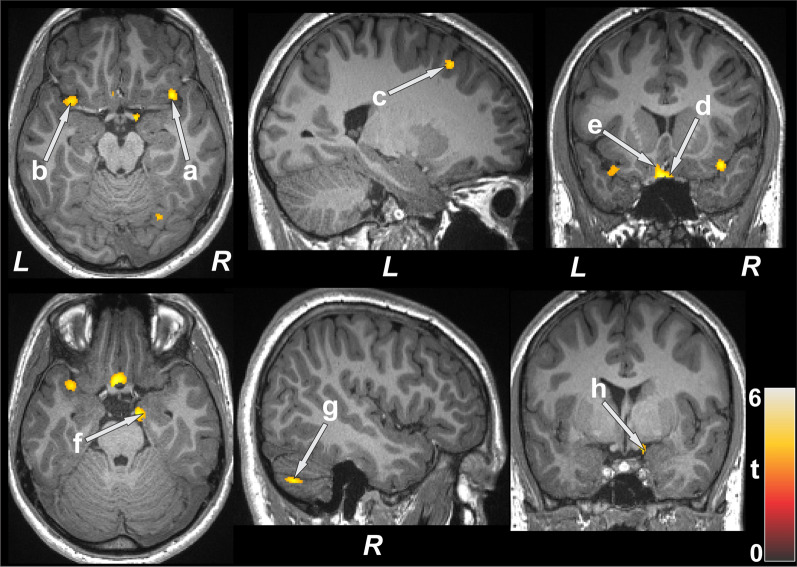
Results: Significantly increased GM-density, indicating tissues injury, emerged in multiple brain regions, including the cerebellum, hippocampus, amygdala, basal forebrain, insula, and frontal and prefrontal cortices. Various brain areas showed significantly reduced T2-relaxation values in CF subjects, indicating predominant acute tissue changes, in the cerebellum, cerebellar tonsil, prefrontal and frontal cortices, insula, and corpus callosum.
Conclusions: Cystic fibrosis subjects show predominant acute tissue changes in areas that control mood, cognition, respiratory, and autonomic functions and suggests that tissue changes may contribute to symptoms resulting from ongoing hypoxia accompanying the condition.
Keywords: Cognition; Gray matter density; Magnetic resonance imaging; Mood; T2-relaxometery.
© 2021. The Author(s).
Conflict of interest statement
The authors declare that they have no competing interests
Figures

References
-
- About cystic fibrosis. Bethesda: Cystic Fibrosis Foundation.
-
- Cystic Fibrosis Foundation patient registry 2019 annual data report. Bethesda: Cystic Fibrosis Foundation; 2019.
-
- Welsh MJ, Ramsey BW, Accurso F, Cutting GR, et al. Cystic fibrosis. In: Scriver C, Beaudet AL, Sly W, Valle DL, Childs B, Kinzler K, et al., editors. The metabolic and molecular basis of inherited disease. 8. New York: McGraw-Hill Education; 2001. pp. 5121–5188.
