Review
. 2021 Dec:125:154910.
doi: 10.1016/j.metabol.2021.154910. Epub 2021 Oct 8.

Heart failure in diabetes

Affiliations
Review

Heart failure in diabetes

Stanislovas S Jankauskas et al. Metabolism. 2021 Dec.

Abstract

Heart failure and cardiovascular disorders represent the leading cause of death in diabetic patients. Here we present a systematic review of the main mechanisms underlying the development of diabetic cardiomyopathy. We also provide an excursus on the relative contribution of cardiomyocytes, fibroblasts, endothelial and smooth muscle cells to the pathophysiology of heart failure in diabetes. After having described the preclinical tools currently available to dissect the mechanisms of this complex disease, we conclude with a section on the most recent updates of the literature on clinical management.

Keywords: Adrenergic receptors; Aging; BHB; Bioenergetics; Cardiomyocytes; Cardiovascular endocrinology; Diabetes mellitus; Diabetic cardiomyopathy; Diastolic dysfunction; Endothelium; FOXO1; Fibroblasts; Fibrosis; HFpEF; Heart failure; Mitochondria; NADH; Oxidative stress; ROS; Senescence; T1DM; T2DM; VSMC.


Conflict of interest statement

Declaration of competing interest Nothing to declare.

Figures
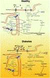
Fig. 1.
Fig. 1.. Myocardial bioenergetics in the healthy and in the diabetic heart.
6-P-gluconolactone: 6-phospho-gluconolactone; Acetyl-CoA: acetyl coenzyme A; ADP: Adenosine diphosphate; ATP: Adenosine triphosphate; CD36/FAT: Cluster of differentiation 36/fatty acid translocase; CPT1: Carnitine palmitoyl transferase I; CPT2: Carnitine palmitoyl transferase II; ETC: Electron transport chain; Fructose-6-P: fructose-6-phosphate; G6PDH: Glucose-6-phosphate dehydrogenase; GAPDH: Glyceraldehyde 3-phosphate dehydrogenase; GFAT: Glutamine fructose-6-phosphate aminotransferase; Glucosamine-6-P: glucosamine-6-phosphate; Glucose-6-P: glucose-6-phosphate; GLUT1: Glucose transporter type 1; GLUT4: Glucose transporter type 4; GPI: Glucose-6-phosphate isomerase; HBP: hexosamine biosynthetic pathway; HK2: hexokinase 2; IMM: inner mitochondrial membrane; NAD: Nicotinamide adenine dinucleotide; NOX: NADPH oxidase; O-GlcNAcylation: O-linked-N-acetylglucosaminylation; OGT: O-linked N-acetylglucosaminyltransferase; OMM: outer mitochondrial membrane; PDH: pyruvate dehydrogenase; RNS: reactive nitrogen species; ROS: reactive oxygen species; TCA: tricarboxylic acid cycle; UDP-GlcNAc: Uridine diphosphate N-acetylglucosamine.
Fig. 2.
Fig. 2.. Main molecular mechanisms underlying cardiac dysfunction in diabetes.
AGEs: Advanced glycation end products; Ang II: Angiotensin II; ASC: Apoptosis-associated speck-like protein containing a caspase-recruitment domain; AT1: Angiotensin II type 1 receptor; ATPase: Adenosine triphosphatase; [Ca2+]i: Intracellular calcium; Casp-1: Caspase-1; CD36/FAT: Cluster of differentiation 36/fatty acid translocase; cGMP: Cyclic guanosine monophosphate; FFA: Free fatty acid; GLUT4: Glucose transporter type 4; ICAM-1: Intercellular adhesion molecule; IL-18: Interleukin 18; IL-1β: Interleukin 1 beta; IL-6: Interleukin 6; IR: Insulin receptor; MMPs: Matrix metalloproteinases; mTOR/S6K: Mechanistic target of rapamycin (mTOR)-ribosomal S6 kinase (S6K) pathway; NF-kB: Nuclear factor kappa-light-chain-enhancer of activated B cells; NLRP3: Nucleotide-binding oligomerization domain like receptor (NLR) pyrin domain containing 3; NO: Nitric oxide; PI3K/Akt: Phosphoinositide-3-kinase (PI3K)-protein kinase B (Akt); PKC: Protein kinase C; PKG: Protein kinase G; Pro-casp1: Pro-caspase-1; PTP: Permeability transition pore; ROS: Reactive oxygen species; SERCA2a: Sarcoplasmic reticulum Ca2+-ATPase 2a; TGF-β: Transforming Growth Factor beta; TNF-α: Tumor Necrosis Factor alpha; VCAM-1: Vascular cell adhesion molecule 1.
Fig. 3.
Fig. 3.. Animal models of diabetic cardiomyopathy.

References

    1. Kansakar U, Varzideh F, Jankauskas SS, Gambardella J, Trimarco B, Santulli G. Advances in the understanding of excitation-contraction coupling: the pulsing quest for drugs against heart failure and arrhythmias. Eur Heart J Cardiovasc Pharmacother. 2021:pvab069 In press. - PMC - PubMed
    1. Balaban RS, Kantor HL, Katz LA, Briggs RW. Relation between work and phosphate metabolite in the in vivo paced mammalian heart. Science. 1986;232:1121–3. - PubMed
    1. Zhao Q, Sun Q, Zhou L, Liu K, Jiao K. Complex regulation of mitochondrial function during cardiac development. J Am Heart Assoc. 2019;8:e012731. - PMC - PubMed
    1. Wang Y, Christopher BA, Wilson KA, Muoio D, McGarrah RW, Brunengraber H, et al. Propionate-induced changes in cardiac metabolism, notably CoA trapping, are not altered by l-carnitine. Am J Physiol Endocrinol Metab. 2018;315 (E622–E33). - PMC - PubMed
    1. Zhang Y, Taufalele PV, Cochran JD, Robillard-Frayne I, Marx JM, Soto J, et al. Mitochondrial pyruvate carriers are required for myocardial stress adaptation. Nat Metab. 2020;2:1248–64. - PMC - PubMed

Publication types

MeSH terms