. 2022 Jan 1;1868(1):166288.
doi: 10.1016/j.bbadis.2021.166288. Epub 2021 Oct 8.

GPR65 (TDAG8) inhibits intestinal inflammation and colitis-associated colorectal cancer development in experimental mouse models

Affiliations

GPR65 (TDAG8) inhibits intestinal inflammation and colitis-associated colorectal cancer development in experimental mouse models

Mona A Marie et al. Biochim Biophys Acta Mol Basis Dis. .

Abstract

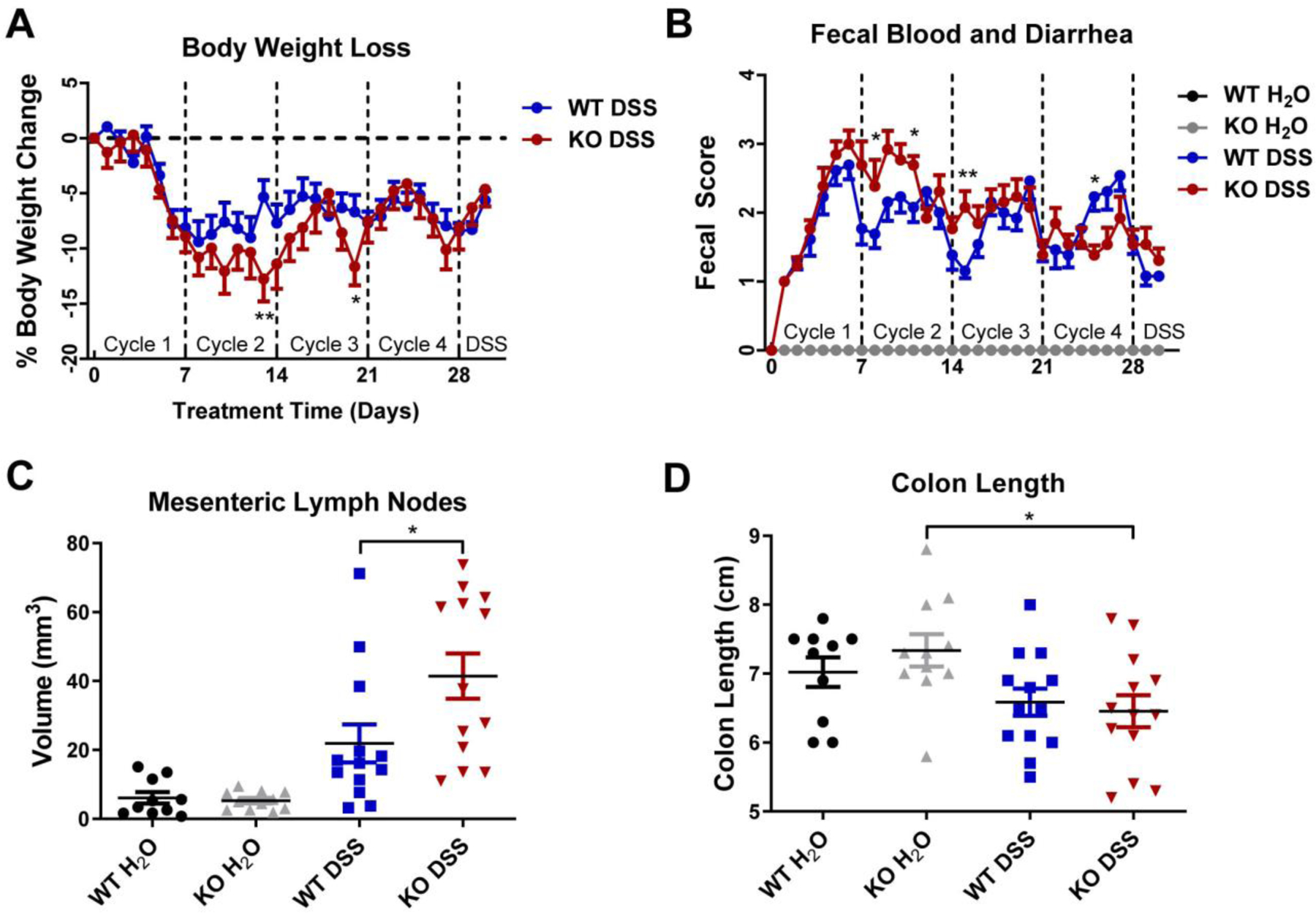
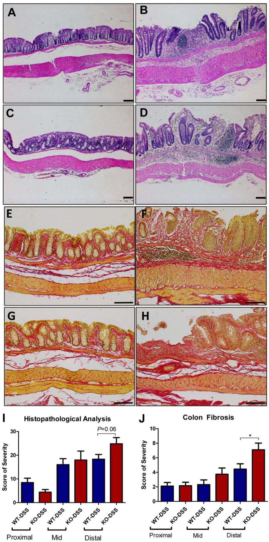
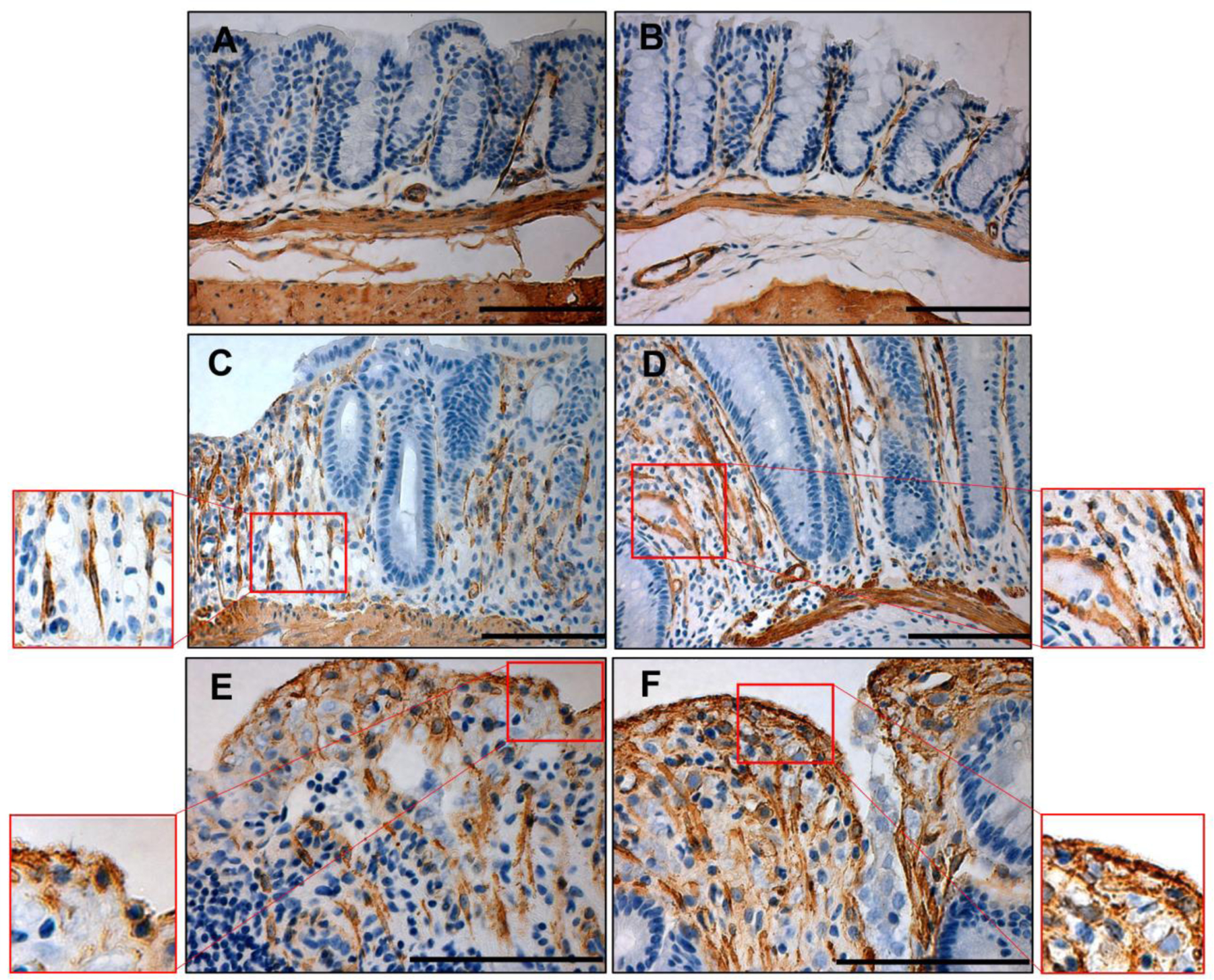
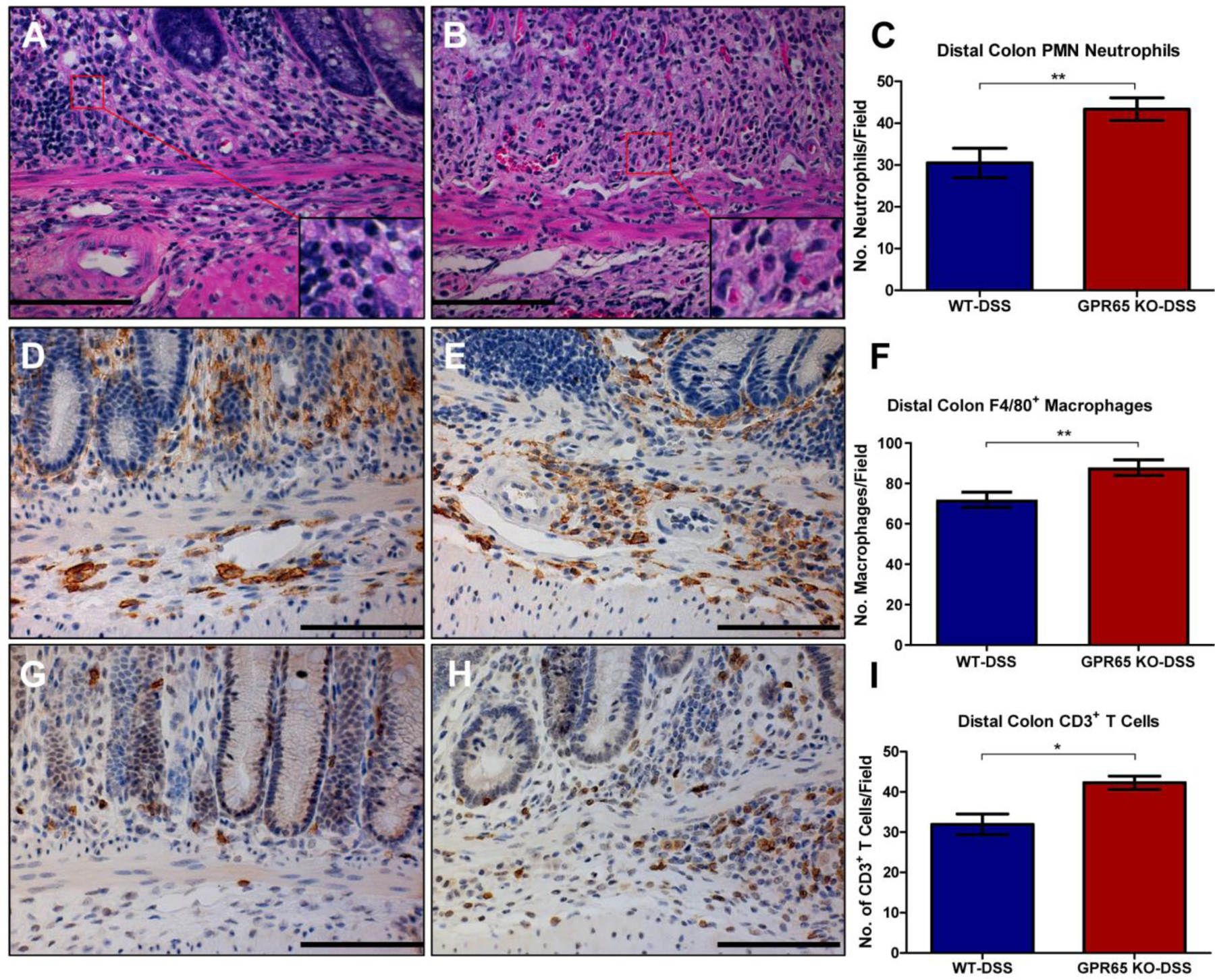
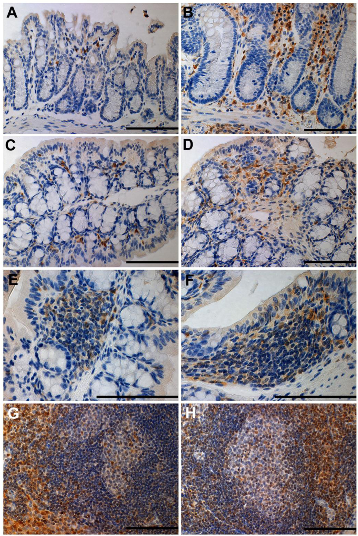
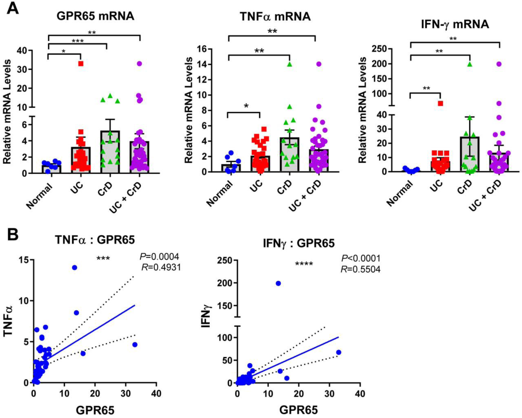
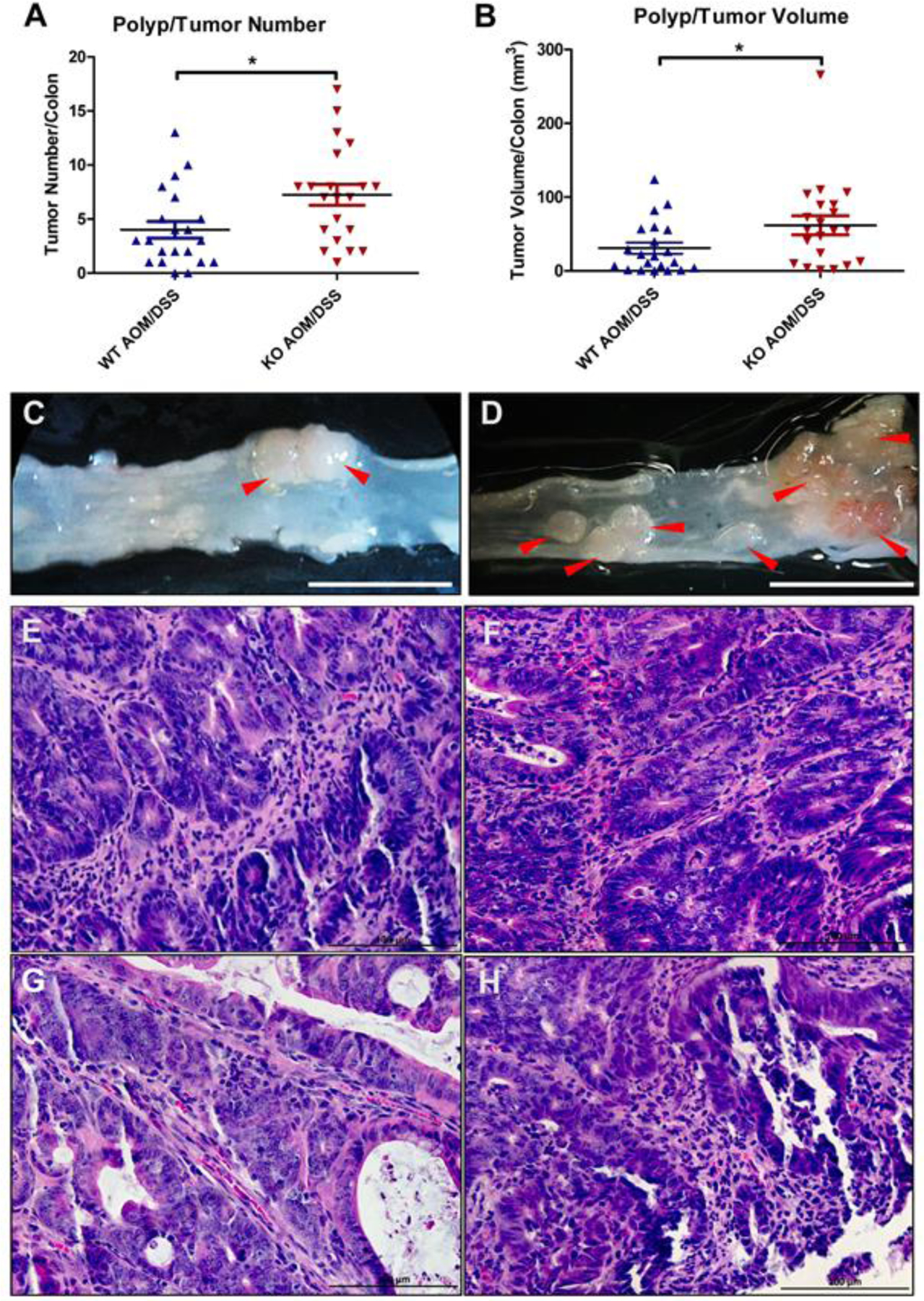
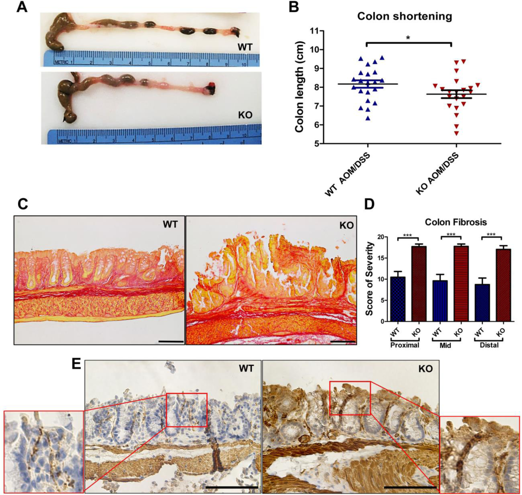
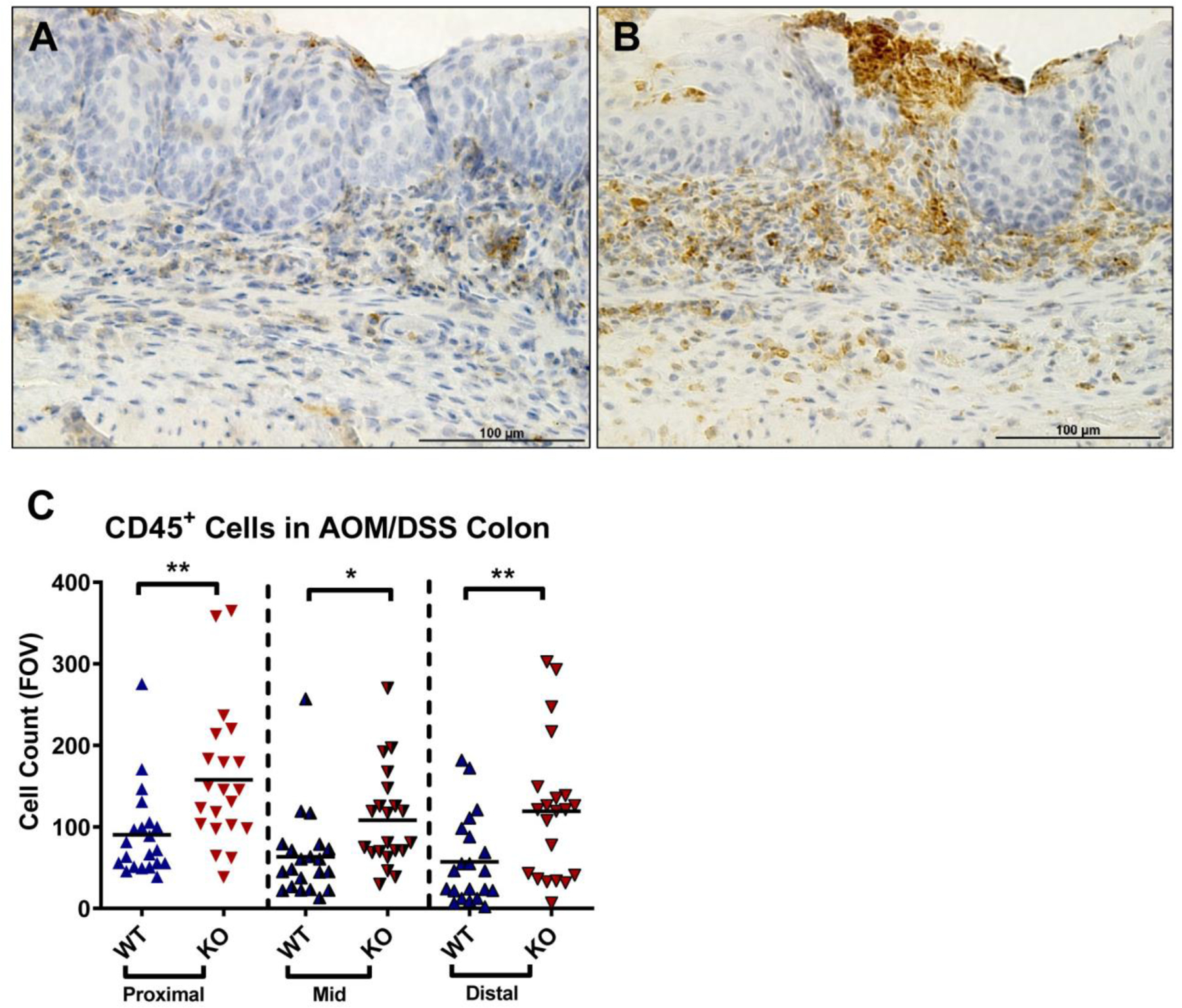
GPR65 (TDAG8) is a proton-sensing G protein-coupled receptor predominantly expressed in immune cells. Genome-wide association studies (GWAS) have identified GPR65 gene polymorphisms as an emerging risk factor for the development of inflammatory bowel disease (IBD). Patients with IBD have an elevated risk of developing colorectal cancer when compared to the general population. To study the role of GPR65 in intestinal inflammation and colitis-associated colorectal cancer (CAC), colitis and CAC were induced in GPR65 knockout (KO) and wild-type (WT) mice using dextran sulfate sodium (DSS) and azoxymethane (AOM)/DSS, respectively. Disease severity parameters such as fecal score, colon shortening, histopathology, and mesenteric lymph node enlargement were aggravated in GPR65 KO mice compared to WT mice treated with DSS. Elevated leukocyte infiltration and fibrosis were observed in the inflamed colon of GPR65 KO when compared to WT mice which may represent a cellular mechanism for the observed exacerbation of intestinal inflammation. In line with high expression of GPR65 in infiltrated leukocytes, GPR65 gene expression was increased in inflamed intestinal tissue samples of IBD patients compared to normal intestinal tissues. Moreover, colitis-associated colorectal cancer development was higher in GPR65 KO mice than WT mice when treated with AOM/DSS. Altogether, our data demonstrate that GPR65 suppresses intestinal inflammation and colitis-associated tumor development in murine colitis and CAC models, suggesting potentiation of GPR65 with agonists may have an anti-inflammatory therapeutic effect in IBD and reduce the risk of developing colitis-associated colorectal cancer.

Keywords: Colorectal cancer; GPR65 (TDAG8); Inflammatory bowel disease (IBD).


Conflict of interest statement

Declaration of interests

The authors declare that they have no known competing financial interests or personal relationships that could have appeared to influence the work reported in this paper.

Conflict of Interest

None.

Figures

Figure 1.
Figure 1.
Disease indicators of chronic colitis induction in wild-type (WT) and GPR65 knockout (KO) mice. The extent of DSS-induced intestinal inflammation was assessed in WT-DSS and GPR65 KO-DSS mice. WT and GPR65 KO mice given regular water served as the control. GPR65 KO-DSS mice presented elevated disease parameters compared to WT-DSS mice. Clinical phenotypes of intestinal inflammation such as (A) body weight loss normalized to control mice and (B) fecal blood and diarrhea were assessed. Macroscopic disease indicators such as (C) mesenteric lymph node expansion and (D) colon shortening were also recorded. Data are presented as the mean ± SEM and statistical significance was determined using the unpaired t-test between WT-DSS and GPR65 KO-DSS groups. WT control (N=10, with 5 males and 5 females), WT-DSS (N=13, with 6 males and 7 females), GPR65 KO control (N=11, with 5 males and 6 females), and GPR65 KO-DSS (N=13, with 6 males and 7 females) mice were used for experiments. (*P < 0.05, **P < 0.01).
Figure 2.
Figure 2.
Histopathological analysis of proximal, middle, and distal colon. Characteristic histopathological features of colitis were assessed to further characterize the degree of intestinal inflammation. GPR65 KO-DSS mice presented elevated disease parameters compared to WT-DSS mice. Representative H&E pictures were taken for (A) WT control, (B) WT-DSS, (C) GPR65 KO control, and (D) GPR65 KO-DSS mice. Representative pictures of Picrosirius red stained tissue sections for fibrosis assessment were taken of (E) WT control, (F) WT-DSS, (G) GPR65 KO control, and (H) GPR65 KO-DSS mice. Graphical representation of (I) total histopathological scores and (J) colonic fibrosis are presented. WT control (N=10), WT-DSS (N=13), GPR65 KO control (N=11), and GPR65 KO-DSS (N=13) mouse tissues were used for histopathological analysis. Scale bar is 100μm. Data are presented as the mean ± SEM and statistical significance was determined using the unpaired t-test between WT-DSS and GPR65 KO-DSS groups. (*P < 0.05).
Figure 3.
Figure 3.
Myofibroblast expansion in distal colon mucosa. SMA+ myofibroblasts were evaluated in distal colon as a cellular basis for increased colonic fibrosis. Representative pictures of (A) WT control, (B) GPR65 KO-control, (C,E) WT-DSS, and (D,F) GPR65 KO-DSS. Scale bar is 100μm.
Figure 4.
Figure 4.
Leukocyte infiltrates in distal colon. Polymorphonuclear (PMN) neutrophils, F4/80+ macrophages, and CD3+ T cells were counted in the distal colon. GPR65 KO-DSS mice had increased neutrophils, macrophages, and T cells in distal colon when compared to WT-DSS mice. (A) Representative pictures of WT-DSS (left) and GPR65 KO-DSS (right) mouse (A-B) neutrophils, (D-E) macrophages, and (G-H) T cells, respectively. Graphical representation of (C) neutrophils, (F) macrophages, (I) and T cells. Scale bar is 100μm. Data are presented as the mean ± SEM and statistical significance was determined using the unpaired t-test between WT-DSS and GPR65 KO-DSS groups. (*P < 0.05, **P <0.01).
Figure 5.
Figure 5.
GFP signal in the intestine and intestinal associated lymphoid tissues. GFP knock-in signal under the control of GPR65 promoter serves as a surrogate marker for endogenous GPR65 expression in GPR65 KO mice. GFP signal could be detected in GPR65 KO mouse (A) distal colon mucosa, (C) proximal colon transverse folds, (E) isolated lymphoid follicles (ILFs), and (G) mesenteric lymph nodes (MLNs). GFP signal could be detected in DSS-treated GPR65 KO (B) intestinal mucosa, (D) transverse folds, (F) ILFs, and (H) MLNs. Based on cellular morphology and localization, GFP signal was observed in intestinal and MLN macrophages, lymphocytes, and neutrophils. Scale bar is 100μm.
Figure 6.
Figure 6.
GPR65 and inflammatory gene expression in human ulcerative colitis and Crohn’s disease. GPR65 mRNA is increased in ulcerative colitis (UC) and Crohn’s disease (CrD) compared to non-inflamed intestinal tissues and positively correlates with TNFα and IFNγ gene expression. (A) GPR65, TNFα and IFNγ mRNA levels in UC and CrD intestinal tissues. (B) Correlation of GPR65 mRNA expression with TNFα and IFNγ mRNA levels. Data are presented as the mean ± SEM and statistical significance was determined using the Mann-Whitney test between control and diseased intestinal tissues. Correlation of gene expression was determined by the linear regression analysis. (*P < 0.05, **P < 0.01, ***P < 0.001, ****P < 0.0001).
Figure 7.
Figure 7.
Effects of GPR65 on tumorigenesis in the colitis associated colorectal cancer (CAC) mouse model. Tumor burden is increased in the colons of GPR65 KO-AOM/DSS mice (N=21, with 11 males and 10 females) compared to WT-AOM/DSS mice (N=21, with 11 males and 10 females). Red arrows indicate polyps and tumors. (A) Polyp/tumor number, (B) polyp/tumor volume, (C-D) representative pictures of distal colons bearing polyps/tumors in (C) WT and (D) GPR65 KO, and (E-H) histopathological representation of dysplasia and adenocarcinoma in (E, G) WT and (F, H) GPR65 KO AOM/DSS mouse colons. Data are presented as the mean ± SEM and statistical significance was determined using the unpaired t-test between WT and GPR65 KO AOM/DSS mice. (*P <0.05).
Figure 8.
Figure 8.
Colon shortening, fibrosis, and myofibroblast expansion in AOM/DSS mice. The colons of GPR65 KO-AOM/DSS mice show reduced length, increased fibrosis, and heightened myofibroblast expansion compared to WT AOM/DSS mice. (A, B) Colon length, (C) representative images of pathological fibrosis by Picrosirius Red staining, (D) fibrosis score of severity (WT N=21 and GPR65 KO N=21), and (E) immunohistochemistry of α-SMA+ myofibroblasts in mouse colon tissues. Scale bar is 100μm. Data are presented as the mean ± SEM and statistical significance was determined using the unpaired t-test between WT AOM/DSS and GPR65 KO-AOM/DSS mice (*P < 0.05, ***P < 0.001).
Figure 9.
Figure 9.
Leukocyte infiltration in colons of AOM/DSS mice. Leukocyte infiltration in colon tissues is increased in GPR65 KO-AOM/DSS mice when compared to WT AOM/DSS mice. CD45+ immunohistochemistry of leukocytes in (A) WT and (B) GPR65 KO AOM/DSS mouse colon sections, and (C) cell count of leukocyte infiltration in distal, middle, and proximal colon segments (WT N=21 and GPR65 KO N=21). Scale bar is 100μm. Data are presented as the mean ± SEM and statistical significance was determined using the unpaired t-test between WT and GPR65 KO AOM/DSS mice (*P < 0.05, **P < 0.01).

Similar articles

Cited by

References

    1. Hussman JP, Beecham AH, Schmidt M, Martin ER, McCauley JL, Vance JM, Haines JL, Pericak-Vance MA, GWAS analysis implicates NF-kappaB-mediated induction of inflammatory T cells in multiple sclerosis, Genes Immun 17 (2016) 305–312. - PMC - PubMed
    1. Hardin M, Cho M, McDonald ML, Beaty T, Ramsdell J, Bhatt S, van Beek EJ, Make BJ, Crapo JD, Silverman EK, Hersh CP, The clinical and genetic features of COPD-asthma overlap syndrome, Eur Respir J (2014). - PMC - PubMed
    1. Al-Mossawi MH, Chen L, Fang H, Ridley A, de Wit J, Yager N, Hammitzsch A, Pulyakhina I, Fairfax BP, Simone D, Yi Y, Bandyopadhyay S, Doig K, Gundle R, Kendrick B, Powrie F, Knight JC, Bowness P, Unique transcriptome signatures and GM-CSF expression in lymphocytes from patients with spondyloarthritis, Nat Commun 8 (2017) 1510. - PMC - PubMed
    1. Jostins L, Ripke S, Weersma RK, Duerr RH, McGovern DP, Hui KY, Lee JC, Schumm LP, Sharma Y, Anderson CA, Essers J, Mitrovic M, Ning K, Cleynen I, Theatre E, Spain SL, Raychaudhuri S, Goyette P, Wei Z, Abraham C, Achkar JP, Ahmad T, Amininejad L, Ananthakrishnan AN, Andersen V, Andrews JM, Baidoo L, Balschun T, Bampton PA, Bitton A, Boucher G, Brand S, Buning C, Cohain A, Cichon S, D’Amato M, De Jong D, Devaney KL, Dubinsky M, Edwards C, Ellinghaus D, Ferguson LR, Franchimont D, Fransen K, Gearry R, Georges M, Gieger C, Glas J, Haritunians T, Hart A, Hawkey C, Hedl M, Hu X, Karlsen TH, Kupcinskas L, Kugathasan S, Latiano A, Laukens D, Lawrance IC, Lees CW, Louis E, Mahy G, Mansfield J, Morgan AR, Mowat C, Newman W, Palmieri O, Ponsioen CY, Potocnik U, Prescott NJ, Regueiro M, Rotter JI, Russell RK, Sanderson JD, Sans M, Satsangi J, Schreiber S, Simms LA, Sventoraityte J, Targan SR, Taylor KD, Tremelling M, Verspaget HW, De Vos M, Wijmenga C, Wilson DC, Winkelmann J, Xavier RJ, Zeissig S, Zhang B, Zhang CK, Zhao H, Silverberg MS, Annese V, Hakonarson H, Brant SR, Radford-Smith G, Mathew CG, Rioux JD, Schadt EE, Daly MJ, Franke A, Parkes M, Vermeire S, Barrett JC, Cho JH, Host-microbe interactions have shaped the genetic architecture of inflammatory bowel disease, Nature 491 (2012) 119–124. - PMC - PubMed
    1. Lahue KG, Lara MK, Linton AA, Lavoie B, Fang Q, McGill MM, Crothers JW, Teuscher C, Mawe GM, Tyler AL, Mahoney JM, Krementsov DN, Identification of novel loci controlling inflammatory bowel disease susceptibility utilizing the genetic diversity of wild-derived mice, Genes and immunity 21 (2020) 311–325. - PMC - PubMed

Publication types

MeSH terms