. 2022 Jan:251:109209.
doi: 10.1016/j.cbpc.2021.109209. Epub 2021 Oct 8.

Examining the effect of manganese on physiological processes: Invertebrate models

Affiliations

Examining the effect of manganese on physiological processes: Invertebrate models

Cecilia Pankau et al. Comp Biochem Physiol C Toxicol Pharmacol. 2022 Jan.

Abstract

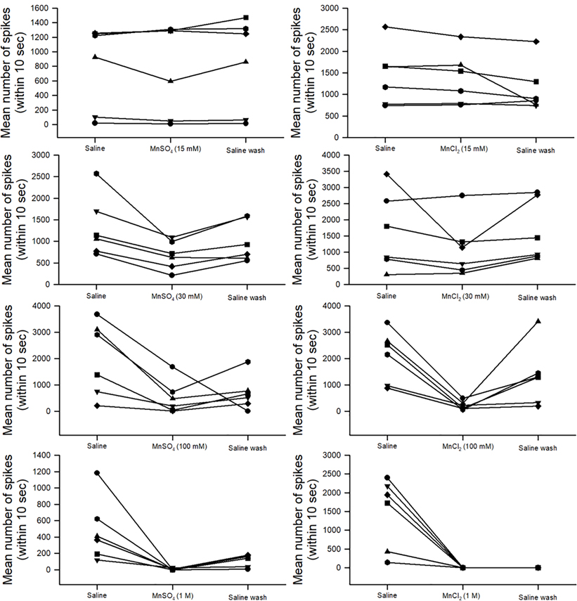
Manganese (Mn2+ as MnSO4 &/or MnCl2) is a common and essential element for maintaining life in plants and animals and is found in soil, fresh waters and marine waters; however, over exposure is toxic to organisms. MnSO4 is added to soil for agricultural purposes and people are exposed to Mn2+ in the mining industry. Hypermanganesemia in mammals is associated with neurological issues mimicking Parkinson's disease (PD) and appears to target dopaminergic neural circuits. However, it also seems that hypermanganesemia can affect many aspects of health besides dopaminergic synapses. We examined the effect on development, behavior, survival, cardiac function, and glutamatergic synaptic transmission in the Drosophila melanogaster. In addition, we examined the effect of Mn2+ on a sensory proprioceptive organ and nerve conduction in a marine crustacean and synaptic transmission at glutamatergic neuromuscular junctions of freshwater crayfish. A dose-response effect of higher Mn2+ retards development, survival and cardiac function in larval Drosophila and survival in larvae and adults. MnSO4 as well as MnCl2 blocks stretch activated responses in primary proprioceptive neurons in a dose-response manner. Mn2+ blocks glutamatergic synaptic transmission in Drosophila as well as crayfish via presynaptic action. This study is relevant in demonstrating the effects of Mn2+ on various physiological functions in order to learn more about acute and long-term consequences Mn2+ exposure.

Keywords: Cardiac; Crab; Crayfish; Drosophila; Manganese; Neuromuscular junction; Sensory; Survival.


Conflict of interest statement

Declaration of competing interests

The authors declare that they have no known competing financial interests or personal relationships that could have appeared to influence the work reported in this paper.

Figures

Figure 1:
Figure 1:
The filleted larva preparation used for measurement of heart rate when exposed to MnSO4 and MnCl2. Heartbeats were counted by manual inspection through a dissecting microscope before and after switching to the compound of interest. Heart rates were measured from the caudal end of the preparation near the point where the two tracheal tubes bifurcate.
Figure 2:
Figure 2:
The first or second walking leg of the crab was used to expose the Pd organ and associated nerve. (A) The joint was initially bent at 90 degrees and then extended out to 180 degrees within 1 second and held for at least another 9 seconds. (B) The entire 10 seconds was then used for analysis in the number of spikes that occurred while being bathed in different solutions.
Figure 3:
Figure 3:
Recording the evoked compound action potential (CAP) of the Pd nerve in a crab first walking leg. The Pd nerve is dissected out of the leg from the Pd organ to the autotomy plane of the leg. The proximal end is used to record the Pd activity and induced CAP in different bathing media. The distal end of the nerve close to the Pd organ was pulled into a suction electrode to induce CAPs in the Pd nerve. The preparation is pinned to a Sylard lined dish to hold the dissected leg in place. A ground wire is placed in the bathing media.
Figure 4:
Figure 4:
A schematic diagram of the recording arrangement of a 3rd instar larva for obtaining evoked and spontaneous synaptic excitatory junction potentials from m6 muscle fiber.
Figure 5:
Figure 5:
The opener muscle of the crayfish walking leg with facilitated synaptic responses in the excitatory junction potentials from the distal muscle fibers. The synaptic responses depicted show the facilitation which occurs over 25 stimuli. The arrows indicate the stimuli given to the nerve.
Figure 6:
Figure 6:
Survival of adults exposed to food tainted with MnSO4 or MnCl2 as compared to controls. Controls were fed standard cornmeal food while others had food tainted with 5, 15 or 30 mM MnSO4 or MnCl2. Twenty adults were placed into each condition and observed daily.
Figure 7:
Figure 7:
Larval behaviors after 24 hours of exposure to tainted food of 15 or 30 mM MnSO4 or MnCl2 as compared to standard media. The mouth hook movements per minute were significantly reduced with both 15 and 30 mM of MnSO4 or MnCl2. The larvae were transferred to a dilute solution of yeast mixed with distilled water for the assay. Crawling behavior, as indexed in body wall contractions per minute, indicated that being exposed to food of 30 mM for MnSO4 or MnCl2 reduced the rate. However only the 15 mM MnSO4 resulted in a significantly lower rate than controls. The rates are expressed as a mean +/− SEM and significance is p<0.05 ANOVA, N>20 larvae for each condition.
Figure 8:
Figure 8:
The effect of MnSO4 or MnCl2 exposure on heart rate of in situ hearts in 3rd instar larvae. The early 3rd instar larvae is dissected and filleted in order to expose the heart tube to the bathing saline and flush away any endogenous compounds. The heart rate is counted in saline and then the media is changed to saline or to a concentration of either MnSO4 or MnCl2, and this is followed by flushing the preparation with fresh saline without any Mn2+ and the heart rate is counted again. At all concentrations of MnSO4 or MnCl2, the rate dropped significantly upon exposure and the extent in the decreased rate is concentration-dependent. The heart rate did not recover even after 2 minutes following flushing with fresh saline for the 30 mM concentrations for either MnSO4 or MnCl2. There was no significant effect for saline exchanged with saline only. Each line represents an individual larva, and each larva was only examined once.
Figure 9:
Figure 9:
Percent differences in heart rate from saline to exposure of MnSO4 or MnCl2. The percent differences are compared for each condition to the initial rate in saline for each individual larva. The “Mn to saline” is the condition of flushing away of Mn to fresh saline and the percent difference is from the initial saline condition.
Figure 10:
Figure 10:
Activity of the Pd nerve with displacement of the Pd joint from the flexed position to an extended position. The mean number of spikes (averaged of three displacements in each bathing media) for 10 seconds with the 1st second moving the joint from a flex position to an extended position for the remaining 9 seconds (static position). Six preparations were used for each concentration (15, 30, 100 mM and 1M) of MnSO4 or MnCl2. Each line is an individual preparation which was used only once.
Figure 11:
Figure 11:
Activity of the Pd nerve with displacement of the Pd joint from the flexed position to an extended position with repeated saline-only bath exchanges. (A) Six preparations were used while measuring the mean number of spikes (average of three displacements for each time the bathing media was exchanged) over 10 seconds with the 1st second being the movement of the joint from a flexed position to an extended position, and the remaining 9 seconds consisting of the extended static position. Each line is an individual preparation which was used only once. (B) The mean percent change from the initial saline exposure to the following bath exchange is depicted along with each individual preparation as dots.
Figure 12:
Figure 12:
The activity of the stretch-activated channels in the Pd organ and electrical conduction along the Pd nerve. (A1) The activity of the Pd nerve is induced by static-position-sensitive neurons firing while the joint is pinned in a given position (flexed). While recording the activity in the Pd nerve, a stimulation (Stim) is given every 2 seconds by the second suction electrode to induce compound action potentials (CAPs). The CAP from such stimulation is shown in A2. The stimulus artifact (SA) is seen followed by the CAP in the rectangular box. (B1) Exposure to 100 mM MnSO4 decreased the stretch-activated channels (SACs) induced activity in the Pd nerve, but not the ability of the nerve to conduct experimentally induced CAPs (B2). (C1) Return of the sensory response from the SACs appears with the flushing away of Mn2+, but the CAPs are still present (C2).
Figure 13:
Figure 13:
The effects of high concentration of MnCl2 or mannitol on the activity of the Pd organ during joint movement and induction of compound action potential (CAP). (A) The activity before and during exposure to 1 M MnCl2. The recording was continuous while adding the MnCl2, which resulted in the activity of the Pd organ to stop immediately. The two large deflections in the trace are due to swishing the bathing media over the nerve. (B) The activity during the movement and hold paradigm before (B1) and during 1 M mannitol exposure (B2). In separate experiments, the CAP was induced before (C1) and during 1 M mannitol exposure (C2). The dotted circle outlines the CAP while the stimulus artifact (SA) is the electrical event in the media from the stimulation.
Figure 14:
Figure 14:
The mean percent change in the number of spikes before and during exposure to 1 M sucrose with displacement of the Pd joint for 10 seconds. There was no significant difference in the activity due to exposure of sucrose.
Figure 15:
Figure 15:
The effect of exposure to MnSO4 (2.5 mM) to synaptic transmission at the larval D. melanogaster neuromuscular junction. (A) Evoked excitatory junction potentials (EJPs) (0.5 Hz stimulation rate) reduced rapidly upon exposure to MnSO4 and slowly recovers upon washing away the MnSO4. (B) The spontaneous quantal events are still present while evoked EJPs are depressed.
Figure 16:
Figure 16:
The effect of exposure to MnSO4 (2.5 mM) on synaptic transmission at the neuromuscular junction of the opener muscle in the walking leg of crayfish. Evoked excitatory junction potentials (EJPs) (60 Hz stimulation rate for 30 pulses) are reduced rapidly upon exposure to MnSO4 and partially recover after washing away the MnSO4.

References

    1. Aiello EL 1990. Nervous control of gill ciliary activity in Mytilus edulis. Stefano GB (Ed.), Neurobiology of Mytilus edulis, vol. 10, Manchester University Press, Manchester: (1990), pp. 189–208.
    1. Alexandrowicz JS, 1972. The comparative anatomy of leg propriocetors in some decapod Crustacea. J. Mar. Biol. Assoc. U.K. 52(3), 605–634.
    1. Baden SP, Eriksson SP, 2006. Role, routes and effects of manganese in crustaceans. Oceanography and Marine Biol. 44, 61–83.
    1. Badre NH, Cooper RL, 2008. Reduced calcium channel function in Drosophila disrupts associative learning in larva, and behavior in adults. Inter. J. Zool. Res. 4(3), 152–164.
    1. Baek SY, Lee MJ, Jung HS, Kim HJ, Lee CR, Yoo C, Lee JH, Lee H, Yoon CS, Kim YH, Park J, Kim W, Jeon BS, Kim Y, 2003. Effect of manganese exposure on MPTP neurotoxicities. Neurotoxicol. 24, 657–665. - PubMed

MeSH terms

LinkOut - more resources