. 2023 Mar;30(3):605-617.
doi: 10.1038/s41418-021-00888-8. Epub 2021 Oct 9.

M6A RNA methylation-mediated RMRP stability renders proliferation and progression of non-small cell lung cancer through regulating TGFBR1/SMAD2/SMAD3 pathway

Affiliations

M6A RNA methylation-mediated RMRP stability renders proliferation and progression of non-small cell lung cancer through regulating TGFBR1/SMAD2/SMAD3 pathway

Hang Yin et al. Cell Death Differ. 2023 Mar.

Erratum in

Abstract

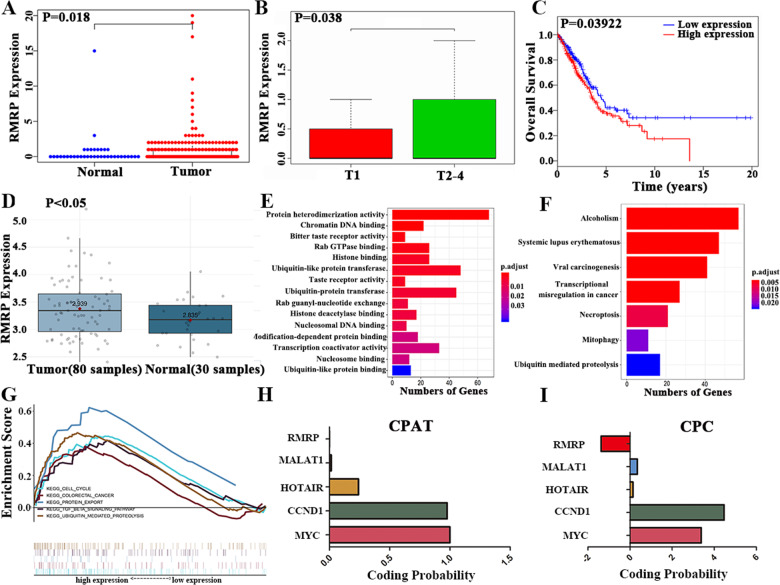
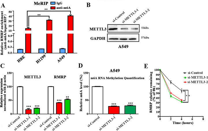
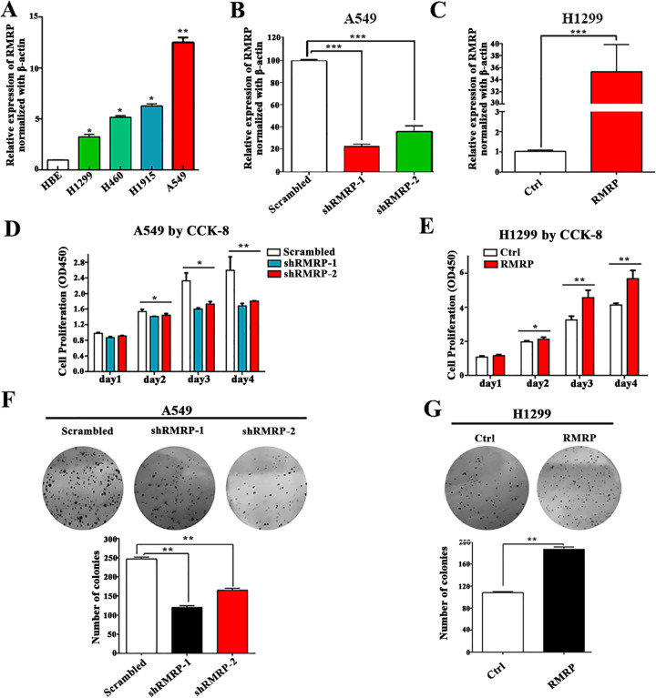
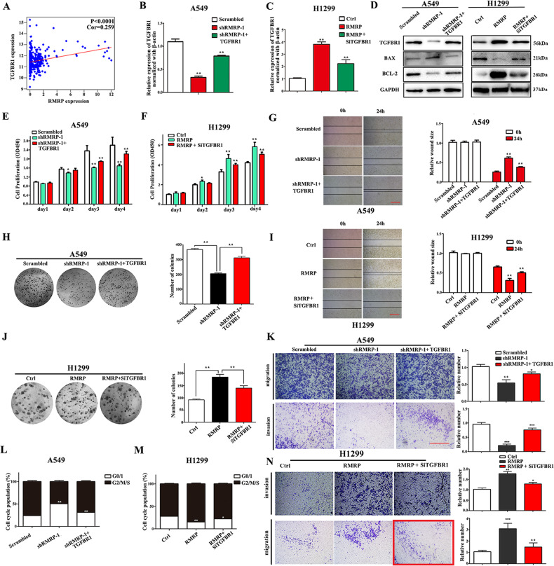
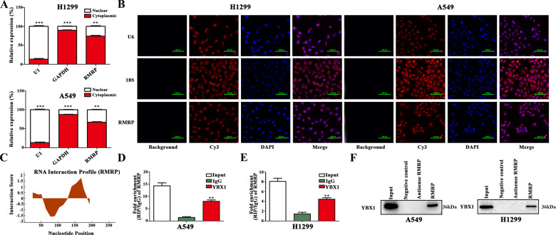
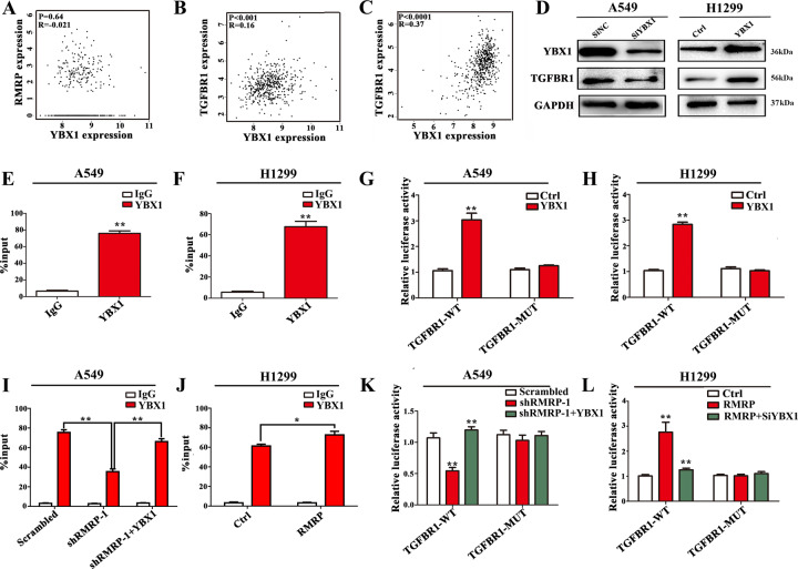
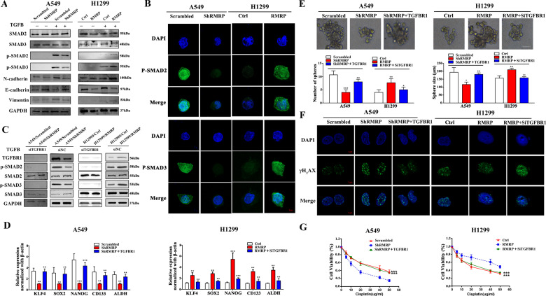
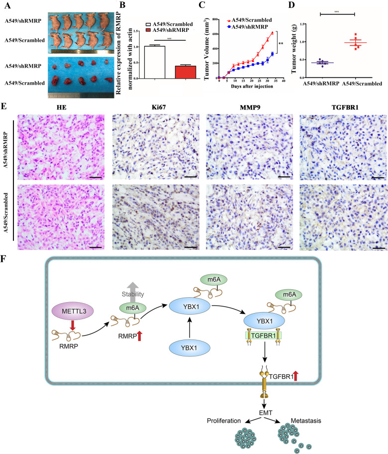
Non-small cell lung cancer (NSCLC) has the highest mortality rate among all malignancies worldwide. The role of long noncoding RNAs (lncRNAs) in the progression of cancers is a contemporary research hotspot. Based on an integrative analysis of The Cancer Genome Atlas database, we identified lncRNA-RNA Component of Mitochondrial RNA Processing Endoribonuclease (RMRP) as one of the most highly upregulated lncRNAs that are associated with poor survival in NSCLC. Furthermore, N(6)-methyladenosine (m6A) was highly enriched within RMRP and enhanced its RNA stability. In vitro and in vivo experiments showed that RMRP promoted NSCLC cell proliferation, invasion, and migration. In terms of mechanism, RMRP recruited YBX1 to the TGFBR1 promotor region, leading to upregulation of the transcription of TGFBR1. The TGFBR1/SMAD2/SMAD3 pathway was also regulated by RMRP. In addition, RMRP promoted the cancer stem cells properties and epithelial mesenchymal transition, which promote the resistance to radiation therapy and cisplatin. Clinical data further confirmed a positive correlation between RMRP and TGFBR1. In short, our work reveals that m6A RNA methylation-mediated RMRP stability renders proliferation and progression of NSCLC through regulating TGFBR1/SMAD2/SMAD3 pathway.


Conflict of interest statement

The authors declare no competing interests.

Figures

Fig. 1
Fig. 1. Integrated analysis of NSCLC reveals that lncRNA-RMRP is a potential biomarker for NSCLC patients.
A. RMRP was significantly upregulated in NSCLC tissues (n = 535) as compared to normal lung tissues (n = 59) (Wilcoxon signed-rank test). B. RMRP was associated with tumor size in NSCLC patients (n = 335; Wilcoxon signed-rank test). C High RMRP expression was associated with worse survival rates by Kaplan–Meier method (n = 504). D RMRP was upregulated in NSCLC tissues (n = 80) compared to normal lung tissues (n = 30) in GSE43458, as shown in the Gene Expression Omnibus (GEO) database, which was analyzed by the online tool Lung Cancer Explorer (https://lce.biohpc.swmed.edu/lungcancer/). E Gene Ontology (GO) results showed that RMRP functions in protein heterodimerization activity, chromatin DNA binding, bitter taste receptor activity, Rab GTPase binding, histone binding, ubiquitin-like protein transferase activity, taste receptor activity, ubiquitin-protein transferase activity, Rab guanyl-nucleotide exchange factor activity, histone deacetylase binding, nucleosomal DNA binding, modification-dependent protein binding, transcription coactivator activity, nucleosome binding, and ubiquitin-like protein binding. F The KEGG results showed that RMRP was enriched in pathways of Alcoholism, Systemic lupus erythematosus, viral carcinogenesis, transcriptional misregulation in cancer, Necroptosis, Mitophagy—animal and Ubiquitin mediated proteolysis. G The GSEA research showed RMRP was enriched in the cell cycle, colorectal cancer, protein export, TGFB pathway, and ubiquitin-mediated proteolysis pathway. H The protein-coding potential of RMRP through the CPAT database. I The protein-coding potential of RMRP through the CPC database. LncRNA-MALAT1, LncRNA-HOTAIR, CCND1, and MYC were also explored as control.
Fig. 2
Fig. 2. The m6A level of RMRP was upregulated in NSCLC cells than lung epithelial cells.
A The m6A methylation level of RMRP in human normal lung epithelial cells (HBE) and NSCLC cells (H1299 and A549) were determined by MeRIP-qPCR assays. The input RNA fraction Ct value was used to account for RNA sample preparation differences; negative control groups (IgG) were used to adjust background fraction (n = 3). B Results of Western blot assay showing METTL3 protein expression in A549 cells transfected with two different short interference RNAs for METTL3 (si-METTL3-1 and si-METTL3-2) or negative controls (si-Control) (n = 3). C Results of qRT-PCR assays showing the mRNA levels of METTL3 and RMRP in the treated A549 cells (n = 3). β-actin was used as the reference for normalization. D The m6A level in the treated A549 cells (n = 3). E Reduction of RMRP RNA stability in METTL3-knockdown A549 cells as compared to control. Cells were treated with 5 μg/mL actinomycin D and RNA was isolated at 0 h, 2 h, and 4 h (n = 3). Data presented as mean ± SD from at least three independent experiments. *P < 0.05, **P < 0.01, ***P < 0.001.
Fig. 3
Fig. 3. RMRP promotes NSCLC cell proliferation.
A Results of quantitative RT-PCR (qRT-PCR) analysis showing RMRP expression in NSCLC cell lines (H1299, H460, H1915, A549) and human lung epithelial cell line (HBE). β-actin was used as the endogenous control (n = 3). B Results of qRT-PCR analysis showing RMRP expression in A549 cells transfected with two different shRNAs against RMRP (shRMRP-1 and shRMRP-2) or scrambled control (Scrambled). β-actin was used as the endogenous control (n = 3). C Results of qRT-PCR analysis showing RMRP expression in H1299 cells transfected with RMRP-overexpressing plasmid (RMRP) or matched Controls (Ctrl). β-actin was used as the endogenous control (n = 3). D CCK-8 assays of A549 cells transfected with shRMRP compared with those transfected with Scrambled (n = 3). E CCK-8 assays of H1299 cells transfected with RMRP or Ctrl (n = 3). F Representative images (Up) and quantification (Down) of colony formation assays in A549 cells transfected with shRMRP or Scrambled (n = 3). G Representative images (Up) and quantification (Down) of colony formation assays in H1299 cells transfected with RMRP or Ctrl (n = 3). Data presented as mean ± SD from at least three independent experiments. *P < 0.05, **P < 0.01, ***P < 0.001.
Fig. 4
Fig. 4. TGFBR1 is a critical target by which RMRP promotes proliferation, invasion, and migration.
A The TCGA database of lung adenocarcinoma showed a positive correlation between the expressions of TGFBR1 and RMRP (n = 594). B Results of qRT-PCR analysis showing the expression of TGFBR1 mRNA in A549 cells after being transfected with shRMRP or TGFBR1-overexpressing plasmid (shRMRP + TGFBR1) as well as their respective negative controls. C Results of qRT-PCR analysis showing the expression of TGFBR1 mRNA in H1299 cells after being transfected with RMRP or short interference RNAs for TGFBR1 (RMRP + SiTGFBR1) as well as their respective negative controls. D Western blot analysis of the treated A549 and H1299 cells using the indicated antibodies (n = 3). E CCK-8 assays of the treated A549 cells (n = 3). F CCK-8 assays of the treated H1299 cells (n = 3). G Representative images (Left) and quantification (Right) of the wound-healing assays of the treated A549 cells (n = 3, Scale bar, 200 μm). H Representative images (Left) and quantification (Right) of colony formation assay of the treated A549 cells (n = 3). I Representative images (Left) and quantification (Right) of the wound-healing assays in the treated H1299 cells (n = 3). J Representative images (Left) and quantification (Right) of colony formation assay of the treated H1299 cells (n = 3). K Representative images (Left) and quantification (Right) of the transwell assays including invasion and migration of the treated A549 cells (n = 3). L The cell cycle assays of the treated A549 cells (n = 3). M The cell cycle assays of the treated H1299 cells (n = 3). N Representative images (Left) and quantification (Right) of the transwell assays including invasion and migration of the treated H1299 cells (n = 3, Scale bar, 200 μm). Data presented as mean ± SD from at least three independent experiments. *P < 0.05, **P < 0.01, ***P < 0.001.
Fig. 5
Fig. 5. RMRP is associated with the transcription factor YBX1.
A qRT-PCR assays of the relative expression levels after the fractionation of nuclear and cytoplasmic RNA of H1299 and A549 cells. GAPDH was used as a cytoplasmic marker, and U1 was used as a nuclear marker (n = 3). B Representative RNA-FISH images showing the subcellular location of RMRP in H1299 and A549 cells. 18S and U6 were used as cytoplasmic and nuclear markers, respectively (Scale bar, 100 μm) (n = 3). C The analysis of RNA interaction profile showed the putative binding sequence of RMRP to YBX1 protein by catRAPID. D, E RNA immune precipitation (RIP) experiment was performed using the YBX1 and IgG antibodies to probe A549 and H1299 cells extracts, and the levels of the co-precipitated RNAs were determined using qRT-PCR (n = 3). F Total protein was extracted from A549 and H1299 cells and utilized in biotinylated RNA pull-down assay. Proteins that were associated with RMRP were explored by Western blot with anti-YBX1 antibody (n = 3). Data presented as mean ± SD from at least three independent experiments. *P < 0.05, **P < 0.01, ***P < 0.001.
Fig. 6
Fig. 6. RMRP promotes TGFBR1 transcription by recruiting YBX1 to the TGFBR1 promoter.
A The expression of YBX1 was not correlated with RMRP in TCGA-LUAD database by GEPIA (http://gepia.cancer-pku.cn/) using Pearson Correlation Coefficient. B The expression of YBX1 showed a positive correlation with TGFBR1 in TCGA-Lung adenocarcinoma (LUAD) database by GEPIA using Pearson Correlation Coefficient. C The expression of YBX1 showed a positive correlation with TGFBR1 in TCGA-Kidney Renal Clear Cell Carcinoma (KIRC) database by GEPIA using Pearson Correlation Coefficient. D Results of Western blot assays: knockdown of YBX1 inhibited the expression of TGFBR1, while overexpression of YBX1 increased the expression of TGFBR1 (n = 3). E, F YBX1-bound complex showed remarkable enrichment of the TGFBR1 promoter by ChIP and quantitative RT-PCR analysis in A549 and H1299 cells. IgG was used as a negative control. Enrichment was quantified relative to input controls (n = 3). G, H Luciferase activity assays were performed in A549 and H1299 cells transfected with wide type (TGFBR1-WT) or mutant-type (TGFBR1-MUT) TGFBR1 promoter-containing pGL3 reporter vector. These cells were further transfected with YBX1 (YBX1) or control (Ctrl). After 48 h, firefly luciferase activity was detected and normalized to Renilla luciferase activity (n = 3). I, J ChIP and quantitative RT-PCR analysis in the treated A549 and H1299 cells. IgG was used as a negative control. Enrichment was quantified relative to input controls (n = 3). K, L Luciferase activity assays in the treated A549 and H1299 cells were further transfected with TGFBR1-MUT or TGFBR1-WT. After 48 h, firefly luciferase activity was detected and normalized to Renilla luciferase activity, respectively (n = 3). Data presented as mean ± SD from three independent experiments. Student’s t test was used for statistical analysis: *P < 0.05; **P < 0.01; ***P < 0.001.
Fig. 7
Fig. 7. RMRP regulated the TGFBR1/SMAD2/ SMAD3 pathway in NSCLC.
A A549/Scrambled, A549/shRMRP, H1299/Ctrl, and H1299/RMRP cells under the TGFB treatment were collected and subjected to Western blot analysis using the indicated antibodies. The TGFB treatment was stimulated with 5 ng/mL TGFB for 1 h (n = 3). B Representative immunofluorescence images of anti-p-SMAD2/3 (green) and DAPI (blue) staining of the treated NSCLC cells. The cells were also cultured under the TGFB treatment and were collected for immunofluorescence analysis (n = 3) (Scale bar, 5 μm). C A549/Scrambled, A549/shRMRP, H1299/Ctrl, and H1299/RMRP cells were treated with TGFBR1 knockdown (siTGFBR1) or siRNA negative controls (siNC). The samples under the TGFB treatment were collected and subjected to Western blot analysis using the indicated antibodies (n = 3). D Related cells were collected and analyzed by qRT-PCR (n = 3). β-actin was used as the reference for normalization. E The number of tumorspheres was counted, and the morphology was observed under a light microscope. Upper panel: Representative pictures of tumorspheres. Lower panel: Tumorspheres quantification results including the number and size of spheres. (Scale bar, 100 μm). F The treated A549 and H1299 cells were subjected to irradiation (6 Gy). Subsequently, immunofluorescence analysis was performed after 1 h (n = 3, Scale bar, 5 μm). G Results of CCK-8 assays showing cell viability after incubation with different concentrations of cisplatin for 24 h. *P < 0.05; **P < 0.01; ***P < 0.001. Data represent three independent experiments.
Fig. 8
Fig. 8. Knockdown of RMRP limited tumor growth in vivo.
A A549 cells with stable expression of shRMRP (A549/shRMRP) or scrambled control (A549/Scrambled) were transplanted into the axilla of nude mice to construct tumor growth model. Representative images of nude mice and tumors. B A549/Scrambled and A549/shRMRP group samples were collected and analyzed by qRT-PCR (n = 3). C Tumor volumes in the A549/Scrambled and A549/shRMRP groups. D Tumor weight in the A549/Scrambled and A549/shRMRP groups. E Representative images of haematoxylin and eosin (H&E) staining, Ki-67, MMP9, and TGFBR1 expression by immunohistochemical analysis in xenograft tumors derived from A549/Scrambled and A549/shRMRP groups. F Flowchart of our experiments. Our study suggests the m6A mark improved the stability of methylated RMRP transcripts by decreasing the RNA degradation rate, which may partially account for the upregulation of RMRP in NSCLC. Furthermore, RMRP promotes TGFBR1 transcription by recruiting YBX1 to TGFBR1 promoter.

References

    1. DeSantis CE, Miller KD, Goding Sauer A, Jemal A, Siegel RL. Cancer statistics for African Americans, 2019. CA Cancer J Clin. 2019;69:211–33.. doi: 10.3322/caac.21555. - DOI - PubMed
    1. Jamal-Hanjani M, Wilson GA, McGranahan N, Birkbak NJ, Watkins TBK, Veeriah S, et al. Tracking the evolution of non-small-cell lung cancer. N Engl J Med. 2017;376:2109–21.. doi: 10.1056/NEJMoa1616288. - DOI - PubMed
    1. Zhou M, Wang H, Zeng X, Yin P, Zhu J, Chen W, et al. Mortality, morbidity, and risk factors in China and its provinces, 1990–2017: a systematic analysis for the Global Burden of Disease Study 2017. Lancet. 2019;394:1145–58. - PMC - PubMed
    1. Beermann J, Piccoli MT, Viereck J, Thum T. Non-coding RNAs in development and disease: background, mechanisms, and therapeutic approaches. Physiol Rev. 2016;96:1297–325. doi: 10.1152/physrev.00041.2015. - DOI - PubMed
    1. Yin H, Wang X, Zhang X, Wang Y, Zeng Y, Xiong Y, et al. Integrated analysis of long noncoding RNA associated-competing endogenous RNA as prognostic biomarkers in clear cell renal carcinoma. Cancer Sci. 2018;109:3336–49.. doi: 10.1111/cas.13778. - DOI - PMC - PubMed

Publication types

MeSH terms