. 2021 Oct 11;21(1):445.
doi: 10.1186/s12887-021-02890-y.

Neuroimaging findings of posterior reversible encephalopathy syndrome (PRES) following haematopoietic stem cell transplantation in paediatric recipients

Affiliations

Neuroimaging findings of posterior reversible encephalopathy syndrome (PRES) following haematopoietic stem cell transplantation in paediatric recipients

Ali Önder Atça et al. BMC Pediatr. .

Abstract

Background: Haematopoietic stem cell transplantation (HSCT) is used worldwide in various malignant and nonmalignant childhood diseases, including haematologic, genetic, autoimmune and metabolic disorders, and is the only curative treatment for many of these illnesses. The survival rates of many childhood diseases have been increased due to HSCT treatment. However, associated complications are still important for management. Central nervous system (CNS) complications in paediatric HSCT recipients can be associated with high morbidity and significantly contribute to mortality. Posterior reversible encephalopathy syndrome (PRES) is one of the most common CNS complications in patients with neurological symptoms following HSCT. Magnetic resonance imaging (MRI) is the modality of choice and shows typical bilateral vasogenic oedema at the posterior parts of the cerebral hemispheres; however, various atypical imaging manifestations can also occur. In this study, we retrospectively examined CNS complications in our paediatric HSCT recipients with a focus on the typical and atypical neuroimaging manifestations of PRES following HSCT.

Methods: We retrospectively reviewed the medical records of 300 consecutive paediatric HSCT recipients from January 2014 to November 2018. A total of 130 paediatric HSCT recipients who experienced neurological signs and symptoms and were evaluated with neuroimaging studies following HSCT were enrolled in the study. The timing of CNS complications was defined according to immune status, including the pre-engraftment period (< 30 days after HSCT), the early postengraftment period (30-100 days after HSCT), and the late postengraftment period (> 100 days after HSCT), which were defined as phases 1, 2 and 3, respectively.

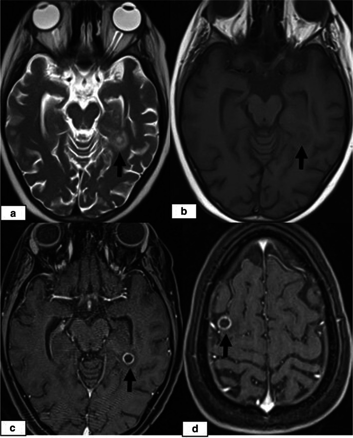
Results: Overall, 130 paediatric HSCT recipients experienced neurological signs and symptoms and therefore underwent neuroimaging examinations. Among these 130 patients, CNS complications were present in 23 patients (17.6%, 23/130), including 13 (56.5%) females and 10 (43.5%) males with a median age of 8.0 years (range, 8 months to 18.0 years). Among these 23 patients, 14 cases of PRES (60.9%), 5 (21.7%) cases of leukoencephalopathy, 3 cases of acute subdural haemorrhage (ASDH) (13%) and 1 (4.3%) case of fungal CNS infection were identified by neuroimaging. On MRI, typical parietooccipital vasogenic oedema was present in 78.5% of the PRES cases (11/14). The following atypical neuroimaging manifestations were observed: isolated involvement of the bilateral frontal lobes in 1 case, isolated cerebellar vermis involvement in 1 case, and isolated basal ganglia involvement in 1 case. Restricted diffusion associated with cytotoxic damage was demonstrated in 2 of 14 cases, one of which also showed subacute cytotoxic injury with ADC pseudonormalization.

Conclusion: Paediatric HSCT recipients presenting with CNS signs and symptoms should be evaluated by neuroimaging studies for timely diagnosis and early management. PRES is the most common CNS complication and may present with atypical MRI manifestations, which should not dissuade a PRES diagnosis in appropriate clinical settings.

Keywords: CNS complications; Haematopoietic stem cell transplantation; MRI; PRES; Posterior reversible encephalopathy syndrome.


Conflict of interest statement

none.

Figures

Fig. 1
Fig. 1
An 11-year-old boy presenting with seizures and visual disturbances 3 months after undergoing allogeneic HSCT for acute myeloid leukaemia. a Axial and b) coronal FLAIR images demonstrating bilateral symmetrical parietal (a) and occipital (b) cortico-subcortical hyperintensity (black arrows). Note the prominent involvement of cortical grey matter (1a, white arrow). c DWI and d) an ADC map showing a high signal (arrows) representing the T2 shine-through effect but not true restricted diffusion. e Follow-up coronal FLAIR image demonstrating complete resolution of vasogenic oedema in the occipital lobes after 3 months
Fig. 2
Fig. 2
A 17-year-old girl presenting with an altered level of consciousness 6 months after undergoing allogeneic HSCT for thalassemia major. a Axial T2W image showing bilateral frontal cortico-subcortical hyperintensity (arrows). b DWI and c) an ADC map showing a high signal (arrows) representing the T2 shine-through effect but not true restricted diffusion. d Axial T2W image demonstrating complete resolution of vasogenic oedema after 3 months
Fig. 3
Fig. 3
A 7-year-old girl presenting with seizures and headache 37 days after undergoing allogeneic HSCT for haemophagocytic lymphohistiocytosis. a Axial T2W image showing abnormal cerebellar hyperintensity (arrows). b DWI and c) ADC images showing diffusion restriction (arrows). She died 3 weeks after PRES onset due to acute pulmonary GVHD
Fig. 4
Fig. 4
A 17-year-old girl presenting with an altered level of consciousness 6 months after undergoing allogeneic HSCT for thalassemia major. a Coronal FLAIR image showing hyperintensity in the bilateral basal ganglia (arrows). b DWI showing increased signal intensity in the basal ganglia. c ADC map showing normal signal intensity, which was considered to be indicative of ADC pseudonormalization. d Follow-up coronal FLAIR image after 6 months demonstrating persistent hyperintensity associated with volume loss, suggesting cytotoxic injury
Fig. 5
Fig. 5
An 8-year-old girl presenting with headache, nausea and vomiting 5 months after undergoing allogeneic HSCT for ALL. a Axial CT of the head did not reveal any abnormalities. b Axial T2W image showing increased signal intensity in the periventricular white matter (b, arrows). d Follow-up axial T2W image 3 months after the first presentation shows persistent increased signal intensity in the periventricular white matter (c, arrows)
Fig. 6
Fig. 6
A 9-month-old female girl presenting with nausea and vomiting 25 days after undergoing allogeneic HSCT for SCID. a Axial CT image showing acute hyperdense right frontoparietal SDH (arrow). b Follow-up axial CT image 1 month after the onset of acute SDH showing complete resolution
Fig. 7
Fig. 7
A 9-year-old girl presenting with headache and impaired consciousness 5 months after undergoing allogeneic HSCT for aplastic anaemia. a Axial T2W image showing a cystic lesion with peripheral oedema in the left medial temporal region (arrow). b Axial T1w image showing hypointensity of the lesion. c, d Postcontrast axial T1W images showing ring enhancement of the same lesion and another similar lesion at the frontal convexity (d, arrow) compatible with a brain abscess

References

    1. Nishiguchi T, Mochizuki K, Shakudo M, Takeshita T, Hino M, Inoue Y. CNS complications of hematopoietic stem cell transplantation. Am J Roentgenol. 2009;192:1003–1011. doi: 10.2214/AJR.08.1787. - DOI - PubMed
    1. Yoshida S, Hayakawa K, Yamamoto A, Kuroda H, Imashuku S. The central nervous system complications of bone marrow transplantation in children. Eur Radiol. 2008;18:2048–2059. doi: 10.1007/s00330-008-1000-3. - DOI - PubMed
    1. Chen BT, Ortiz AO, Dagis A, Torricelli C, Parker P, Openshaw H. Brain imaging findings in symptomatic patients after allogeneic haematopoietic stem cell transplantation: correlation with clinical outcome. Eur Radiol. 2012;22:2273–2281. doi: 10.1007/s00330-012-2490-6. - DOI - PubMed
    1. Bartynski WS, Zeigler ZR, Shadduck RK, Lister J. Pretransplantation conditioning influence on the occurrence of cyclosporine or FK-506 neurotoxicity in allogeneic bone marrow transplantation (original research) AJNR Am J Neuroradiol. 2004;25(2):261–269. - PMC - PubMed
    1. Straathof K, Anoop P, Allwood Z, Silva J, Nikolajeva O, Chiesa R, et al. Long-term outcome following cyclosporine-related neurotoxicity in paediatric allogeneic haematopoietic stem cell transplantation (original research) Bone Marrow Transplant. 2017;52(1):159–162. doi: 10.1038/bmt.2016.232. - DOI - PMC - PubMed

LinkOut - more resources