Review
. 2021 Dec:145:116452.
doi: 10.1016/j.trac.2021.116452. Epub 2021 Oct 2.

Point-of-care COVID-19 diagnostics powered by lateral flow assay

Affiliations
Review

Point-of-care COVID-19 diagnostics powered by lateral flow assay

Yaofeng Zhou et al. Trends Analyt Chem. 2021 Dec.

Abstract

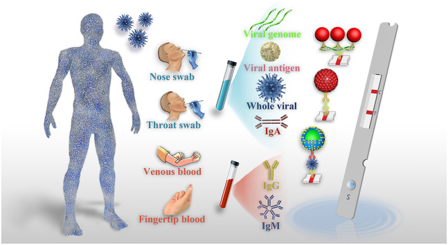
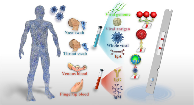
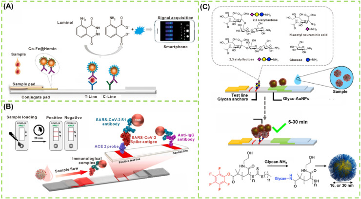
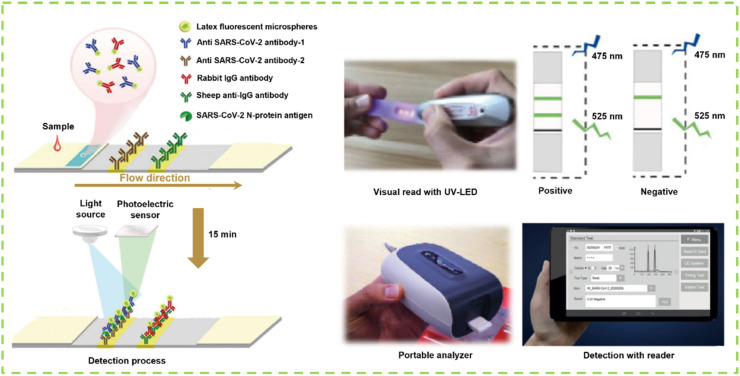
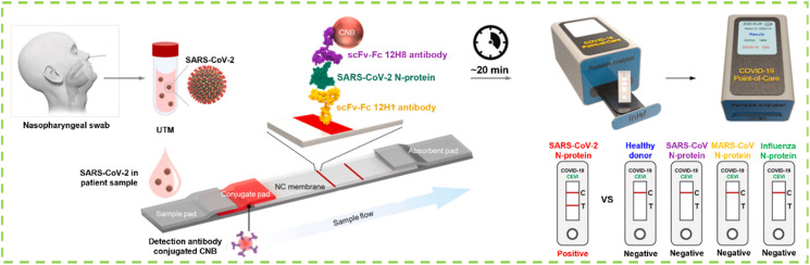
Since its first discovery in December 2019, the global coronavirus disease 2019 (COVID-19) pandemic caused by the novel coronavirus (SARS-CoV-2) has been posing a serious threat to human life and health. Diagnostic testing is critical for the control and management of the COVID-19 pandemic. In particular, diagnostic testing at the point of care (POC) has been widely accepted as part of the post restriction COVID-19 control strategy. Lateral flow assay (LFA) is a popular POC diagnostic platform that plays an important role in controlling the COVID-19 pandemic in industrialized countries and resource-limited settings. Numerous pioneering studies on the design and development of diverse LFA-based diagnostic technologies for the rapid diagnosis of COVID-19 have been done and reported by researchers. Hundreds of LFA-based diagnostic prototypes have sprung up, some of which have been developed into commercial test kits for the rapid diagnosis of COVID-19. In this review, we summarize the crucial role of rapid diagnostic tests using LFA in targeting SARS-CoV-2-specific RNA, antibodies, antigens, and whole virus. Then, we discuss the design principle and working mechanisms of these available LFA methods, emphasizing their clinical diagnostic efficiency. Ultimately, we elaborate the challenges of current LFA diagnostics for COVID-19 and highlight the need for continuous improvement in rapid diagnostic tests.

Keywords: COVID-19; Immunoassay; Lateral flow assay; Rapid diagnostic test; SARS-CoV-2.


Conflict of interest statement

The authors declare that they have no known competing financial interests or personal relationships that could have appeared to influence the work reported in this paper.

Figures

Image 1
Graphical abstract
Scheme 1
Scheme 1
Schematic representation for the POC diagnostics of COVID-19 powered by LFA.
Fig. 1
Fig. 1
LFA for detection of viral RNA: (A) The genome information of SARS-CoV-2 and its corresponding protein structure [36]; (B) Working principle and flow of RT-PCR-enhanced LFA sensor for multiple-detection of N, ORF3a, and RdRP gene to diagnose SARS-CoV-2 [53]; (C) The work principle of RT-RPA combined LFA in detecting SARS-CoV-2 [38]; (D) Schematic of SARS-CoV-2 DETECTR workflow, including conventional RNA extraction, RT-LAMP preamplification, CRISPR-Cas12-based enhancement for E gene, N gene and RNase P, and LFA-based visual detection [48]; (E) RT-RPA and CRISPR co-enhanced SHERLOCK detector in detecting SARS-CoV-2 [79]; (F) The principle of S9.6 antibody-based signal amplification for amplifying LFA [50].
Fig. 2
Fig. 2
LFA for detection of antibodies: (A) Design and fabrication of a LNP-based LFIA for detection of IgG [105]; (B) AuNP-based LFIA for co-detection of SARS-CoV-2 IgM and IgG for diagnosis of CVID-19 [110]; (C) Schematic of chemiluminescence LFIA for detection of IgA [113]; (D) QD nanobeads as the fluorescence probes to fabricate LFIA for detection of SARS-CoV-2 total antibody [131].
Fig. 3
Fig. 3
(A) Schematic illustration of the principle and process of PEI-CISG-enhanced LFIA for detection of SARS-CoV-2 total antibody; (B) LFA and PEI-CISG-enhanced LFA in detecting diluted SARS-CoV-2 positive serum samples; (C) The performance of PEI-CISG-enhanced LFA in eliminating false negative results in diagnosis of SARS-CoV-2 [132].
Fig. 4
Fig. 4
LFA-based sensors for detection of SARS-CoV-2 S protein: (A) Working principle of nanozyme-based LFA sensor for detection of SARS-CoV-2 S protein [142]; (B) Schematic of ACE2 receptor and antibody co-fabricated LFA for detection of SARS-CoV-2 S protein [143]; (C) Molecular structure of glycan capture units, the formation of glyconanoparticles, and the design concept of glyconanoparticles-based LFA [144].
Fig. 5
Fig. 5
The whole detection process of fluorescent microsphere-based double-antibody sandwiched LFIA for the diagnosis of SARS-CoV-2 including oropharyngeal swab sampling, visual read with the UV-LED, and quantitative detection by the portable detector [152].
Fig. 6
Fig. 6
Schematic diagram of the fabrication of phage display technology-assisted LFIA using scFv-Fc fusion proteins as capture antibody (12H1) and detection antibody (12H8) with CNBs as signal reporters for the diagnosis of SARS-CoV-2 [155].

References

    1. Shrock E., Fujimura E., Kula T., Timms R.T., Lee I.-H., Leng Y., Robinson M.L., Sie B.M., Li M.Z., Chen Y. Science. 2020;370:4250.
    1. Zhang X., Tan Y., Ling Y., Lu G., Liu F., Yi Z., Jia X., Wu M., Shi B., Xu S. Nature. 2020;583:437. - PubMed
    1. Schett G., Manger B., Simon D., Caporali R. Nat. Rev. Rheumatol. 2020;160:465. - PMC - PubMed
    1. Le T.T., Andreadakis Z., Kumar A., Román R.G., Tollefsen S., Saville M., Mayhew S. Nat. Rev. Drug Discov. 2020;19:305. - PubMed
    1. Cao X. Nat. Rev. Immunol. 2020;20:269. - PMC - PubMed