. 2021 Sep 24:14:745219.
doi: 10.3389/fnmol.2021.745219. eCollection 2021.

Treatment With Nepicastat Decreases Contextual Traumatic Memories Persistence in Post-traumatic Stress Disorder

Affiliations

Treatment With Nepicastat Decreases Contextual Traumatic Memories Persistence in Post-traumatic Stress Disorder

Raquel Martinho et al. Front Mol Neurosci. .

Abstract

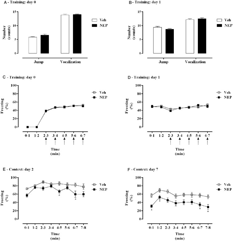
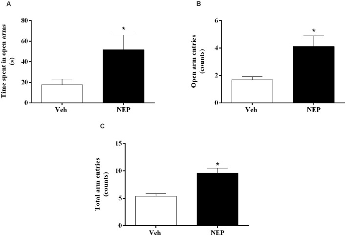
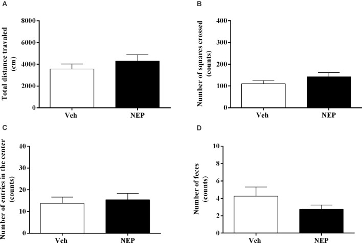
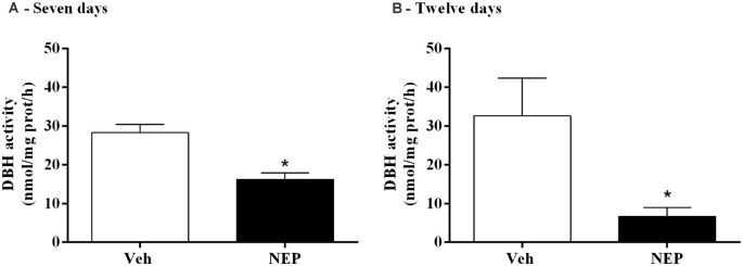
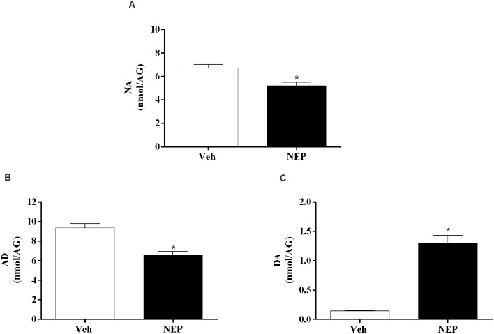
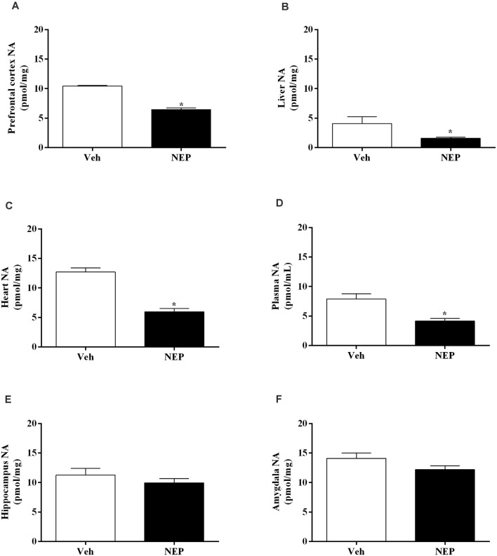
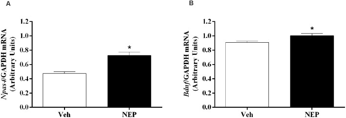
Post-traumatic stress disorder (PTSD) is a common anxiety mental disorder and can be manifested after exposure to a real or perceived life-threatening event. Increased noradrenaline and adrenaline in plasma and urine have been documented in PTSD. Dopamine-β-hydroxylase (DBH) catalyzes the conversion of dopamine to noradrenaline and consequently, DBH inhibition reduces catecholamines. Our aim was to evaluate if nepicastat treatment decreases PTSD signs in an animal model. Wild-type (129x1/SvJ) female mice were submitted to PTSD induction protocol. DBH-inhibitor nepicastat (30 mg/kg) or vehicle (0.2% HPMC) were administered once daily since day 0 until day 7 or 12. The percentage of freezing was calculated on days 0, 1, 2, and 7, and behavioral tests were performed. Quantification of nepicastat in plasma and DBH activity in the adrenal gland was evaluated. Catecholamines were quantified by HPLC with electrochemical detection. mRNA expression of Npas4 and Bdnf in hippocampus was evaluated by qPCR.Mice in the PTSD-group and treated with nepicastat showed a decrease in freezing, and an increase in the time spent and entries in open arms in elevated plus maze test. In mice treated with nepicastat, adrenal gland DBH activity was decreased, and catecholamines were also decreased in plasma and tissues. On day 7, in mice treated with nepicastat, there was an increase of Npas4 and Bdnf mRNA expression in the hippocampus.In conclusion, DBH inhibitor nepicastat has an effect consistent with a decrease in the persistence of traumatic memories and anxiety-like behavior in this PTSD mice model. The disruption of traumatic memories through interference with the formation, consolidation, retrieval, and/or expression processes may be important to decrease PTSD symptoms and signs. The increase in Npas4 and Bdnf mRNA expression in the hippocampus may be important to develop a weaker traumatic contextual memory after nepicastat treatment.

Keywords: contextual traumatic memory; dopamine β-hydroxylase; nepicastat; noradrenaline; post-traumatic stress disorder.


Conflict of interest statement

CF-L and CC are employed by BIAL—Portela & Ca, S.A. (S. Mamede Coronado, Portugal). The remaining authors declare that the research was conducted in the absence of any commercial or financial relationships that could be construed as a potential conflict of interest.

Figures

Figure 1
Figure 1
Schematic representation of the experimental design: treatments, behavioral protocols, and samples collection. (A) First experimental protocol and (B) second experimental protocol. i.a., immediately after.
Figure 2
Figure 2
(A,B) Shock responsivity and (C–F) freezing behavior during induction of post-traumatic stress disorder (PTSD) on (A,C) day 0, (B,D) day 1, (E) day 2, and (F) day 7. Values are means ± SEM of 13–15 mice per group from both experimental protocols. Veh, mice in the PTSD-group and treated with vehicle; NEP, mice in the PTSD-group and treated with nepicastat; ↑ = 3 footshocks delivered with a duration of 10 s and a 10 s interval; *, significantly different from correspondent values in mice in the PTSD-group and treated with vehicle (p < 0.05).
Figure 3
Figure 3
(A) Time spent in open arms, (B) open arm entries, and (C) total arm entries of the elevated plus maze test, on day 9 after post-traumatic stress disorder (PTSD) induction. Values are means ± SEM of eight mice per group. Veh, mice in the PTSD-group and treated with vehicle; NEP, mice in the PTSD-group and treated with nepicastat; *, significantly different from correspondent values in mice in the PTSD-group and treated with vehicle (p < 0.05).
Figure 4
Figure 4
(A) Total distance traveled and number of (B) squares crossed, (C) entries in the center, and (D) feces of the open field test, on day 11 after post-traumatic stress disorder (PTSD) induction. Values are means ± SEM of eight mice per group. Veh, mice in the PTSD-group and treated with vehicle; NEP, mice in the PTSD-group and treated with nepicastat.
Figure 5
Figure 5
DBH activity in the adrenal gland after treatment with nepicastat (A) 7 and (B) 12 days after post-traumatic stress disorder (PTSD) induction. Values are means ± SEM of 5–8 mice per group. Veh, mice in the PTSD-group and treated with vehicle; NEP, mice in the PTSD-group and treated with nepicastat; *, significantly different from correspondent values in mice in the PTSD-group and treated with vehicle (p < 0.05).
Figure 6
Figure 6
Concentration of (A) noradrenaline (NA), (B) adrenaline (AD), and (C) dopamine (DA) in the adrenal gland (AG) expressed as nmol/AG on day 7 after post-traumatic stress disorder (PTSD) induction. Values are means ± SEM of 5–7 mice per group. Veh, mice in the PTSD-group and treated with vehicle; NEP, mice in the PTSD-group and treated with nepicastat; *, significantly different from correspondent values in mice in the PTSD-group and treated with vehicle (p < 0.05).
Figure 7
Figure 7
Noradrenaline (NA) concentration in the (A) prefrontal cortex, (B) liver, (C) heart, (D) plasma, (E) hippocampus, and (F) amygdala in mice induced with post-traumatic stress disorder (PTSD). Values are means ± SEM of eight mice per group. Veh, mice in the PTSD-group and treated with vehicle; NEP, mice in the PTSD-group and treated with nepicastat; *, significantly different from correspondent values in mice in the PTSD-group and treated with vehicle (p < 0.05).
Figure 8
Figure 8
Hippocampus mRNA expression of (A) Npas4 and (B) Bdnf on day 7 of post-traumatic stress disorder (PTSD) induction. Values are means ± SEM of 5–7 mice per group. Results of mRNA are expressed as arbitrary units (AUs) after normalization for Glyceraldehyde 3-phosphate dehydrogenase (GAPDH). Veh, mice in the PTSD-group and treated with vehicle; NEP, mice in the PTSD-group and treated with nepicastat; *, significantly different from correspondent values in mice in the PTSD-group and treated with vehicle (p < 0.05).

Similar articles

Cited by

References

    1. Alberini C. M. (2005). Mechanisms of memory stabilization: are consolidation and reconsolidation similar or distinct processes? Trends Neurosci. 28, 51–56. 10.1016/j.tins.2004.11.001 - DOI - PubMed
    1. Alves E., Lukoyanov N., Serrao P., Moura D., Moreira-Rodrigues M. (2016). Epinephrine increases contextual learning through activation of peripheral β2-adrenoceptors. Psychopharmacology 233, 2099–2108. 10.1007/s00213-016-4254-5 - DOI - PubMed
    1. American Psychiatric Association . (2013). Diagnostic and Statistical Manual of Mental Disorders, Fifth Edition: (DSM-5). Washington, DC: American Psychiatric Association.
    1. Arnsten A. F. (2000). Through the looking glass: differential noradenergic modulation of prefrontal cortical function. Neural Plast. 7, 133–146. 10.1155/NP.2000.133 - DOI - PMC - PubMed
    1. Arnsten A. F. T. (2007). Catecholamine and second messenger influences on prefrontal cortical networks of “representational knowledge”: a rational bridge between genetics and the symptoms of mental illness. Cereb. Cortex 17, i6–i15. 10.1093/cercor/bhm033 - DOI - PubMed