. 2021 Sep 24:12:713595.
doi: 10.3389/fphar.2021.713595. eCollection 2021.

Bifidobacterium Strains Present Distinct Effects on the Control of Alveolar Bone Loss in a Periodontitis Experimental Model

Affiliations

Bifidobacterium Strains Present Distinct Effects on the Control of Alveolar Bone Loss in a Periodontitis Experimental Model

Natali Shimabukuro et al. Front Pharmacol. .

Abstract

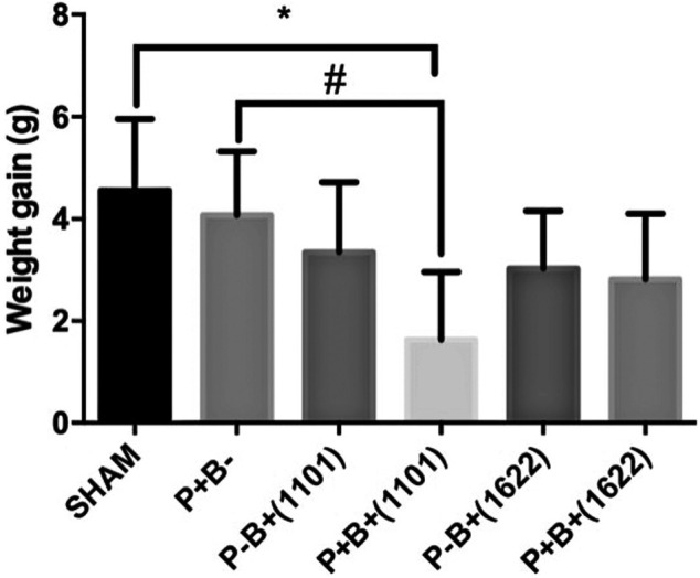
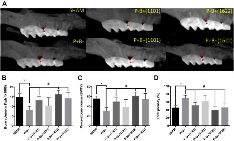
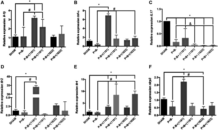
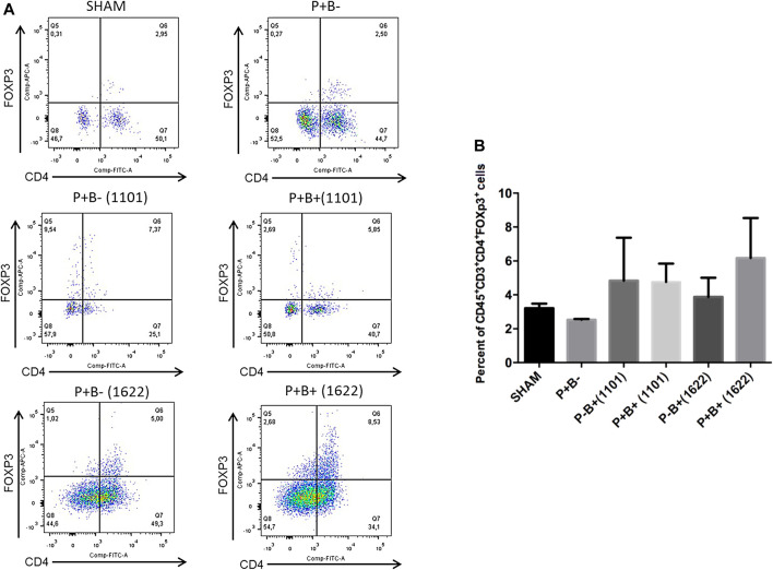
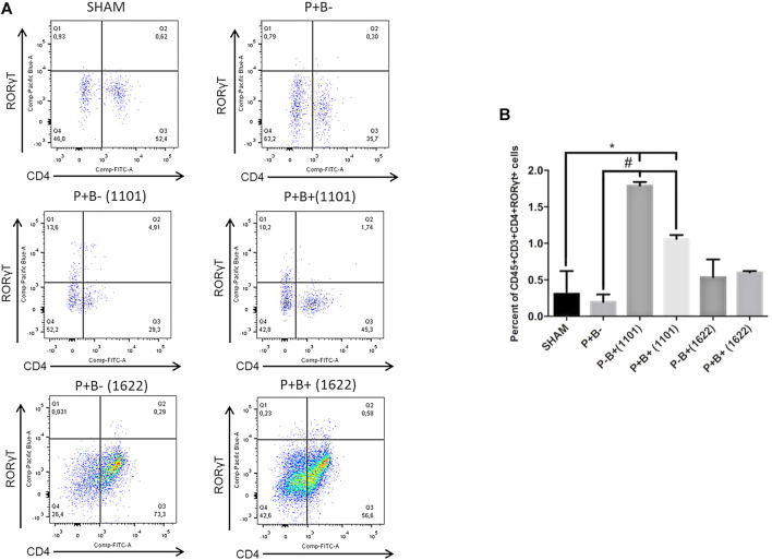
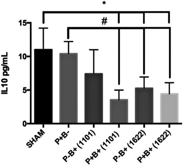
Periodontitis is an inflammatory disease induced by a dysbiotic oral microbiome. Probiotics of the genus Bifidobacterium may restore the symbiotic microbiome and modulate the immune response, leading to periodontitis control. We evaluated the effect of two strains of Bifidobacterium able to inhibit Porphyromonas gingivalis interaction with host cells and biofilm formation, but with distinct immunomodulatory properties, in a mice periodontitis model. Experimental periodontitis (P+) was induced in C57Bl/6 mice by a microbial consortium of human oral organisms. B. bifidum 1622A [B+ (1622)] and B. breve 1101A [B+ (1101)] were orally inoculated for 45 days. Alveolar bone loss and inflammatory response in gingival tissues were determined. The microbial consortium induced alveolar bone loss in positive control (P + B-), as demonstrated by microtomography analysis, although P. gingivalis was undetected in oral biofilms at the end of the experimental period. TNF-α and IL-10 serum levels, and Treg and Th17 populations in gingiva of SHAM and P + B- groups did not differ. B. bifidum 1622A, but not B. breve 1101A, controlled bone destruction in P+ mice. B. breve 1101A upregulated transcription of Il-1β, Tnf-α, Tlr2, Tlr4, and Nlrp3 in P-B+(1101), which was attenuated by the microbial consortium [P + B+(1101)]. All treatments downregulated transcription of Il-17, although treatment with B. breve 1101A did not yield such low levels of transcripts as seen for the other groups. B. breve 1101A increased Th17 population in gingival tissues [P-B+ (1101) and P + B+ (1101)] compared to SHAM and P + B-. Administration of both bifidobacteria resulted in serum IL-10 decreased levels. Our data indicated that the beneficial effect of Bifidobacterium is not a common trait of this genus, since B. breve 1101A induced an inflammatory profile in gingival tissues and did not prevent alveolar bone loss. However, the properties of B. bifidum 1622A suggest its potential to control periodontitis.

Keywords: Bifidobacterium; P. gingivalis; immune modulation; periodontitis; probiotics.


Conflict of interest statement

The authors declare that the research was conducted in the absence of any commercial or financial relationships that could be construed as a potential conflict of interest.

Figures

FIGURE 1
FIGURE 1
Mean and SD of weight gain in grams after 45 days of experimental period of C57Bl/6 mice submitted to different treatments: SHAM (negative control), P + B- (positive control), P-B+ (1101) (B. breve 1101A), P + B+ (1101) (microbial consortium + B. breve 1101A), P-B+ (1622) (B. bifidum 1622A) and P + B+ (1622) (Microbial consortium + B. bifidum 1622A). *Statistically significant difference in relation to negative control (SHAM), # Statistically significant difference in relation to positive control (P + B-). ANOVA, Tukey’s multiple comparison, p <0.05%. Data representative of two independent experiments (n = 8 mice/per group).
FIGURE 2
FIGURE 2
Alveolar bone analysis determined by Microtomography in the interproximal region of first and second M at the right maxilla of C57Bl/6 mice submitted to different treatments for 45 days: SHAM (negative control), P + B- (positive control), P-B+ (1101) (B. breve 1101A), P + B+ (1101) (microbial consortium + B. breve 1101A), P-B+ (1622) (B. bifidum 1622A) and P + B+ (1622) (microbial consortium + B. bifidum 1622A). (A) Representative images of alveolar bone. All data were obtained in the region between the red points. Data on Alveolar bone volume (ABV) (Average and sd) determined in pixels3 (B), Percentage of alveolar bone volume (Average and sd) and (C) Percentage of total porosity (Average and sd) (D) of the different groups. * Statistically significant difference in relation to negative control (SHAM), # Statistically significant difference in relation to positive control (P + B-). ANOVA, Tukey’s multiple comparison, p <0.05%. Data representative of two independent experiments (n = 8 mice/per group).
FIGURE 3
FIGURE 3
Bifidobacteria alter transcription of genes encoding cytokines and receptors for PAMPS in gingival tissues. Relative transcription of Il-1β (A) and Tnf-α (B), Il-17 (C), Tlr2 (D), Tlr4 (E) and Nrlp3 (F), determined by RT-qPCR in gingival tissues of C57Bl/6 mice submitted to different treatments for 45 days of experimental period: SHAM (negative control), P + B- (positive control), P-B + (1101) (B. breve 1101A), P + B+ (1101) (microbial consortium + B. breve 1101A), P-B+ (1622) (B. bifidum 1622A) and P + B+ (1622) (microbial consortium + B. bifidum 1622A). * Statistically significant difference in relation to negative control (SHAM), # Statistically significant difference in relation to positive control (P + B-). & Statistically significant difference in relation to P-B+ (1101). ANOVA, Tukey’s multiple comparison, p <0.05%. Data representative of two independent experiments (n = 8 mice/per group).
FIGURE 4
FIGURE 4
Treg populations remained unchanged in gingival tissues of C57Bl/6 mice submitted to different treatments: SHAM (negative control), P + B- (microbial consortium), P-B+ (1101) (B. breve 1101A), P + B+ (1101) (microbial consortium + B. breve 1101A), P-B+ (1622) (B. bifidum 1622A) and P + B+ (1622) (microbial consortium + B. bifidum 1622A). In (A) representative flow cytometry diagram showing the gating of CD4+, FoxP3+ Treg cells. In (B) Average percentages of CD4+, FoxP3+ Treg cells. No differences among the groups. ANOVA, Tukey’s multiple comparison, p>0.05%. Facs plots represent the results of one of two independent experiments with similar results (n = 2 pooled samples from 4 mice/per group).
FIGURE 5
FIGURE 5
Oral administration of B. breve 1101A increases Th17 population in the gingival tissue. Th17 population in the gingival tissue of C57Bl/6 mice submitted to different treatments: SHAM (negative control), P + B- (microbial consortium), P-B+ (1101) (B. breve 1101A), P + B+ (1101) (microbial consortium + B. breve 1101A), P-B+ (1622) (B. bifidum 1622A) and P + B+ (1622) (microbial consortium + B. bifidum 1622A). In (A) representative flow cytometry diagram showing the gating of CD4+, RORγt+ Th17 cells. In (B) Average percentages of CD4+, RORγt+ Th17 cells. *Statistically significant difference in relation to negative control (SHAM), # Statistically significant difference in relation to positive control (P + B-). ANOVA, Tukey’s multiple comparison, p<0.05%. Facs plots represent the results of one of two independent experiments with similar results (n = 2 pooled samples from 4 mice/per group).
FIGURE 6
FIGURE 6
Serum levels (pg/ml) of IL-10 in C57Bl/6 mice submitted to different treatments for 45 days: SHAM (negative control), P + B- (positive control), P-B+ (1101) (B. breve 1101A), P + B+ (1101) (microbial consortium + B. breve 1101A), P-B+ (1622) (B. bifidum 1622A) and P + B+ (1622) (Microbial consortium + B. bifidum 1622A). * Statistically significant difference in relation to negative control (SHAM), # Statistically significant difference in relation to positive control (P + B-). ANOVA, Tukey’s multiple comparison, p <0.05%. Data representative of two independent experiments (n = 8 mice/per group).

Similar articles

Cited by

References

    1. Abderrazak A., Syrovets T., Couchie D., El Hadri K., Friguet B., Simmet T., et al. (2015). NLRP3 Inflammasome: From a Danger Signal Sensor to a Regulatory Node of Oxidative Stress and Inflammatory Diseases. Redox Biol. 4, 296–307. 10.1016/j.redox.2015.01.008 - DOI - PMC - PubMed
    1. Albuquerque-Souza E., Balzarini D., Ando-Suguimoto E. S., Ishikawa K. H., Simionato M. R. L., Holzhausen M., et al. (2019). Probiotics Alter the Immune Response of Gingival Epithelial Cells Challenged by Porphyromonas gingivalis . J. Periodontal Res. 54 (2), 115–127. 10.1111/jre.12608 - DOI - PubMed
    1. Amano A., Kuboniwa M., Nakagawa I., Akiyama S., Morisaki I., Hamada S. (2000). Prevalence of Specific Genotypes of Porphyromonas gingivalis fimA and Periodontal Health Status. J. Dent Res. 79 (9), 1664–1668. 10.1177/00220345000790090501 - DOI - PubMed
    1. Aral K., Milward M. R., Kapila Y., Berdeli A., Cooper P. R. (2020). Inflammasomes and Their Regulation in Periodontal Disease: A Review. J. Periodontal Res. 55 (4), 473–487. 10.1111/jre.12733 - DOI - PubMed
    1. Aral K., Milward M. R., Cooper P. R. (2021). Inflammasome Dysregulation in Human Gingival Fibroblasts in Response to Periodontal Pathogens. Oral Dis.. [Epub ahead of print]. 10.1111/odi.13760 - DOI - PubMed

LinkOut - more resources