

Protective Role of Vitamin B1 in Doxorubicin-Induced Cardiotoxicity in Rats: Focus on Hemodynamic, Redox, and Apoptotic Markers in Heart
- PMID: 34630136
- PMCID: PMC8494423
- DOI: 10.3389/fphys.2021.690619
Protective Role of Vitamin B1 in Doxorubicin-Induced Cardiotoxicity in Rats: Focus on Hemodynamic, Redox, and Apoptotic Markers in Heart
Abstract
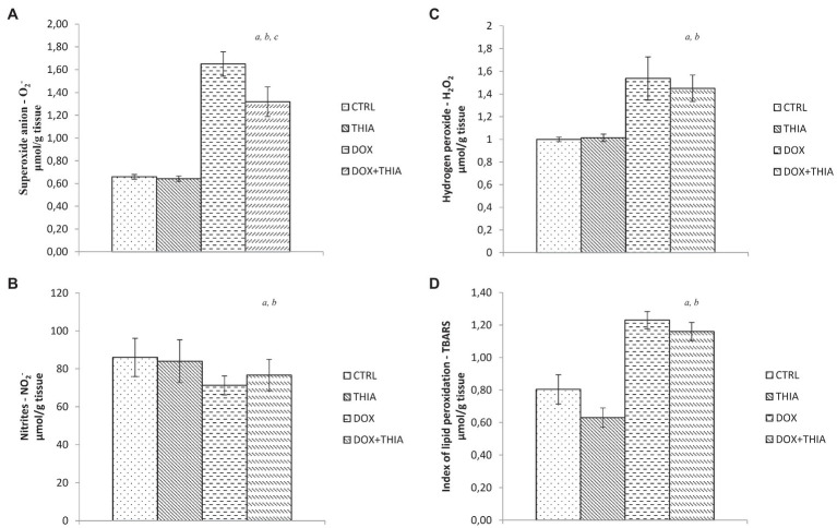
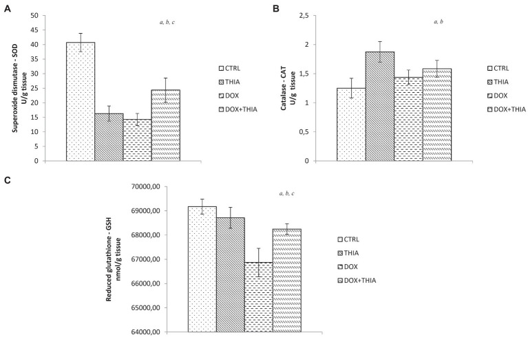
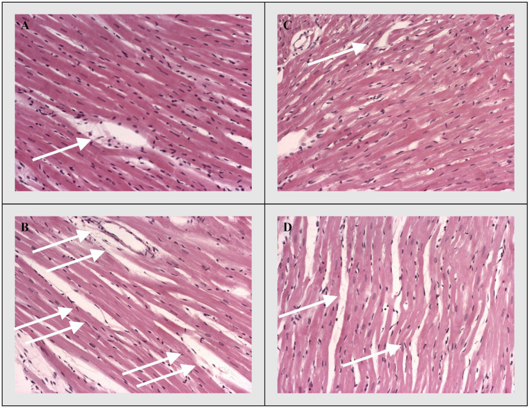
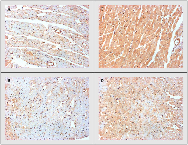
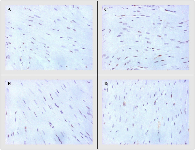
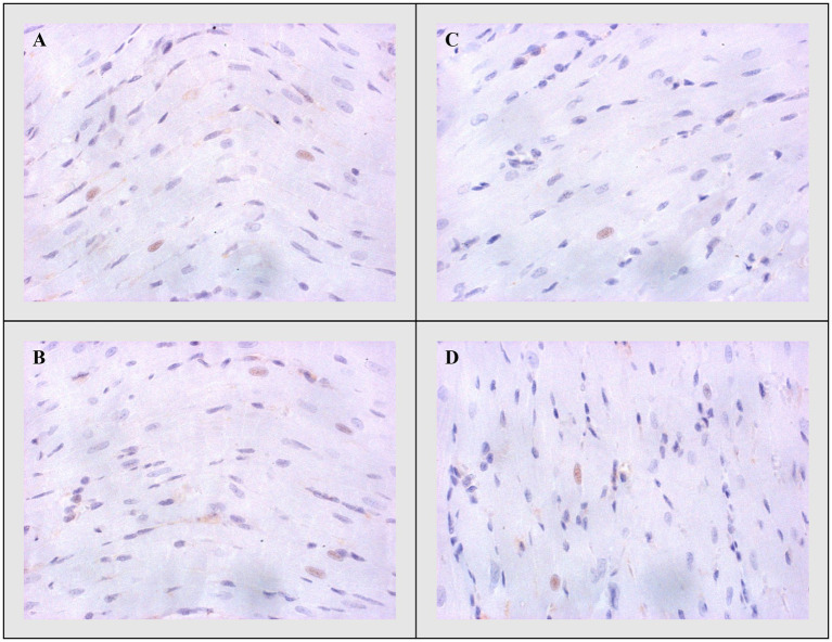
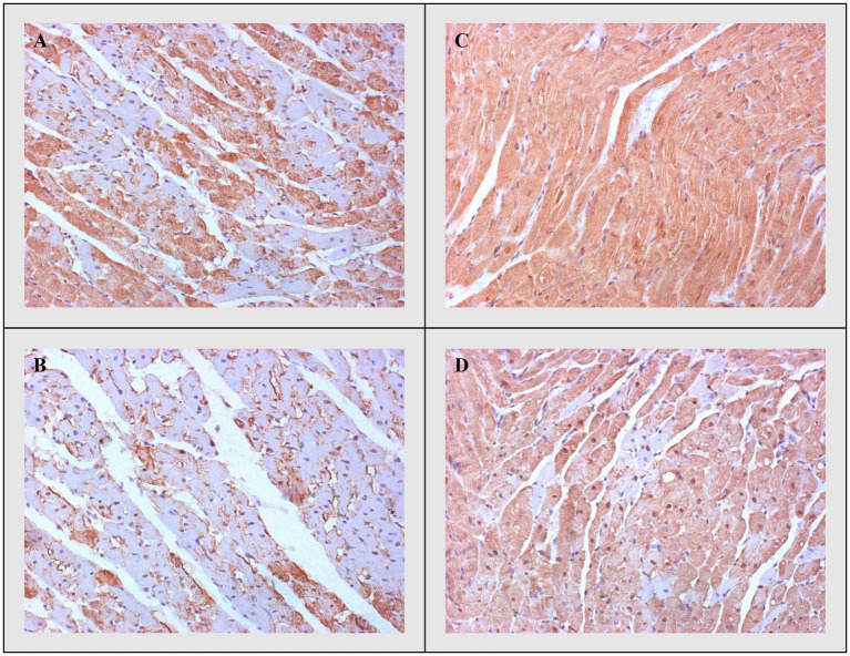
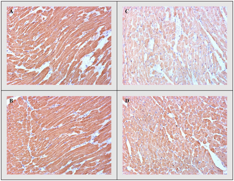
Up until now, the specific mechanisms involved in doxorubicin (DOX)-induced cardiotoxicity have not been fully elucidated. Since thiamine deficiency is associated with myocardial dysfunction and it may lead to cardiomyopathy, we aimed to investigate whether thiamine (Vitamin B1) treatment provides cardioprotection and modulates DOX mediated subchronic cardiotoxicity as well as to determine possible mechanisms of its effects. The study involved 48 Wistar albino rats divided into four groups: healthy non-treated rats and healthy rats treated with thiamine and DOX rats without treatment and DOX rats treated with thiamine. DOX was applied as a single i.p.injection (15mg/kg), while thiamine treatment lasted 7days (25mg/kg/dayi.p.). Before and after the treatment hemodynamic changes were monitored in vivo by echocardiography. When the protocol was completed, animals were sacrificed and rat hearts were isolated in order to evaluate parameters of cardiac oxidative stress [superoxide anion radical-O2 -, hydrogen peroxide-H2O2, nitric oxide-NO-, index of lipid peroxidation-thiobarbituric acid (TBA) reactive substances (TBARS), superoxide dismutase - SOD, catalase (CAT), and reduced glutathione-GSH] and apoptosis (Bax, Bcl-2, caspases). DOX treatment significantly reduced the ejection fraction, while thiamine treatment led to its minor increase in the DOX-treated group. In that sense, heart oxidative stress markers were significantly increased in DOX-treated rats, while therapeutic dose of thiamine decreased the levels of free radicals. Our study demonstrated the promising ameliorative effects of thiamine against DOX-induced cardiotoxicity through modulation of oxidative stress, suppression of apoptosis, and possibility to improve myocardial performance and morphometric structure of rats` hearts.
Keywords: apoptosis; cardiotoxicity; doxorubicin; heart oxidative stress; left ventricular function; rat; thiamine.
Copyright © Rankovic, Draginic, Jeremic, Samanovic, Stojkov, Mitrovic, Jeremic, Radonjic, Srejovic, Bolevich, Svistunov, Jakovljevic and Turnic.
Conflict of interest statement
The authors declare that the research was conducted in the absence of any commercial or financial relationships that could be construed as a potential conflict of interest.
Figures
Similar articles
-
The Effects of Thiamine Hydrochloride on Cardiac Function, Redox Status and Morphometric Alterations in Doxorubicin-Treated Rats.Cardiovasc Toxicol. 2020 Apr;20(2):111-120. doi: 10.1007/s12012-019-09536-7. Cardiovasc Toxicol. 2020. PMID: 31270735
-
α-Linolenic acid attenuates doxorubicin-induced cardiotoxicity in rats through suppression of oxidative stress and apoptosis.Acta Biochim Biophys Sin (Shanghai). 2013 Oct;45(10):817-26. doi: 10.1093/abbs/gmt082. Epub 2013 Jul 28. Acta Biochim Biophys Sin (Shanghai). 2013. PMID: 23896563
-
Chrysin alleviates acute doxorubicin cardiotoxicity in rats via suppression of oxidative stress, inflammation and apoptosis.Eur J Pharmacol. 2014 Apr 5;728:107-18. doi: 10.1016/j.ejphar.2014.01.065. Epub 2014 Feb 6. Eur J Pharmacol. 2014. PMID: 24509133
-
All-trans-retinoic acid ameliorates doxorubicin-induced cardiotoxicity: in vivo potential involvement of oxidative stress, inflammation, and apoptosis via caspase-3 and p53 down-expression.Naunyn Schmiedebergs Arch Pharmacol. 2018 Jan;391(1):59-70. doi: 10.1007/s00210-017-1437-5. Epub 2017 Oct 30. Naunyn Schmiedebergs Arch Pharmacol. 2018. PMID: 29085977
-
Hesperidin alleviates doxorubicin-induced cardiotoxicity in rats.J Egypt Natl Canc Inst. 2009 Jun;21(2):175-84. J Egypt Natl Canc Inst. 2009. PMID: 21057569
Cited by
-
The Pivotal Role of Thiamine Supplementation in Counteracting Cardiometabolic Dysfunctions Associated with Thiamine Deficiency.Int J Mol Sci. 2025 Mar 27;26(7):3090. doi: 10.3390/ijms26073090. Int J Mol Sci. 2025. PMID: 40243711 Free PMC article. Review.
-
Liraglutide Treatment Restores Cardiac Function After Isoprenaline-Induced Myocardial Injury and Prevents Heart Failure in Rats.Life (Basel). 2025 Mar 12;15(3):443. doi: 10.3390/life15030443. Life (Basel). 2025. PMID: 40141787 Free PMC article.
-
Lady's Bedstraw as a Powerful Antioxidant for Attenuation of Doxorubicin-Induced Cardiotoxicity.Antioxidants (Basel). 2023 Jun 15;12(6):1277. doi: 10.3390/antiox12061277. Antioxidants (Basel). 2023. PMID: 37372007 Free PMC article.
-
From Chemistry to Pharmacology: Exploring the Anti-Inflammatory and Antioxidant Potential of Novel Dexketoprofen Amide Derivatives.Antioxidants (Basel). 2025 Jun 27;14(7):796. doi: 10.3390/antiox14070796. Antioxidants (Basel). 2025. PMID: 40722899 Free PMC article.
-
Pediatric Cardio-Oncology: Screening, Risk Stratification, and Prevention of Cardiotoxicity Associated with Anthracyclines.Children (Basel). 2024 Jul 22;11(7):884. doi: 10.3390/children11070884. Children (Basel). 2024. PMID: 39062333 Free PMC article. Review.
References
-
- Atar S., Ye Y., Lin Y., Freeberg S. Y., Nishi S. P., Rosanio S., et al. . (2006). Atorvastatin-induced cardioprotection is mediated by increasing inducible nitric oxide synthase and consequent S-nitrosylation of cyclooxygenase-2. Am. J. Physiol. Heart Circ. Physiol. 290, H1960–H1968. doi: 10.1152/ajpheart.01137.2005, PMID: - DOI - PubMed
-
- Auclair C., Voisin E. (1985). “Nitroblue tetrazolium reduction,” in Handbook of Methods for Oxygen Radical Research. ed. Greenvvald R. A. (Boca Raton: CRC Press, Une; ), 123–132.
-
- Bertinchant J. P., Polge A., Juan J. M., Oliva-Lauraire M. C., Giuliani I., Marty-Double C., et al. . (2003). Evaluation of cardiac troponin I and T levels as markers of myocardial damage in doxorubicin-induced cardiomyopathy rats, and their relationship with echocardiographic and histological findings. Clin. Chim. Acta 329, 39–51. doi: 10.1016/S0009-8981(03)00013-5, PMID: - DOI - PubMed
LinkOut - more resources
Full Text Sources
Research Materials
Miscellaneous