

In vivo Measurement of Intraosseous Vascular Haemodynamic Markers in Human Bone Tissue Utilising Near Infrared Spectroscopy
- PMID: 34630158
- PMCID: PMC8497693
- DOI: 10.3389/fphys.2021.738239
In vivo Measurement of Intraosseous Vascular Haemodynamic Markers in Human Bone Tissue Utilising Near Infrared Spectroscopy
Abstract
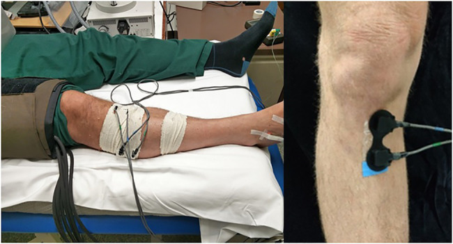
Objective: Poor vascular health is associated with reduced bone strength and increased risk of fragility fracture. However, direct measurement of intraosseous vascular health is difficult due to the density and mineral content of bone. We investigated the feasibility of using a commercially available continuous wave near infrared spectroscopy (NIRS) system for the investigation of vascular haemodynamics in human bone in vivo. Approach: An arterial occlusion (AO) protocol was developed for obtaining haemodynamic measurements of the proximal tibia and lateral calf, including assessment of the protocol's intra operator reproducibility. For 36 participants, intraosseous haemodynamics derived by NIRS were compared to alternative tests of bone health based on dual x-ray absorptiometry (DXA) testing and MRI. Main Results: Near infrared spectroscopy markers of haemodynamics of the proximal tibia demonstrated acceptable reproducibility, comparable with reproducibility assessments of alternative modalities measuring intraosseous haemodynamics, and the use of NIRS for measuring muscle. Novel associations have been demonstrated between haemodynamic markers of bone measured with NIRS and body composition and bone mineral density (BMD) measurements obtained with both DXA and MRI. Significance: Near infrared spectroscopy provides inexpensive, non-invasive, safe, and real time data on changes in oxygenated and deoxygenated haemoglobin concentration in bone at the proximal tibia. This study has demonstrated the potential for NIRS to contribute to research investigating the pathophysiological role of vascular dysfunction within bone tissue, but also the limitations and need for further development of NIRS technology.
Keywords: bone; haemodynamic analysis; near infrared spectroscopy; tibia; vascular physiology.
Copyright © 2021 Meertens, Knapp, Strain, Casanova, Ball, Fulford and Thorn.
Conflict of interest statement
The authors declare that the research was conducted in the absence of any commercial or financial relationships that could be construed as a potential conflict of interest.
Figures


Similar articles
-
Measuring tibial hemodynamics and metabolism at rest and after exercise using near-infrared spectroscopy.Appl Physiol Nutr Metab. 2021 Nov;46(11):1354-1362. doi: 10.1139/apnm-2021-0135. Epub 2021 May 21. Appl Physiol Nutr Metab. 2021. PMID: 34019778
-
Near-infrared spectroscopy (NIRS): a non-invasive in vivo methodology for analysis of brain vascular and metabolic activities in real time in rodents.Curr Vasc Pharmacol. 2007 Oct;5(4):305-21. doi: 10.2174/157016107782023398. Curr Vasc Pharmacol. 2007. PMID: 17979797
-
Assessment of Bone Mineral Density at the Distal Femur and the Proximal Tibia by Dual-Energy X-ray Absorptiometry in Individuals With Spinal Cord Injury: Precision of Protocol and Relation to Injury Duration.J Clin Densitom. 2018 Jul-Sep;21(3):338-346. doi: 10.1016/j.jocd.2017.05.006. Epub 2017 Jun 26. J Clin Densitom. 2018. PMID: 28662973
-
The Case for Measuring Long Bone Hemodynamics With Near-Infrared Spectroscopy.Front Physiol. 2020 Dec 18;11:615977. doi: 10.3389/fphys.2020.615977. eCollection 2020. Front Physiol. 2020. PMID: 33391034 Free PMC article. Review.
-
DXA in vivo BMD methodology: an erroneous and misleading research and clinical gauge of bone mineral status, bone fragility, and bone remodelling.Bone. 2007 Jul;41(1):138-54. doi: 10.1016/j.bone.2007.02.022. Epub 2007 Mar 1. Bone. 2007. PMID: 17481978 Review.
Cited by
-
Near-Infrared Spectroscopy Allows for Monitoring of Bone Fracture Healing via Changes in Oxygenation.J Funct Biomater. 2024 Dec 19;15(12):384. doi: 10.3390/jfb15120384. J Funct Biomater. 2024. PMID: 39728184 Free PMC article.
References
-
- Alneami A. I. (2015). Measuring Blood Perfusion in Bone Using NIRS (Bone Optical Spectroscopy). Boston, Massachusetts: Northeastern University.
-
- Bakker A., Smith B., Ainslie P., Smith K. (2012). Near-Infrared Spectroscopy, in Applied Aspects of Ultrasonography in Humans. London, UK: IntechOpen.
-
- Binzoni T., Blanchi S., Fasel J. H., Bounameaux H., Hiltbrand E., Delpy D. (2002). Human tibia bone marrow blood perfusion by non-invasive near infrared spectroscopy: a new tool for studies on microgravity. J. Gravit. Physiol. 9, P183–P184. PMID: - PubMed
LinkOut - more resources
Full Text Sources