. 2021 Nov;22(5):1291.
doi: 10.3892/etm.2021.10726. Epub 2021 Sep 13.

Interleukin-17 activates JAK2/STAT3, PI3K/Akt and nuclear factor-κB signaling pathway to promote the tumorigenesis of cervical cancer

Affiliations

Interleukin-17 activates JAK2/STAT3, PI3K/Akt and nuclear factor-κB signaling pathway to promote the tumorigenesis of cervical cancer

Yanfei Bai et al. Exp Ther Med. 2021 Nov.

Abstract

Interleukin (IL)-17 has been regarded as a significant factor in inflammation. In addition, IL-17 is known to be involved in the progression of cancers; however, the function of IL-17 in cervical cancer remains unclear. In the present study, cell viability was detected by Cell Counting Kit-8 assay. Quantitative PCR and western blotting were performed to detect gene and protein expression levels, respectively, in cancer cells or tissues. Ki-67 staining was used to evaluate cell proliferation. Wound-healing assay was used to detect cell migration. Moreover, Transwell assay was performed to investigate the invasion of cervical cancer cells. The results revealed that IL-17 significantly promoted the proliferation of cervical cancer cells. Additionally, IL-17 notably enhanced the migration and invasion of cervical cancer cells in vitro. IL-17 promoted the progression of cervical cancer via the activation of JAK2/STAT3 and PI3K/Akt/NF-κB signaling. In conclusion, IL-17 was a key regulator during the progression of cervical cancer through the JAK2/STAT3 and PI3K/Akt/nuclear factor-κB signaling pathway, which may serve as a novel target for the treatment of cervical cancer.

Keywords: JAK2/STAT3; PI3K/Akt; cervical cancer; interleukin-17; nuclear factor-κB.


Conflict of interest statement

The authors declare that they have no competing interests.

Figures

Figure 1
Figure 1
IL-17 is upregulated in cervical cancer tissues. (A) Reverse transcription-quantitative PCR was used to detect the expression level of IL-17 in cervical cancer tissue or adjacent normal tissues. **P<0.01 via paired Student's t-test. (B) Enzyme-linked immunosorbent assay was performed to detect the level of IL-17 in the serum of patients with cervical cancer or healthy individuals. **P<0.01 via unpaired Student's t-test. IL, interleukin.
Figure 2
Figure 2
JAK2/STAT3, NF-κB, VEGF and PI3K are involved in tumorigenesis of cervical cancer. (A) Protein expression levels of JAK2, p-JAK2, STAT3, p-STAT3, p-p65, Akt, p-Akt and VEGF in tissues detected by western blotting. Relative expression levels of (B) p-JAK2 (normalized to JAK2), (C) p-STAT3 (normalized to STAT3), (E) p-p65 (normalized to p65) and (F) p-Akt (normalized to Akt) were quantified. Relative expression level of (D) VEGF was normalized to GAPDH. **P<0.01 vs. control. The data were analyzed using paired Student's t-test. NF-κB, nuclear factor-κB; VEGF, vascular endothelial growth factor; p-, phosphorylated.
Figure 3
Figure 3
IL-17 promotes the proliferation of cervical cancer cells. Cells were treated with different concentrations of IL-17 for 24-96 h. Then, (A) cell viability was detected by Cell Counting Kit-8 assay. (B-D) HeLa cells were transfected with JAK2 siRNA or STAT siRNA for 24 h. Then, reverse transcription-quantitative PCR and western blotting were performed to detect the transfection efficiency. (E) HeLa cells were untreated or with IL-17, IL-17 plus siNC or IL-17 plus siSTAT3 for 24 h. Then, cell proliferation was detected by Ki-67 staining. Red fluorescence indicates Ki-67. Blue fluorescence indicates DAPI. Scale bar, 100 µm. **P<0.01 vs. control; ##P<0.01 vs. IL-17. IL, interleukin; siRNA, small interfering RNA; NC, negative control.
Figure 4
Figure 4
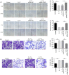
IL-17 significantly promotes the migration and invasion of cervical cancer cells. (A) Migration of HeLa cells was detected by wound-healing assay. Scale bar, 100 µm. (B) Invasion of HeLa cells was tested by Transwell assay. Scale bar, 100 µm. **P<0.01. IL, interleukin; siRNA, small interfering RNA; NC, negative control.
Figure 5
Figure 5
IL-17 promotes the tumorigenesis of cervical cancer via upregulation of IL-17A and IL-17F. (A and B) HeLa cells were transfected with JAK2 siRNA or STAT3 siRNA. The expression levels of IL-17A and IL-17F were detected in HeLa cells by western blotting. The relative expression levels were quantified by normalization to GAPDH. **P<0.01. IL, interleukin; siRNA, small interfering RNA; NC, negative control.
Figure 6
Figure 6
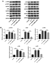
IL-17 promotes the progression of cervical cancer through JAK2/STAT3, PI3K/Akt and NF-κB signaling. (A and B) HeLa cells were transfected with JAK2 siRNA. Then, the protein expression of p65, p-p65 (normalized to p65), STAT3, p-STAT3 (normalized to STAT3), Akt, p-Akt (normalized to Akt), JAK2, p-JAK2 (normalized to JAK2) and VEGF in HeLa cells were detected by western blotting. The relative expression of VEGF was quantified normalized to GAPDH. **P<0.01. IL, interleukin; NF-κB, nuclear factor NF-κB; p-, phosphorylated-; siRNA, small interfering RNA; VEGF, vascular endothelial growth factor; NC, negative control.
Figure 7
Figure 7
STAT3 siRNA reversed the effect of IL-17 on JAK2/STAT3, PI3K/Akt and NF-κB signaling. (A and B) HeLa cells were transfected with STAT3 siRNA. Then, the protein expression of p65, p-p65 (normalized to p65), STAT3, p-STAT3 (normalized to STAT3), Akt, p-Akt (normalized to Akt), JAK2, p-JAK2 (normalized to JAK2) and VEGF in HeLa cells were detected by western blotting. The relative expression of VEGF was quantified normalized to GAPDH. **P<0.01. IL, interleukin; NF-κB, nuclear factor NF-κB; p-, phosphorylated-; siRNA, small interfering RNA; VEGF, vascular endothelial growth factor; NC, negative control.

References

    1. Tsikouras P, Zervoudis S, Manav B, Tomara E, Iatrakis G, Romanidis C, Bothou A, Galazios G. Cervical cancer: Screening, diagnosis and staging. J BUON. 2016;21:320–325. - PubMed
    1. Benard VB, Thomas CC, King J, Massetti GM, Doria-Rose VP, Saraiya M. Vital signs: Cervical cancer incidence, mortality, and screening-United States, 2007-2012. MMWR Morb Mortal Wkly Rep. 2014;63:1004–1009. Centers for Disease C and Prevention (CDC) - PMC - PubMed
    1. Zeng SY, Liang MR, Li LY, Li L, Jiang W, Zhong ML. Application of transvaginal external fascia trachelectomy in the treatment of CIN and micro-invasive cervical cancer. Zhonghua Zhong Liu Za Zhi. 2013;35:543–546. (In Chinese) - PubMed
    1. Gilham C, Sargent A, Kitchener HC, Peto J. HPV testing compared with routine cytology in cervical screening: Long-term follow-up of ARTISTIC RCT. Health Technol Assess. 2019;23:1–44. doi: 10.3310/hta23280. - DOI - PMC - PubMed
    1. Hong DK, Kim SA, Lim KT, Lee KH, Kim TJ, So KA. Clinical outcome of high-grade cervical intraepithelial neoplasia during pregnancy: A 10-year experience. Eur J Obstet Gynecol Reprod Biol. 2019;236:173–176. doi: 10.1016/j.ejogrb.2019.03.023. - DOI - PubMed