

Comprehensive review on the pathophysiology, clinical variants and management of pemphigus (Review)
- PMID: 34630689
- PMCID: PMC8495539
- DOI: 10.3892/etm.2021.10770
Comprehensive review on the pathophysiology, clinical variants and management of pemphigus (Review)
Abstract
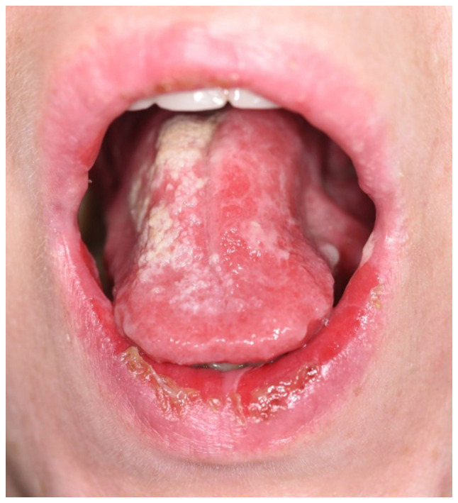
Pemphigus represents a group of chronic inflammatory disorders characterized by autoantibodies that target components of desmosomes, leading to the loss of intercellular adhesion between keratinocytes and causing intraepithelial blistering. The pemphigus group consists of four main clinical types with several variants: pemphigus vulgaris (with pemphigus vegetans and pemphigus herpetiformis as variants), pemphigus foliaceus, paraneoplastic pemphigus and IgA pemphigus (with two clinical variants: intraepidermal neutrophilic IgA dermatosis and subcorneal pustular dermatosis). Genetic factors are involved in the pathogenesis, with HLA-DR4 (DRB1*0402) and HLA-DRw6 (DQB1*0503) allele more common in patients with pemphigus vulgaris, HLA class II DRB1*0344 and HLA Cw*1445 correlated with paraneoplastic pemphigus, and HLA-DRB1*04:01, HLA-DRB1*04:06, HLA-DRB1*01:01, HLA-DRB1*14, associated with a higher risk of developing pemphigus foliaceus. Autoantibodies are conducted against structural desmosomal proteins in the skin and mucous membranes, mainly desmogleins, desmocollins and plakins. Cell-mediated immunity may also play a role, especially in paraneoplastic pemphigus. Patients may present erythema, blisters, erosions, and ulcers that may affect the skin, as well as mucosal surfaces of the oral cavity, eyes, nose, leading to severe complaints including pain, dysphagia, and fetor. Oral mucosal postbullous erosive lesions are frequently the first sign of disease in pemphigus vulgaris and in paraneoplastic pemphigus, without skin involvement, making the diagnosis difficult. Treatment options classically include immunosuppressive agents, such as corticosteroids and corticosteroid-sparing agents such as azathioprine, mycophenolate mofetil, cyclophosphamide, methotrexate or dapsone. Newer therapies focus on blocking cell signaling events induced by pathogenic autoantibodies and/or targeting specific autoantibodies. The disease evolution is conditioned by the treatment with maximum doses of corticosteroids and the side effects associated with long-term immunosuppressive therapy, which is why patients need a multidisciplinary approach in following the treatment. In this review, we provide a comprehensive overview of the epidemiology, pathophysiology, clinical aspect, diagnosis and management of the main intraepidermal blistering diseases from the pemphigus group.
Keywords: IgA pemphigus; paraneoplastic pemphigus; pemphigus foliaceus; pemphigus herpetiformis; pemphigus vegetans; pemphigus vulgaris.
Copyright: © Costan et al.
Conflict of interest statement
The authors declare that they have no competing interests and they have no financial relationships to disclose.
Figures


References
-
- Bickle K, Roark TR, Hsu S. Autoimmune bullous dermatoses: A review. Am Fam Physician. 2002;65:1861–1870. - PubMed
Publication types
LinkOut - more resources
Full Text Sources
Research Materials
Miscellaneous