Case Reports
. 2021 Oct 2;16(12):3643-3646.
doi: 10.1016/j.radcr.2021.08.075. eCollection 2021 Dec.

Mucoepidermoid carcinoma of the lacrimal gland in a patient with the CRTC1-MAML2 fusion gene

Affiliations
Case Reports

Mucoepidermoid carcinoma of the lacrimal gland in a patient with the CRTC1-MAML2 fusion gene

Kensaku Makino et al. Radiol Case Rep. .

Abstract

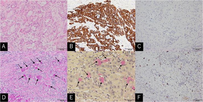
Mucoepidermoid carcinoma (MEC) of the lacrimal gland (LG) is a rare entity. A 47-year-old woman was aware of periorbital swelling for 3 months. At presentation, the patient showed periorbital swelling in the right eye. CT scan showed an isodense mass in the anterior superolateral part of the orbit. MRI delineated the mass as enhancing, extra-conal tumor appearing isointense on T1-weighted sequences, and to be of mixed intensity on T2-weighted sequences. The tumor was totally resected. Microscopically, the tumor tissue was comprised of squamous, epithelioid cells, and cells with plump and clear cytoplasm. Necrosis, neural invasion, or mitotic figures were not observed. Immunohistochemical examination revealed intense staining for cytokeratin 7. A subset of the cells was positively stained with periodic acid-Schiff and mucicarmine stains. Genetic analysis revealed the presence of the CRTC1-MAML2 fusion. The CRTC1-MAML2 fusion may be a useful indicator for the prognosis and planning of adjuvant therapy.

Keywords: CRTC1-MAML2 fusion gene; Lacrimal gland tumor; Mucoepidermoid carcinoma.


Figures

Fig 1
Fig. 1
Non–contrast axial (A) and coronal (B) CT scans showing an isodense mass occupying the superolateral part of the right anterior orbit and posterolateral aspect of the eyeball (A, asterisk). Note that erosive changes are not found in the orbital wall adjacent to the mass (B, arrows).
Fig 2
Fig. 2
Non–contrast T1-weighted images in the axial (A) and coronal (D) planes. Non–contrast T2-weighted images in the axial plane (B). Post-contrast T1-weighted images in the axial (C) and coronal (E) planes. MRI images show an intensely enhancing, extra-conal tumor appearing isointense on T1-weighted sequences and to be of mixed intensity on T2-weighted sequences (A-E, asterisk), with the displaced lateral rectus muscle in the inferomedial direction (D and E, arrow). ON, optic nerve; SRM, superior rectus muscle.
Fig 3
Fig. 3
Intraoperative photos showing the following: 1. completion of lateral orbitotomy, 2. early stage of tumor resection, 3. tumor being dissected from the lacrimal gland, and 4. completion of tumor resection. A, anterior; I, inferior; LG, lacrimal gland; P, posterior; PO, periorbita; S, superior; T, tumor; TP, titanium plate; Arrows in 3: threads hung to the lateral rectus muscle near the attachment site of the eyeball that allow intraoperative identification of the muscle when pulled.
Fig 4
Fig. 4
Photomicrographs of the tumor tissue are mainly comprised of squamous, epithelioid cells and cells that have plump and clear cytoplasms. The latter cells proliferate in alveolar patterns or form gland-like structures. Necrosis and mitotic figures are not found (A: hematoxylin and eosin stain). Immunohistochemical examination showing intense staining for cytokeratin 7 (B) and negative staining for cytokeratin 20 (C). Some tumor cells with clear cytoplasms are stained with periodic acid–Schiff (D, arrows) and mucicarmine (E, arrows) stains. The MIB-1 labelling index is accounted to be 8% (F).
Fig 5
Fig. 5
Results of immunoelectrophoresis (A) and direct sequencing (B) showing the presence of the CRTC1-MAML2 fusion (A, yellow arrows and B) and the absence of the CRTC3-MAML2 fusion (A, white arrows) (Color version of the figure is available online.)

References

    1. Tailor TD, Gupta D, Dalley RW, Keene CD, Anzai Y. Orbital neoplasms in adults: clinical, radiologic, and pathologic review. Radiographics. 2013;33(6):1739–1758. - PubMed
    1. Jung WS, Ahn KJ, Park MR, Kim JY, Choi JJ, Kim BS. The radiological spectrum of orbital pathologies that involve the lacrimal gland and the lacrimal fossa. Korean J Radiol. 2007;8(4):336–342. - PMC - PubMed
    1. Shields CL, Shields JA. Lacrimal gland tumors. Int Ophthalmol Clin. 1993;33(3):181–188. - PubMed
    1. Sofinski SJ, Brown BZ, Rao N, Wan WL. Mucoepidermoid carcinoma of the lacrimal gland. Case report and review of the literature. Ophthalmic Plast Reconstr Surg. 1986;2(3):147–151. - PubMed
    1. Yuksel D, Kosker M, Saribas F, Simsek S. Surgical treatment of mucoepidermoid carcinoma of the lacrimal sac. Semin Ophthalmol. 2014;29(2):70–72. - PubMed

Publication types