

Melanocytic or Not? Dermoscopy and Reflectance Confocal Microscopy for Lesions Difficult to Diagnose: A Cross-Sectional Diagnostic Accuracy Study
- PMID: 34631270
- PMCID: PMC8480465
- DOI: 10.5826/dpc.1104a127
Melanocytic or Not? Dermoscopy and Reflectance Confocal Microscopy for Lesions Difficult to Diagnose: A Cross-Sectional Diagnostic Accuracy Study
Abstract
Background: Different techniques for non-invasive skin examination and early diagnosis of skin lesions are available nowadays, being dermoscopy and reflectance confocal microscopy (RCM) the most diffused ones. Several studies supported the complementary use of dermoscopy and RCM that improves diagnostic accuracy when dealing with melanocytic lesions.
Objectives: To analyze RCM diagnostic accuracy in the differential diagnosis between melanocytic and non-melanocytic lesions.
Methods: This is a cohort selected cross-sectional study conducted at the Dermatology Unit of the University of Campania L. Vanvitelli, Naples, Italy, from 2012 to 2020. We searched the image database for all excised lesions for which the clinical and dermatoscopic differential diagnosis was between melanocytic and non-melanocytic and for which an RCM examination was performed. Sensitivity, specificity, and diagnostic accuracy values were estimated.
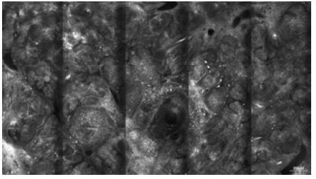
Results: The study included 53 cases that were found to have disagreement between clinical, histological and RCM diagnosis, of which, in 31 cases the differential diagnosis was melanocytic vs non-melanocytic lesion. The RCM reached a specificity of 87% (95% CI: 0.73-1) and a sensitivity of 62.5% (95% CI: 0.29-0.96) in the present sample. Diagnostic accuracy was 80.6% (95% CI: 0.67-0.94).
Conclusion: RCM has a high specificity in differentiating between difficult-to-diagnose melanocytic and non-melanocytic lesions.
Keywords: dermoscopy; diagnosis; melanocytic; reflectance confocal microscopy; skin cancer.
©2021 Scharf et al.
Conflict of interest statement
Competing interests: None.
Figures
Similar articles
-
Role of In Vivo Reflectance Confocal Microscopy in the Analysis of Melanocytic Lesions.Acta Dermatovenerol Croat. 2018 Apr;26(1):64-67. Acta Dermatovenerol Croat. 2018. PMID: 29782304 Review.
-
Reflectance confocal microscopy for diagnosing cutaneous melanoma in adults.Cochrane Database Syst Rev. 2018 Dec 4;12(12):CD013190. doi: 10.1002/14651858.CD013190. Cochrane Database Syst Rev. 2018. PMID: 30521681 Free PMC article.
-
Dynamic dermoscopic and reflectance confocal microscopic changes of melanocytic lesions excised during follow up.J Am Acad Dermatol. 2022 May;86(5):1049-1057. doi: 10.1016/j.jaad.2021.03.081. Epub 2021 Apr 3. J Am Acad Dermatol. 2022. PMID: 33823198
-
Seborrheic keratoses mimicking melanoma unveiled by in vivo reflectance confocal microscopy.Skin Res Technol. 2018 May;24(2):285-293. doi: 10.1111/srt.12427. Epub 2018 Jan 24. Skin Res Technol. 2018. PMID: 29363175
-
The diagnostic accuracy of dermoscopy and reflectance confocal microscopy for amelanotic/hypomelanotic melanoma: a systematic review and meta-analysis.Br J Dermatol. 2020 Aug;183(2):210-219. doi: 10.1111/bjd.18722. Epub 2019 Dec 22. Br J Dermatol. 2020. PMID: 31747045
Cited by
-
Advancements in Basal Cell Carcinoma Diagnosis: Non-Invasive Imaging and Multimodal Approach.J Clin Med. 2023 Dec 20;13(1):39. doi: 10.3390/jcm13010039. J Clin Med. 2023. PMID: 38202046 Free PMC article.
-
Confocal Assessment of Pigmented-Mucosal Lesions: A Monocentric, Retrospective Evaluation of Lip and Genital Area.Dermatol Pract Concept. 2024 Jan 1;14(1):e2024028. doi: 10.5826/dpc.1401a28. Dermatol Pract Concept. 2024. PMID: 38364417 Free PMC article.
-
Editorial: Multidisciplinary Approach to the Diagnosis and Therapy of Skin Neoplasms.Front Oncol. 2022 Jul 14;12:939200. doi: 10.3389/fonc.2022.939200. eCollection 2022. Front Oncol. 2022. PMID: 35912237 Free PMC article. No abstract available.
-
Inflammatory Linear Verrucous Epidermal Nevus Assessment With in Vivo Reflectance Confocal Microscopy: an Easy Option to Consider.Dermatol Pract Concept. 2023 Apr 1;13(2):e2023107. doi: 10.5826/dpc.1302a107. Online ahead of print. Dermatol Pract Concept. 2023. PMID: 37196255 Free PMC article. No abstract available.
References
LinkOut - more resources
Full Text Sources