

Optical Coherence Tomography Angiography of Early Stage 1a Retinal Hemangioblastoma in Von-Hippel-Lindau
- PMID: 34631390
- PMCID: PMC8476344
- DOI: 10.15586/jkcvhl.v8i3.158
Optical Coherence Tomography Angiography of Early Stage 1a Retinal Hemangioblastoma in Von-Hippel-Lindau
Abstract
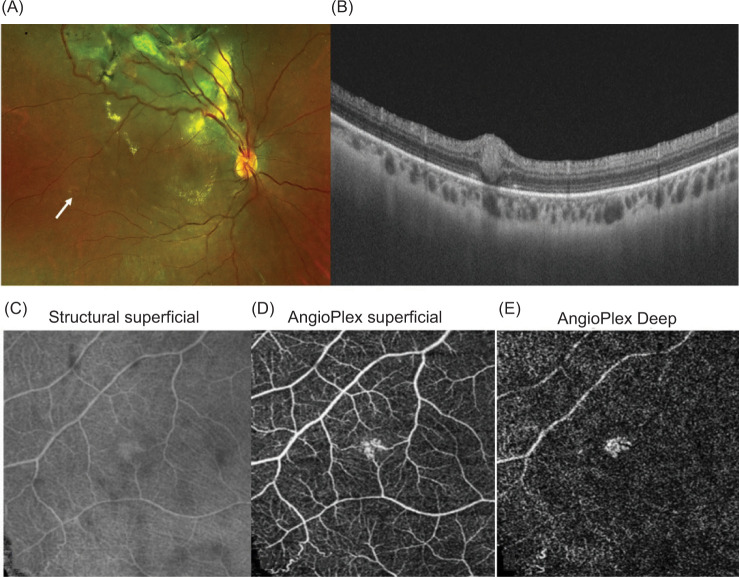
Von-Hippel-Lindau (VHL) syndrome is characterized by focal vasoproliferative tumors of retinal capillaries called retinal capillary hemangioblastomas (RCH). These tumors are initially small and can be easily missed if not looked for carefully. As they grow, these tumors are more demanding to treat and hence the importance of detecting them early and treating them. Herein, we describe and review the optical coherence tomography angiography (OCTA) of the early- stage lesion, which suggested the involvement of superficial and a deeper retinal capillary plexus. In addition, to helping us detect these lesions earlier, OCTA may also help to understand the in vivo changes occurring at an earlier phase.
Keywords: OCTA; VHL; hemangioblastoma; retinal capillary hemangioblastoma.
Copyright: Pradeep V, et al.
Conflict of interest statement
The authors declare no potential conflicts of interest with respect to research, authorship, and/or publication of this article. The manuscript is not being considered for publication elsewhere and has been reviewed and approved by all named authors. The criteria for authorship have been met, and each author believes that the manuscript represents honest work.
Figures
Similar articles
-
Comparison of optical coherence tomography angiography and fundus fluorescein angiography features of retinal capillary hemangioblastoma.Indian J Ophthalmol. 2018 Jun;66(6):872-876. doi: 10.4103/ijo.IJO_1199_17. Indian J Ophthalmol. 2018. PMID: 29786009 Free PMC article.
-
Value of Optical Coherence Tomography Angiography Imaging in Diagnosis and Treatment of Hemangioblastomas in von Hippel-Lindau Disease.Ophthalmic Surg Lasers Imaging Retina. 2016 Oct 1;47(10):935-946. doi: 10.3928/23258160-20161004-07. Ophthalmic Surg Lasers Imaging Retina. 2016. PMID: 27759860
-
Combined therapy guided by multimodal imaging of fifteen retinal capillary hemangioblastomas in a monocular Von Hippel- Lindau syndrome case report.BMC Ophthalmol. 2022 May 6;22(1):205. doi: 10.1186/s12886-022-02409-8. BMC Ophthalmol. 2022. PMID: 35524216 Free PMC article.
-
Von Hippel-Lindau Disease and the Eye.J Ophthalmic Vis Res. 2020 Feb 2;15(1):78-94. doi: 10.18502/jovr.v15i1.5950. eCollection 2020 Jan-Mar. J Ophthalmic Vis Res. 2020. PMID: 32095212 Free PMC article. Review.
-
[Retinal angiomatosis. Ocular manifestation of von Hippel-Lindau disease].Ophthalmologe. 2007 Feb;104(2):107-13. doi: 10.1007/s00347-006-1477-6. Ophthalmologe. 2007. PMID: 17219178 Review. German.
Cited by
-
Role of optical coherence tomography angiography in retinal tumors: A narrative review.Indian J Ophthalmol. 2024 Aug 1;72(8):1082-1090. doi: 10.4103/IJO.IJO_29_24. Epub 2024 Jul 29. Indian J Ophthalmol. 2024. PMID: 39078951 Free PMC article. Review.
References
-
- Apple DJ, Rabb MF. Ocular pathology: Clinical applications and self-assessment. St. Louis: Mosby; 1998.
LinkOut - more resources
Full Text Sources
Research Materials