

Articular cartilage repair & joint preservation: A review of the current status of biological approach
- PMID: 34631411
- PMCID: PMC8488240
- DOI: 10.1016/j.jcot.2021.101602
Articular cartilage repair & joint preservation: A review of the current status of biological approach
Abstract
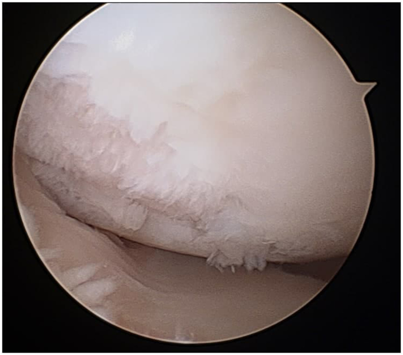
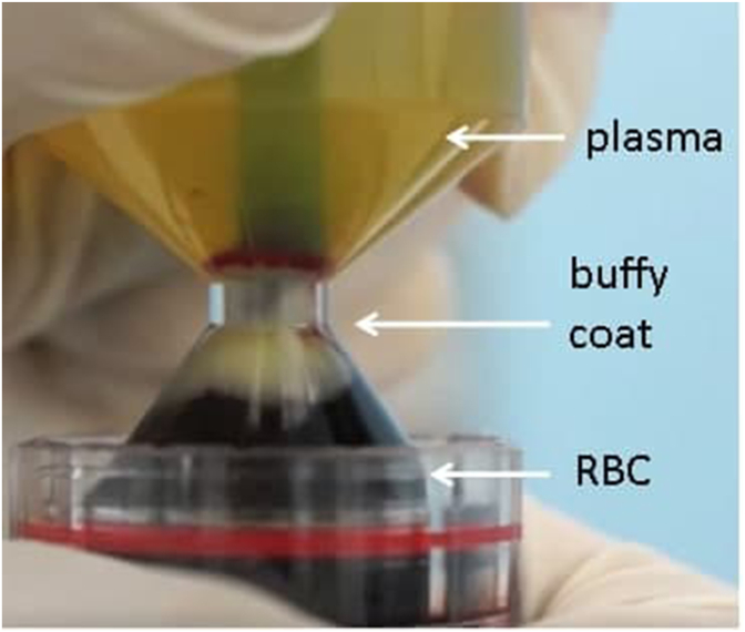
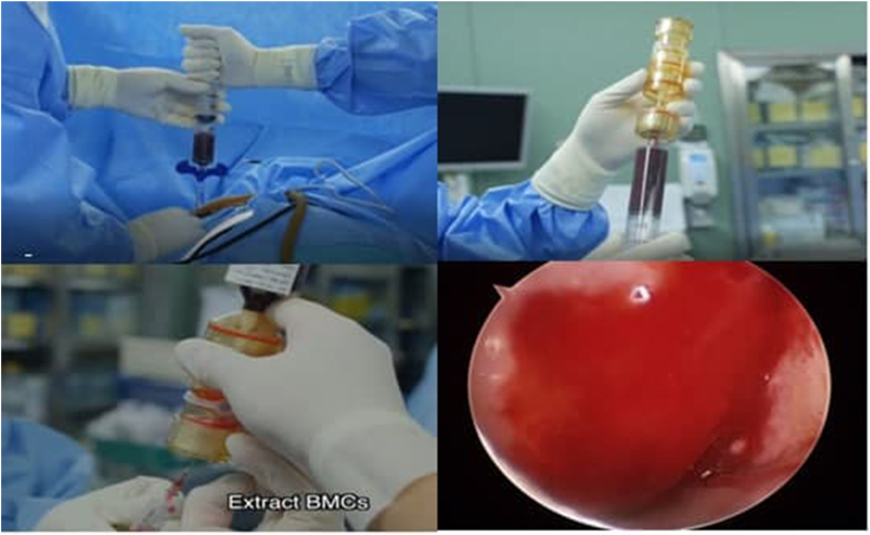
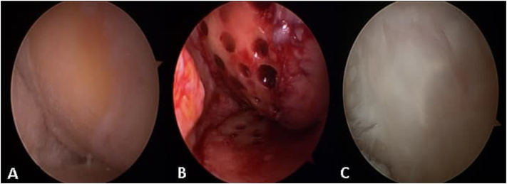
The articular cartilage of the joint is the thin viscoelastic layer of the connective tissue. It has a unique anatomy and physiology, which makes the repair of the articular cartilage damage more difficult and challenging due to its limited healing capacity. Increasing knowledge regarding the importance of articular cartilage for joint preservation has led to increased attention on early identification of cartilage damage as well as degeneration in order to delay osteoarthritis. There are various treatment modalities ranging from preventive management, physical therapy, pharmacological, non-pharmacological and surgical treatments exist in current literature. However most of the studies have limited long term follow up and mainly consists of small case series and case reports. This is an up to date concise review discussing the available management options for articular cartilage damage starting to lifestyle modification to pharmacotherapy, physiotherapy, and osteobiologics till various joint preservation techniques that have been in use currently.
Keywords: Arthroscopy; Articular cartilage lesions; Joint preservation; Orthobiologics; Osteoarthritis.
© 2021 Delhi Orthopedic Association. All rights reserved.
Figures
References
-
- Lories R.J., Luyten F.P. The bone–cartilage unit in osteoarthritis. Nat Rev Rheumatol. 2011;7:43–49. - PubMed
-
- Moran C.J. Restoration of articular cartilage. J Bone Joint Surg Am. 2014;96:336–344. [PubMed: 24553893] - PubMed
-
- Mow V.C., Ratcliffe A., Poole A.R. Cartilage and diarthrodial joints as paradigms for Hierarchical materials and structures. Biomaterials. 1992;13(2):67–97. - PubMed
-
- Roth V., Mow V.C. The intrinsic tensile behavior of the matrix of bovine articular cartilage and its variation with age. J Bone Joint Surg Am. 1980;62:1102–1117. - PubMed
-
- Maquet P.G., Van de Berg A.J., Simonet J.C. Femorotibialweightbearing areas. Experimental determination. J Bone Joint Surg Am. 1975;57:766–771. - PubMed
LinkOut - more resources
Full Text Sources