

Human Three-Dimensional Hepatic Models: Cell Type Variety and Corresponding Applications
- PMID: 34631680
- PMCID: PMC8497968
- DOI: 10.3389/fbioe.2021.730008
Human Three-Dimensional Hepatic Models: Cell Type Variety and Corresponding Applications
Abstract
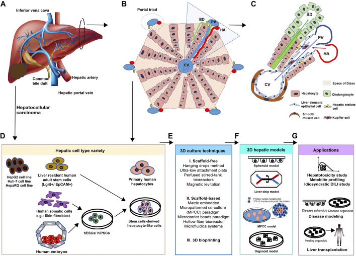
Owing to retained hepatic phenotypes and functions, human three-dimensional (3D) hepatic models established with diverse hepatic cell types are thought to recoup the gaps in drug development and disease modeling limited by a conventional two-dimensional (2D) cell culture system and species-specific variability in drug metabolizing enzymes and transporters. Primary human hepatocytes, human hepatic cancer cell lines, and human stem cell-derived hepatocyte-like cells are three main hepatic cell types used in current models and exhibit divergent hepatic phenotypes. Primary human hepatocytes derived from healthy hepatic parenchyma resemble in vivo-like genetic and metabolic profiling. Human hepatic cancer cell lines are unlimitedly reproducible and tumorigenic. Stem cell-derived hepatocyte-like cells derived from patients are promising to retain the donor's genetic background. It has been suggested in some studies that unique properties of cell types endue them with benefits in different research fields of in vitro 3D modeling paradigm. For instance, the primary human hepatocyte was thought to be the gold standard for hepatotoxicity study, and stem cell-derived hepatocyte-like cells have taken a main role in personalized medicine and regenerative medicine. However, the comprehensive review focuses on the hepatic cell type variety, and corresponding applications in 3D models are sparse. Therefore, this review summarizes the characteristics of different cell types and discusses opportunities of different cell types in drug development, liver disease modeling, and liver transplantation.
Keywords: drug development; hepatic cell types; hepatocyte transplantation; in vitro 3D model; liver disease.
Copyright © 2021 Xu.
Conflict of interest statement
The author declares that the research was conducted in the absence of any commercial or financial relationships that could be construed as a potential conflict of interest.
Figures
Similar articles
-
Availability, Functionality, and Safety as well as Quality Control of Hepatocytes as Seeding Cells in Liver Regenerative Medicine: State of the Art and Challenges.Curr Stem Cell Res Ther. 2023;18(8):1090-1105. doi: 10.2174/1574888X18666230125113254. Curr Stem Cell Res Ther. 2023. PMID: 36698230 Review.
-
Generation of expandable human pluripotent stem cell-derived hepatocyte-like liver organoids.J Hepatol. 2019 Nov;71(5):970-985. doi: 10.1016/j.jhep.2019.06.030. Epub 2019 Jul 9. J Hepatol. 2019. PMID: 31299272
-
Enhanced Metabolizing Activity of Human ES Cell-Derived Hepatocytes Using a 3D Culture System with Repeated Exposures to Xenobiotics.Toxicol Sci. 2015 Sep;147(1):190-206. doi: 10.1093/toxsci/kfv121. Epub 2015 Jun 17. Toxicol Sci. 2015. PMID: 26089346
-
Three-Dimensional Liver Culture Systems to Maintain Primary Hepatic Properties for Toxicological Analysis In Vitro.Int J Mol Sci. 2021 Sep 23;22(19):10214. doi: 10.3390/ijms221910214. Int J Mol Sci. 2021. PMID: 34638555 Free PMC article. Review.
-
Hepatic esterase activity is increased in hepatocyte-like cells derived from human embryonic stem cells using a 3D culture system.Biotechnol Lett. 2018 May;40(5):755-763. doi: 10.1007/s10529-018-2528-1. Epub 2018 Feb 20. Biotechnol Lett. 2018. PMID: 29464570
Cited by
-
Human Multi-Lineage Liver Organoid Model Reveals Impairment of CYP3A4 Expression upon Repeated Exposure to Graphene Oxide.Cells. 2024 Sep 13;13(18):1542. doi: 10.3390/cells13181542. Cells. 2024. PMID: 39329726 Free PMC article.
-
Scaffold-mediated liver regeneration: A comprehensive exploration of current advances.J Tissue Eng. 2024 Oct 13;15:20417314241286092. doi: 10.1177/20417314241286092. eCollection 2024 Jan-Dec. J Tissue Eng. 2024. PMID: 39411269 Free PMC article. Review.
-
Drug Metabolism of Hepatocyte-like Organoids and Their Applicability in In Vitro Toxicity Testing.Molecules. 2023 Jan 7;28(2):621. doi: 10.3390/molecules28020621. Molecules. 2023. PMID: 36677681 Free PMC article.
-
Macrophages clear out necrotic liver lesions: a new magic trick revealed.eGastroenterology. 2023 Sep 22;1(2):e100024. doi: 10.1136/egastro-2023-100024. eCollection 2023 Sep. eGastroenterology. 2023. PMID: 39944003 Free PMC article. No abstract available.
-
A DIY Bioreactor for in Situ Metabolic Tracking in 3D Cell Models via Hyperpolarized 13C NMR Spectroscopy.Anal Chem. 2025 Jan 28;97(3):1594-1602. doi: 10.1021/acs.analchem.4c04183. Epub 2025 Jan 15. Anal Chem. 2025. PMID: 39813686 Free PMC article.
References
Publication types
LinkOut - more resources
Full Text Sources
Other Literature Sources