Review
. 2021 Sep 24:9:741364.
doi: 10.3389/fbioe.2021.741364. eCollection 2021.

Recent Advances and Perspectives on Expanding the Chemical Diversity of Lasso Peptides

Affiliations
Review

Recent Advances and Perspectives on Expanding the Chemical Diversity of Lasso Peptides

Mengjiao Wang et al. Front Bioeng Biotechnol. .

Abstract

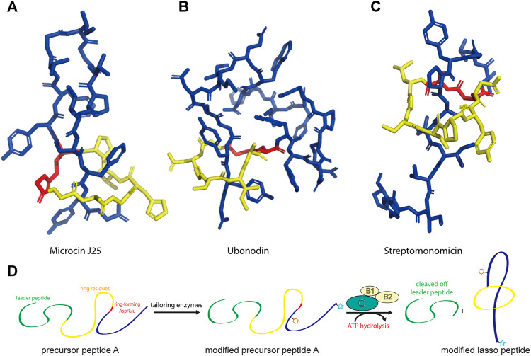
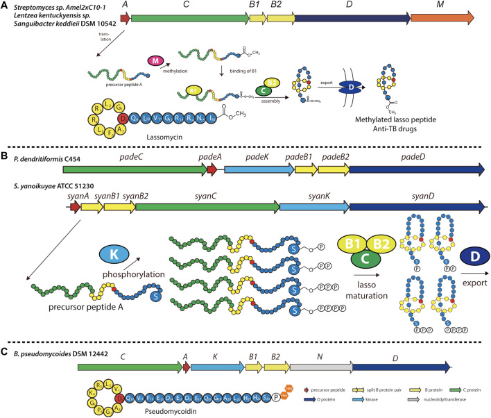
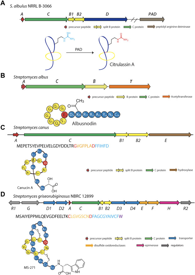
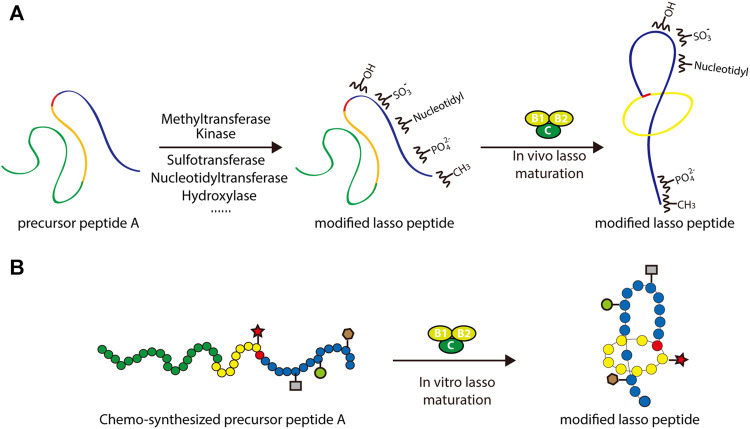
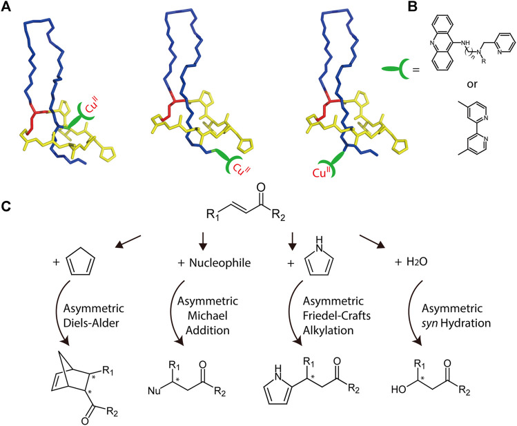
Ribosomally synthesized and post-translationally modified peptides (RiPPs) are a growing family of natural products that exhibit a range of structures and bioactivities. Initially assembled from the twenty proteinogenic amino acids in a ribosome-dependent manner, RiPPs assume their peculiar bioactive structures through various post-translational modifications. The essential modifications representative of each subfamily of RiPP are performed on a precursor peptide by the so-called processing enzymes; however, various tailoring enzymes can also embellish the precursor peptide or processed peptide with additional functional groups. Lasso peptides are an interesting subfamily of RiPPs characterized by their unique lariat knot-like structure, wherein the C-terminal tail is inserted through a macrolactam ring fused by an isopeptide bond between the N-terminal amino group and an acidic side chain. Until recently, relatively few lasso peptides were found to be tailored with extra functional groups. Nevertheless, the development of new routes to diversify lasso peptides and thus introduce novel or enhanced biological, medicinally relevant, or catalytic properties is appealing. In this review, we highlight several strategies through which lasso peptides have been successfully modified and provide a brief overview of the latest findings on the tailoring of these peptides. We also propose future directions for lasso peptide tailoring as well as potential applications for these peptides in hybrid catalyst design.

Keywords: biosynthesis; lasso peptide; natural products; post-translational modification; synthetic biology; tailoring enzymes.


Conflict of interest statement

The authors declare that the research was conducted in the absence of any commercial or financial relationships that could be construed as a potential conflict of interest.

Figures

FIGURE 1
FIGURE 1
Representative structures of lasso peptides: (A) microcin J25 (PDB code 1Q71) (Rosengren et al., 2003), (B) ubonodin (PDB code 6POR) (Cheung-Lee et al., 2020), and (C) streptomonomicin (PDB code 2MW3) (Metelev et al., 2015). The ring is highlighted in yellow, the tail in blue, and the ring-forming Asp/Glu in red. (D) Schematic of the suggested pathway for modified lasso peptide biosynthesis.
FIGURE 2
FIGURE 2
(A) Left, NMR structure of MccJ25 based on PDB code 1Q71 (Rosengren et al., 2003). Right, model of a representative MccJ25 variant showing improved inhibitory activity. Residues targeted for mutation are highlighted. (B) Epitope grafting of MccJ25. From left to right: NMR structures of wild-type MccJ25 (PDB: 1Q71), MccJ25 RGD (PDB code 2MMW) (Knappe et al., 2011), and MccJ25 RGDF (PDB code 2MMT) (Knappe et al., 2011). The surfaces of the four residues at positions 12–15 are depicted in different colors. (C) Peptide drugs containing potential epitopes (highlighted) for lasso peptide engineering. Unless indicated otherwise, lasso peptides are colored as in Figure 1.
FIGURE 3
FIGURE 3
(A) Astexin-1 is displayed on superfolder GFP (sfGFP). The (GSSG)5-Thb-sfGFP part of the protein (Thb = thrombin cleavage site) was modeled in I-TASSER (Yang and Zhang, 2015) and then linked to the structure of astexin-1 (PDB code 2N68) (Zong et al., 2016). (B) Fusion of astexin-1 to the artificial leucine zipper protein A1. Astexin-1 is directly connected to the artificial leucine zipper A1 protein (PDB:4U5T_A). (C) Model for functionalization of protein nanoreactors with lasso peptides (PDB:4PT2). Lasso peptides are colored as in Figure 1.
FIGURE 4
FIGURE 4
(A) Incorporation of ncAAs into MccJ25. The structure of MccJ25 (PDB code 1Q71) (Rosengren et al., 2003) is shown, with positions of incorporation highlighted in green, and the structures of the ncAAs are shown at right. (B) Incorporation of ncAAs into capistruin. The structure of capistruin (PDB code 5OQZ) (Jeanne Dit Fouque et al., 2018) is shown, with positions of ncAA incorporation highlighted in green, and the structures of the ncAAs are shown at right. Met = methionine; Aha = azidohomoalanine; Hpg = homopropargylglycine; Nbk = Nε-5-norbornene-2-yloxycarbonyl-l-lysine; Alk = Nε-Alloc-l-lysine; Bok = Nε-Boc-l-lysine; Ack = Nε-2-azidoethyloxycarbonyl-l-lysine; Pck = Nε-2-propyn-1-yloxycarbonyl-l-lysine. Unless indicated otherwise, lasso peptides are colored as in Figure 1.
FIGURE 5
FIGURE 5
Examples of methylated, phosphorylated, and phosphoglycosylated lasso peptides. (A) Biosynthetic gene cluster and pathway for O-methylated lasso peptides, including lassomycin (primary structure schematic shown below). The precursor peptide is modified at its C-terminal carboxyl group by an O-methyltransferase (encoded by gene M) prior to lasso folding. (B) Biosynthetic gene cluster and pathway for phosphorylated lasso peptides in Firmicutes and Proteobacteria, including paeninodin (from P. dendritiformis C454) and a putative lasso peptide (from S. yanoikuyae ATCC 51230). The precursor peptide can be phosphorylated multiple times at the hydroxy side chain of its C-terminal Ser by an Hpr kinase homolog (encoded by gene K) prior to lasso folding. A circled “P” represents a phosphate group (Zhu et al., 2016b). (C) Biosynthetic gene cluster for the phosphoglycosylated lasso peptide pseudomycoidin (primary structure schematic shown below). A kinase (encoded by gene K) phosphorylates the hydroxyl side chain of the C-terminal Ser, and a nucleotidyltransferase (encoded by gene N) transfers one or two glycosyl groups to that phosphate group. Whether these enzymes act on the precursor peptide or folded lasso peptide is unknown. Lasso peptides are colored as in Figure 1.
FIGURE 6
FIGURE 6
Examples of deiminated, acetylated, hydroxylated, and d-amino acid-containing peptides. (A) Biosynthetic gene cluster and pathway for the deiminated lasso peptide citrulassin A. A peptidyl arginine deiminase (PAD) encoded outside the gene cluster is responsible for the citrulline modification. Whether this enzyme acts on the precursor peptide or folded lasso peptide is unknown. (B) Biosynthetic gene cluster for the acetylated lasso peptide albusnodin (primary structure schematic shown below). An acetyltransferase (encoded by gene T) is responsible for acetylating the primary amine of the side chain of Lys10 prior to lasso folding. (C) Biosynthetic gene cluster for the hydroxylated lasso peptide canucin A. An iron/2-oxoglutarate-dependent hydroxylase (encoded by gene E) introduces a hydroxyl group on the β-carbon of the C-terminal Asp residue prior to lasso folding. (D) Biosynthetic gene cluster for the d-amino acid-containing MS-271 lasso peptide. An epimerase (encoded by the H gene) is responsible for inversion of Cα stereochemistry at the C-terminus prior to lasso folding. Lasso peptides are colored as in Figure 1.
FIGURE 7
FIGURE 7
Two approaches to the rational engineering of lasso peptide analogs. (A) Co-transformation approach using various tailoring enzymes to introduce new structural features into the lasso peptide scaffold. (B) Scheme of chemoenzymatic synthesis of lasso peptide analogs. In both panels, lasso peptides are colored as in Figure 1.
FIGURE 8
FIGURE 8
(A) Schematic representation of potential MccJ25-based asymmetric catalysis using anchoring strategies. The anchoring site should be subjected to optimization. Lasso peptides are colored as in Figure 1. (B) Two potential ligands that may be used for transition metal binding. (C) Potential asymmetric reactions that may be facilitated by the hybrid catalyst.

Similar articles

Cited by

References

    1. Al Musaimi O., Al Shaer D., Albericio F., de la Torre B. (2021). 2020 FDA TIDES (Peptides and Oligonucleotides) Harvest. Pharmaceuticals 14 (2), 145. 10.3390/ph14020145 - DOI - PMC - PubMed
    1. Al Toma R. S., Kuthning A., Exner M. P., Denisiuk A., Ziegler J., Budisa N., et al. (2015). Site-directed and Global Incorporation of Orthogonal and Isostructural Noncanonical Amino Acids into the Ribosomal Lasso Peptide Capistruin. Chembiochem 16 (3), 503–509. 10.1002/cbic.201402558 - DOI - PubMed
    1. Allen C. D., Link A. J. (2016). Self-Assembly of Catenanes from Lasso Peptides. J. Am. Chem. Soc. 138 (43), 14214–14217. 10.1021/jacs.6b09454 - DOI - PMC - PubMed
    1. Arnison P. G., Bibb M. J., Bierbaum G., Bowers A. A., Bugni T. S., Bulaj G., et al. (2013). Ribosomally Synthesized and post-translationally Modified Peptide Natural Products: Overview and Recommendations for a Universal Nomenclature. Nat. Prod. Rep. 30 (1), 108–160. 10.1039/C2NP20085F - DOI - PMC - PubMed
    1. Assrir N., Pavelkova A., Dazzoni R., Ducasse R., Morellet N., Guittet E., et al. (2016). Initial Molecular Recognition Steps of McjA Precursor during Microcin J25 Lasso Peptide Maturation. Chembiochem 17 (19), 1851–1858. 10.1002/cbic.201600306 - DOI - PubMed

LinkOut - more resources