. 2021 Sep 24:9:719351.
doi: 10.3389/fcell.2021.719351. eCollection 2021.

Amlexanox and Forskolin Prevents Isoproterenol-Induced Cardiomyopathy by Subduing Cardiomyocyte Hypertrophy and Maladaptive Inflammatory Responses

Affiliations

Amlexanox and Forskolin Prevents Isoproterenol-Induced Cardiomyopathy by Subduing Cardiomyocyte Hypertrophy and Maladaptive Inflammatory Responses

Gabriel Komla Adzika et al. Front Cell Dev Biol. .

Abstract

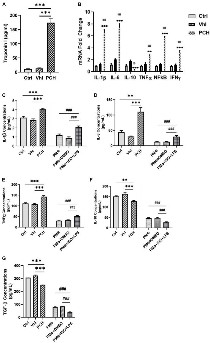
Chronic catecholamine stress (CCS) induces the occurrence of cardiomyopathy-pathological cardiac hypertrophy (PCH), which is characterized by left ventricular systolic dysfunction (LVSD). Recently, mounting evidence has implicated myocardial inflammation in the exacerbation of pathological cardiac remodeling. However, there are currently no well-defined treatment interventions or regimes targeted at both the attenuation of maladaptive myocardial hypertrophy and inflammation during CCS to prevent PCH. G protein-coupled receptor kinase 5 (GRK5) and adenylyl cyclases (ACs)-cAMP mediates both cardiac and inflammatory responses. Also, GRK5 and ACs are implicated in stress-induced LVSD. Herein, we aimed at preventing PCH during CCS via modulating adaptive cardiac and inflammatory responses by inhibiting GRK5 and/or stimulating ACs. Isoproterenol-induced cardiomyopathy (ICM) was modeled using 0.5 mg/100 g/day isoproterenol injections for 40 days. Alterations in cardiac and inflammatory responses were assessed from the myocardia. Similarities in the immunogenicity of cardiac troponin I (cTnI) and lipopolysaccharide under CCS were assessed, and Amlexanox (35 μM/ml) and/or Forskolin (10 μM/ml) were then employed in vitro to modulate adaptive inflammatory responses by inhibiting GRK5 or activating ACs-cAMP, respectively. Subsequently, Amlexanox (2.5 mg/100 g/day) and/or Forskolin (0.5 mg/100 g/day) were then translated into in vivo during CCS to modulate adaptive cardiac and inflammatory responses. The effects of Amlexanox and Forskolin on regulating myocardial systolic functions and inflammatory responses during CCS were ascertained afterward. PCH mice had excessive myocardial hypertrophy, fibrosis, and aggravated LVSD, which were accompanied by massive CD68+ inflammatory cell infiltrations. In vitro, Forskolin-AC/cAMP was effective than Amlexanox-GRK5 at downregulating proinflammatory responses during stress; nonetheless, Amlexanox and Forskolin combination demonstrated the most efficacy in modulating adaptive inflammatory responses. Individually, the translated Amlexanox and Forskolin treatment interventions were ineffective at subduing the pathological remodeling and sustaining cardiac function during CCS. However, their combination was potent at preventing LVSD during CCS by attenuating maladaptive myocardial hypertrophy, fibrosis, and inflammatory responses. The treatment intervention attained its potency mainly via Forskolin-ACs/cAMP-mediated modulation of cardiac and inflammatory responses, coupled with Amlexanox inhibition of GRK5 mediated maladaptive cascades. Taken together, our findings highlight the Amlexanox and Forskolin combination as a potential therapeutic intervention for preventing the occurrence of pathological cardiac hypertrophy during chronic stress.

Keywords: GRK5; adenylyl cyclase; amlexanox; cAMP; chronic catecholamine stress; forskolin; inflammation; isoproterenol-induced cardiomyopathy.


Conflict of interest statement

The authors declare that the research was conducted in the absence of any commercial or financial relationships that could be construed as a potential conflict of interest.

Figures

FIGURE 1
FIGURE 1
Troponin from necrotic cardiomyocytes and lipopolysaccharides (LPS) elicits similar inflammatory responses during chronic catecholamine stress (CCS). (A) Sera concentrations of troponin I were evaluated by ELISA from Control (Ctrl), Vehicle (Vhl), and Pathological cardiac hypertrophy (PCH) groups. ∗∗∗p < 0.001 (B) mRNA expressions of inflammatory markers (IL-1β, IL-6, TNFα, IFNγ, and NF-κB) assessed from the myocardial by qRT-PCR (n = 6 hearts per treatment group). ∗∗p < 0.01, ∗∗∗p < 0.001 vs. Vhl; $$p < 0.01, $$$p < 0.001 vs. Ctrl. (C–G) Comparison with inflammatory cytokines secretion (IL-1β, IL-6, TNFα, IL-10, and TGF-β) between PCH mice (in vivo) and LPS-challenged peritoneal macrophages (PMϕ) (in vitro) during CCS. All ELISA evaluations were performed in triplicates (n = 8 mice per treatment group). ∗∗p < 0.01, ∗∗∗p < 0.001 among in vivo groups; ###p < 0.001 among in vitro groups. Data are expressed as mean ± SEM. Data were analyzed using one-way ANOVA, followed by Tukey’s post hoc analysis. Abbreviations: IL, interleukin; TNFa, tumor necrosis factor-alpha; TGF-β, transforming growth factor-beta; IFNy, interferon-gamma.
FIGURE 2
FIGURE 2
Amlexanox (ALX) and forskolin (FSK) combination inhibits PMϕ proinflammatory responses elicited during CCS, mostly via cyclic adenosine monophosphate (cAMP)-mediated immunoregulation. (A-E) ELISA evaluation of inflammatory cytokines (IL-1β, IL-6, TNFα, IL-10, and TGF-β) secreted by LPS-challenged PMϕ after employing ALX and/or FSK treatment intervention during CCS. (F) cAMP concentrations were assessed to determine the impact of the treatment interventions on ensuring cAMP bioavailability during CCS. The therapeutic groups are ALX treatment, FSK treatment, and ALX and FSK combination treatment. All ELISA evaluations were performed in triplicates (n = 8 mice per treatment group). p < 0.05, ∗∗p < 0.01, ∗∗∗p < 0.001 among the therapeutic groups; ##p < 0.01, ###p < 0.001 LPS + PMϕ + ISO vs. therapeutic groups; &&p < 0.01, &&&p < 0.001 vs. LPS + PMϕ + Vhl. (G) Representative immunofluorescence of G protein-coupled receptor kinase 5 (GRK5) localizations, nuclei (DAPI), and cytoplasmic membrane (CTxB). (H) The plotted values are the GRK5 (nuclear/cytoplasm) expression ratios assessed from each PMϕ (n = 12–15 cells per four mice per treatment group). Color channels were adjusted in the merged images to enhance the visualization of all the respective fluorescence dyes. ∗∗∗p < 0.001 among the therapeutic groups; ##p < 0.01, ###p < 0.001, LPS + PMϕ + ISO vs. therapeutic groups; &&&p < 0.001 vs. LPS + PMϕ + ISO. Data are expressed as mean ± SEM. Data were analyzed using one-way ANOVA, followed by Tukey’s post hoc analysis.
FIGURE 3
FIGURE 3
ALX and FSK combination attenuates left ventricular systolic dysfunction (LVSD) in mice during CCS by normalizing protein expressions. (A) Representative short-axis view M-mode echocardiogram imaging from Ctrl, Vhl, isoproterenol (ISO), ISO + ALX, ISO + FSK, and ISO + ALX + FSK. (BD) Graphical presentations of heart rates, ejection fraction, and fractional shortening assessments from all models. The Vhl mice group is representative of the Ctrl mice group in the graphical presentations due to similarity in their data. The therapeutic groups are ALX treatment, FSK treatment, and ALX and FSK combination treatment (n = 10–11 hearts per treatment group). (EN) Representative Western blots and graphical presentation of βARs, ACs, GRKs, ANP, and BNP, were assessed from all groups (n = 4 hearts per treatment group). Western blots were performed in triplicates, and each protein band in the representative blot is an independent biological sample. (O) Graphical represents of cAMP concentrations evaluated by ELISA (n = 9 mice per treatment group). p < 0.05, ∗∗p < 0.01, ∗∗∗p < 0.001 among the therapeutic groups; &&p < 0.01, &&&p < 0.001 vs. Vhl; #p < 0.05, ##p < 0.01, ###p < 0.001 vs. the therapeutic groups. Data are expressed as mean ± SEM. Data were analyzed using one-way ANOVA, followed by Tukey’s post hoc analysis. Abbreviations: βARs, β-adrenergic receptors; ACs, adenylyl cyclases; GRKs, G-protein-coupled receptor kinases; ANP, atrial natriuretic peptide; BNP, brain natriuretic peptide.
FIGURE 4
FIGURE 4
ALX and FSK combination preserves mice myocardial architecture during CCS. (A) Representative whole hearts from all experimental groups. (B) Representative Masson’s trichrome stained longitudinal section of whole hearts from all groups. (C) Representative Masson’s trichrome stained transverse section of whole hearts from all groups. (D–G) Representative Western blots and graphical presentations cleaved caspase 3, collagen I, and collagen III compared between Vhl and PCH mice, and ALX treatment, FSK treatment, and ALX and FSK combination treatment (n = 4 hearts per treatment group). Western blots were performed in triplicates, and each protein band in the representative blot is an independent biological sample. (H,I) Representative microscopic images of Masson’s trichrome staining and collagen volume fraction (CVF) in the ventricular tissue sections from all groups. The plotted values are the means of the CVF from each mouse (n = 6–8 fields of view per 5–7 sections per 8–16 hearts per group). (J,K) Representative microscopic images of wheat germ agglutinin (WGA) merged with DAPI staining and graphical presentation of measured cardiomyocyte diameters from ventricular tissue sectionings across all groups. The plotted values are the means of the cardiomyocyte sizes from each mouse (n = 10–12 cells per five fields of view per five sections per six to seven hearts per group). Representative cardiomyocytes are marked in yellow boxes, and their zoomed-in (× 5) inserts to show hypertrophy are marked in red boxes. The Vhl mice group is representative of the Ctrl mice group in results illustrations due to similarity in their data. p < 0.05, ∗∗p < 0.01, ∗∗∗p < 0.001 among the therapeutic groups; &&&p < 0.001 vs. Vhl; #p < 0.05, ##p < 0.01, ###p < 0.001 vs. the therapeutic groups. CVF was estimated by dividing the collagen area with the total myocardial area and multiple by 100. Data are expressed as mean ± SEM. Data were analyzed using one-way ANOVA, followed by Tukey’s post hoc analysis.
FIGURE 5
FIGURE 5
ALX and FSK combination prevents proinflammatory responses aggravation in myocardia during CCS by preventing necrosis. (A) Graphical presentations of evaluated sera concentrations of troponin I from groups. The sera troponin I analysis was performed in triplicates (n = 8 mice per treatment group). p < 0.05, ∗∗∗p < 0.001, among the therapeutic groups; &&&p < 0.001 vs. ISO (PCH); ##p < 0.01, ###p < 0.001, vs. the therapeutic groups. (B,C) Representative CD68 IHC staining and graphical presentation of the extent of inflammatory cells infiltration into the myocardial of all groups. The Vhl mice group is representative of the Ctrl mice group in results illustrations due to similarity in their data (n = 6–8 field of view per six to eight sections per six to seven hearts per group). (D–H) Graphical presentations of inflammatory cytokines (IL-1β, IL-6, TNFα, IL-10, and TGF-β) were evaluated by ELISA from all groups. The cytokines analysis was performed in triplicates (n = 7–8 mice per treatment group). p < 0.05, ∗∗p < 0.01, ∗∗∗p < 0.001, among the therapeutic groups; &&&p < 0.001 vs. Vhl; #p < 0.05, ##p < 0.01, ###p < 0.001 vs. the therapeutic groups. Data are expressed as mean ± SEM. Data were analyzed using one-way ANOVA, followed by Tukey’s post hoc analysis.
FIGURE 6
FIGURE 6
Schematics of the pathomechanism of isoproterenol-induced cardiomyopathy and the mechanisms of ALX and FSK combination treatment. (A) During CCS, Gαs-AC is decoupled from βARs, thereby inhibiting cAMP synthesis, which affects cardiac function and immunoregulation in the myocardia adversely. Also, GRK5 upregulates and phosphorylates βARs as well as translocations into the nuclei to induce GPCRs-independent stimuli signaling. The nuclear activity of GRK5 induces the activation of cardiomyocyte hypertrophy transcriptional factors. These result in excessive cardiomyocyte hypertrophy and hyperactive proinflammatory responses. At the same time, cAMP-mediated adaptive immunoregulation is abolished. The synergy of these cascades results in the exacerbation of collagen deposits which pathologically remodels the heart. (B) The administration of ALX and FSK combination facilitates ALX-GRK5 inhibition (expressions and nuclear translocation), which prevents cardiomyocyte hypertrophy; while, FSK-ACs-cAMP modulates adaptive immunoregulation and cardiac inotropic functions. Combining these mechanisms preserves cardiac morphology and prevents left ventricular systolic dysfunction (LVSD) during CCS.

Similar articles

Cited by

References

    1. Abdulkhaleq L. A., Assi M. A., Abdullah R., Zamri-Saad M., Taufiq-Yap Y. H., Hezmee M. N. M. (2018). The crucial roles of inflammatory mediators in inflammation: a review. Vet. World 11 627–635. 10.14202/vetworld.2018.627-635 - DOI - PMC - PubMed
    1. Adzika G. K., Machuki J. O., Shang W., Hou H., Ma T., Wu L., et al. (2019). Pathological cardiac hypertrophy: the synergy of adenylyl cyclases inhibition in cardiac and immune cells during chronic catecholamine stress. J. Mol. Med. 97 897–907. 10.1007/s00109-019-01790-0 - DOI - PubMed
    1. Bopp T., Dehzad N., Reuter S., Klein M., Ullrich N., Stassen M., et al. (2009). Inhibition of cAMP degradation improves regulatory T cell-mediated suppression. J. Immunol. 182 4017–4024. 10.4049/jimmunol.0803310 - DOI - PubMed
    1. Cai Z. L., Shen B., Yuan Y., Liu C., Xie Q. W., Hu T. T., et al. (2020). The effect of HMGA1 in LPS-induced Myocardial Inflammation. Int. J. Biol. Sci. 16 1798–1810. 10.7150/ijbs.39947 - DOI - PMC - PubMed
    1. Chiadak J. D., Arsenijevic T., Verstrepen K., Gregoire F., Bolaky N., Delforge V., et al. (2016). Forskolin Inhibits Lipopolysaccharide-Induced Modulation of MCP-1 and GPR120 in 3T3-L1 Adipocytes through an Inhibition of NFκB. Mediators Inflamm. 2016:1431789. - PMC - PubMed